대한치과재료학회지 42(4) : 279-288, 2015 ISSN:2384-4434 (Print); 2384-3268 (Online) Available online at http://www.kadm.org http://dx.doi.org/10.14815/kjdm.2015.42.4.279
에탄올 전처리가 미백된 상아질과 복합레진의 접착에 미치는 영향
손가은
1
, 권태엽2
, 김영경1*
경북대학교 치의학전문대학원 치과보존학교실1,
경북대학교 치의학전문대학원 치과생체재료학교실 및 경북대학교 생체재료연구소2
<Abstract>
Influence of Ethanol Pretreatment on the Bonding of Resin Composite to Bleached Dentin
Ga-Eun Son 1 , Tae-Yub Kwon 2 , Young Kyung Kim 1*
Department of Conservative Dentistry, School of Dentistry, Kyungpook National University1,
Department of Dental Biomaterials, School of Dentistry; and Institute for Biomaterials Research & Development;
Kyungpook National University2
본 연구에서는 근관치료된 변색치의 미백치료 후 복합레진 수복 시 미백제에 의한 부작용을 줄이기 위하여 사용되는 에탄올 전처리가 미백된 상아질과 복합레진의 접착에 미치는 영향을 평가하고자 사람의 소구치로 상아질 시편 제작 후, sodium perborate와 증류수의 혼합물을 7일간 적용하여 미백과정을 시행하였다. 미백 후 시편을 세 군으로 나누어 미백 직후, 7일간 증류수에 보관 후, 치면을 75% 에탄올 용액으로 3분간 처리 후 각각 접착술식을 시행하였다. 시편을 37℃에서 24시간 동안 물에 담근 후 미세인장결합강도를 측정하고, 파절면을 주사전자현 미경을 이용하여 관찰하였다. 결합강도에 있어서, 에탄올로 전처리 또는 7일간 증류수에 보관한 경우 미백처리 하지 않은 대조군과 유의차를 보이지 않았으나 미백 직후 접착한 경우에는 대조군보다 유의하게 낮았다. 파절양상은 혼합성 파절이 우세하게 나타났으나 미백 직후 접착한 군에서는 주로 접착성 파절이 나타났다. 파절면의 현미경 관찰 시 에탄올로 처리한 군에서 대조군과 유사한 접착계면의 양상을 보였는데 균일한 두께의 혼성층과 길고 굵은 resin tag이 관찰되었다. 본 연구의 결과 미백 후 에탄올을 이용한 치면처리가 미백된 상아질에 대한 복합레진의 결합강도를 유의하게 증가시키는 것으로 보였다.
Key words
: Bleached Dentin, Bond Strength, Ethanol Pretreatment, Nonvital Bleaching* Correspondence: 김영경
대구광역시 중구 달구벌대로 2177(우 41940) 경북대학교 치의 학전문대학원 치과보존학교실
Tel : +82-53-600-7601 E-mail : [email protected]
Received: Sep 09, 2015; Revised: Sep 25, 2015; Accepted: Sep 25, 2015
Ⅰ. INTRODUCTION
Nonvital tooth bleaching has been considered as an effective and conservative esthetic treatment for endodontically-treated discolored teeth. Since adhesive
restoration of access cavity usually follows bleaching, bonding between tooth and restoration material is important to long-term success of endodontic therapy and bleaching treatment (Howell, 1981). However, application of sodium perborate and/or hydrogen peroxide in nonvital bleaching may interfere with resin infiltration into etched enamel/dentin or inhibit polymerization of resins, leading to compromised bonding to bleached tooth surface (Teixeira et al., 2002; Timpawat et al., 2005).
A considerable reduction in bond strength of resin
composite to tooth immediately after bleaching may occur because of the remnant of peroxide in the collagen matrix and dentinal tubules or change in organic content due to protein denaturation (Nour El-din et al., 2006; Bittencourt et al., 2010). Hence, delayed bonding after bleaching is commonly recommended to avoid problems related to decreased bonding. However, from the clinical standpoint, immediate restoration after nonvital bleaching is preferred in completing the restoration of the access cavity to prevent coronal leakage (Khoroushi et al., 2009).
It has been reported that application of ethanol solution, a drying agent to bleached enamel surface prior to bonding significantly reduced the adverse effect of bleaching on the bond strength of resin composite to enamel through its ability to extract water from the hard tissue (Barghi, 1994).
Three-minute treatment with 70% ethanol seems to be an effective and fast-acting adjunct when the bonding procedure is carried out immediately (Kum et al., 2003; Niat et al., 2012). Notwithstanding the proven efficacy of ethanol pretreatment on composite-enamel bonding, relatively little attention has been paid to the effect of ethanol on the access cavity of intracoronally-bleached tooth in which the most part of bonding surface is dentin.
Bonding to dentin has proved to be challenging because of the complex histological structure and variable composition of dentin itself (Zhang et al., 2014).
Therefore, this in vitro study investigated the effect of ethanol pretreatment following nonvital bleaching on composite bonding to dentin. The null hypothesis tested was that the application of ethanol solution on bleached dentin surface prior to bonding would not affect the microtensile bond strength of resin composite to dentin.
Ⅱ. MATERIALS AND METHODS 1. Tooth selection and specimen preparation
Sixteen human maxillary premolars were collected with patients' informed consents and the study was approved by the Institutional Review Board (IRB) of Kyungpook National University Hospital, Daegu, Korea (document number: BMRI 74005-452).
The teeth were cleaned, disinfected in 0.1% thymol solution for 7 d, and stored in distilled water at 4℃ until use. The occlusal enamel and roots of teeth were removed using a low-speed diamond saw (Isomet, Buehler Ltd., Lake Bluff, IL, USA) under water-cooling to form 5-6 mm thick, parallel-sided crown segments. The exposed tooth surface was further wet-polished with a 600-grit silicon carbide paper to create a flat, midcoronal dentin surface (Spyrides et al., 2000).
The specimens were randomly divided into four groups of four specimens each. All specimens except for the control group (no bleaching) were subjected to a bleaching procedure by applying a mixture of sodium perborate (Junsei Chemical Co., Ltd., Tokyo, Japan) and distilled water in a 1:1 ratio onto the dentin surface for 7 d, capped with the customized tray to prevent loss of the bleaching agent from the tooth surface during the storage in 100%
relative humidity at 37℃ (Elkhatib et al., 2003).
The dentin surfaces of group IB (immediate bonding) were restored immediately after bleaching using the bonding procedure. For group DB (delayed bonding), the teeth were immersed in distilled water for 7 d and subjected to bonding. For group ET (ethanol treatment), 75% ethanol solution was applied onto the bleached dentin surface for 3 min (Table 1).
In the bonding steps, all tooth surfaces were etched with
32% phosphoric acid (Uni-Etch, Bisco Inc., Schaumburg, IL,
USA) for 15 s. After rinsing and drying, adhesive (One-Step,
Bisco Inc.) was applied to the etched surface according to
the manufacturer's instructions and light-cured for 10 s with
a LED light-curing unit (Bluephase, Ivoclar Vivadent Inc.,
Amherst, NY, USA; 1000 mW/cm
2irradiance). Resin
Table 1.
Group codes according to bleaching and surface pretreatment
Group Nonvital bleaching Surface pretreatment
Control None None
IB Sodium perborate + distilled water None (immediate bonding)
DB Sodium perborate + distilled water None (delayed bonding)
ET Sodium perborate + distilled water 3-min application of 75% ethanol solution before composite bonding
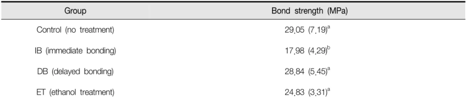
Table 2.
Microtensile bond strengths of resin composite to bleached dentin (
n= 20)
Group Bond strength (MPa)
Control (no treatment) 29.05 (7.19)a
IB (immediate bonding) 17.98 (4.29)b
DB (delayed bonding) 28.84 (5.45)a
ET (ethanol treatment) 24.83 (3.31)a
composite (AELITETM All-purpose body, A2 shade, Bisco Inc.) was built up in three 2-mm increments. Each composite layer was light-cured for 20 s and final cure was done for 40 s. All teeth of four groups were stored in distilled water at 37℃ for 24 h before sectioning.
2. Microtensile bond strength testing and failure mode analysis
Each bonded specimen was longitudinally sectioned into 1 mm-thick slabs with the low-speed diamond saw (Isomet, Buehler Ltd.). Each slab was fixed on a glass platform with sticky wax and serially sectioned into 1 mm
2(± 0.1 mm
2) sticks, in accordance with the ‘non-trimming’ version of the microtensile test (Shono et al., 1999). The exact dimensions of each stick were measured using a digital caliper to calculate the precise cross-sectional area.
For microtensile bond strength testing, five sticks were
sectioned from the center of each bonded specimen (Hiraishi et al., 2009). Thus, the use of four teeth resulted in a total of 20 sticks for each of the groups tested. The bonded composite-dentin sticks were attached to a testing device with cyanoacrylate glue (Zap-It, DVA, Corona, CA, USA). The device was attached to a microtensile bond tester (Bisco Inc.) and loaded in tension at a crosshead speed of 1 mm/min until failure. Premature failures were not considered in the data analysis (Briso et al., 2014).
After fracturing, all specimens were examined under the
stereomicroscope (SZ4045, Olympus, Tokyo, Japan) at a
magnification of 25× and failure modes were classified into
one of following modes: (a) adhesive failure between
dentin and the adhesive resin layer; (b) cohesive failure
in dentin; (c) cohesive failure in resin composite; (d) mixed
failure involving the combinations of cohesive failures in
adhesive, composite or dentin. Each type of failure mode
was expressed as a percentage of the total number of
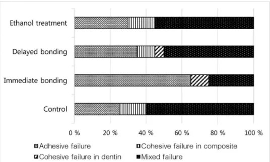
Figure 1. Distribution of failure pattern (%) in the test groups
specimens in that group.
3. Examination of the fractured surfaces using scanning electron microscopy (SEM)
To observe the hybrid layer of bonded specimens, representative fractured sticks (dentin sides, n = 2) were selected from each group with tensile bond strengths that were close to the mean bond strength of that group.
Bonded interfaces were polished using No. 400-, No. 600-, and No. 1,200-grades of silicon carbide papers (3M, St.
Paul, MN, USA). After sonication in distilled water for 5 min, specimens were demineralized in 1N HCl for 5 min, deproteinized in 5.25% NaOCl for 5 min. and then sequentially dehydrated in ascending grades of ethanol (25%, 50%, 75%, and 95% for 20 min each, and 100% for 60 min) (Ferreira et al., 2011). After a 24 h air drying, the specimens were sputter-coated with gold and examined under a field emission-scanning electron microscope (FE-SEM, SU8220, Hitachi, Tokyo, Japan).
4. Statistical analysis
The normally distributed (as determined by the Kolmogorov-Smirnov test) bond strength was analyzed with one-way analysis of variance (ANOVA) followed by multiple comparisons with Tukey’s test at the 0.05 significance level. The statistical analysis was performed using SPSS 22.0 for Windows (SPSS Inc., Chicago, IL, USA).
Ⅲ. RESULTS
Table 2 summarizes microtensile bond strengths of composite resin to bleached dentin surface treated with different regimens prior to bonding. One-way ANOVA showed statistically significant differences among the test groups (p < 0.001). Group IB showed a significantly lower microtensile bond strength than the control group (p <
0.001) and the groups DB and ET statistically similar values to the control group (p > 0.05).
Figure 1 shows the failure pattern distribution (%) as
assigned using the light microscope. Mixed failure mode
was predominant in all groups except for in the IB group
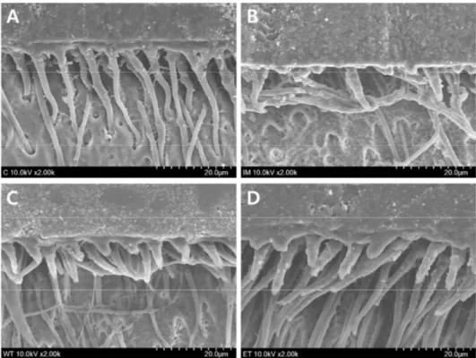
Figure 2. Representative SEM micrographs showing hybrid layer and resin penetration in the fractured dentin sides. (A) non-bleached control; (B) immediate bonding; (C) delayed bonding; (D) ethanol treatment (2000×).