• 검색 결과가 없습니다.

Acute Malocclusion Caused by Articular Disc Perforation: A Case Report JOMP

N/A
N/A
Protected

Academic year: 2021

Share "Acute Malocclusion Caused by Articular Disc Perforation: A Case Report JOMP"

Copied!
5
0
0

로드 중.... (전체 텍스트 보기)

전체 글

(1)

pISSN 2288-9272 eISSN 2383-8493 J Oral Med Pain 2018;43(4):142-146 https://doi.org/10.14476/jomp.2018.43.4.142

Acute Malocclusion Caused by Articular Disc Perforation:

A Case Report

Hyun Nam 1 , Young-Joo Shim 1,2 , Jin-Kyu Kang 1,2

1 Department of Orofacial Pain and Oral Medicine, Wonkwang University Daejeon Dental Hospital, Daejeon, Korea

2 Wonkwang Dental Research Institute, Wonkwang University, Iksan, Korea

Received November 9, 2018 Revised December 7, 2018 Accepted December 12, 2018

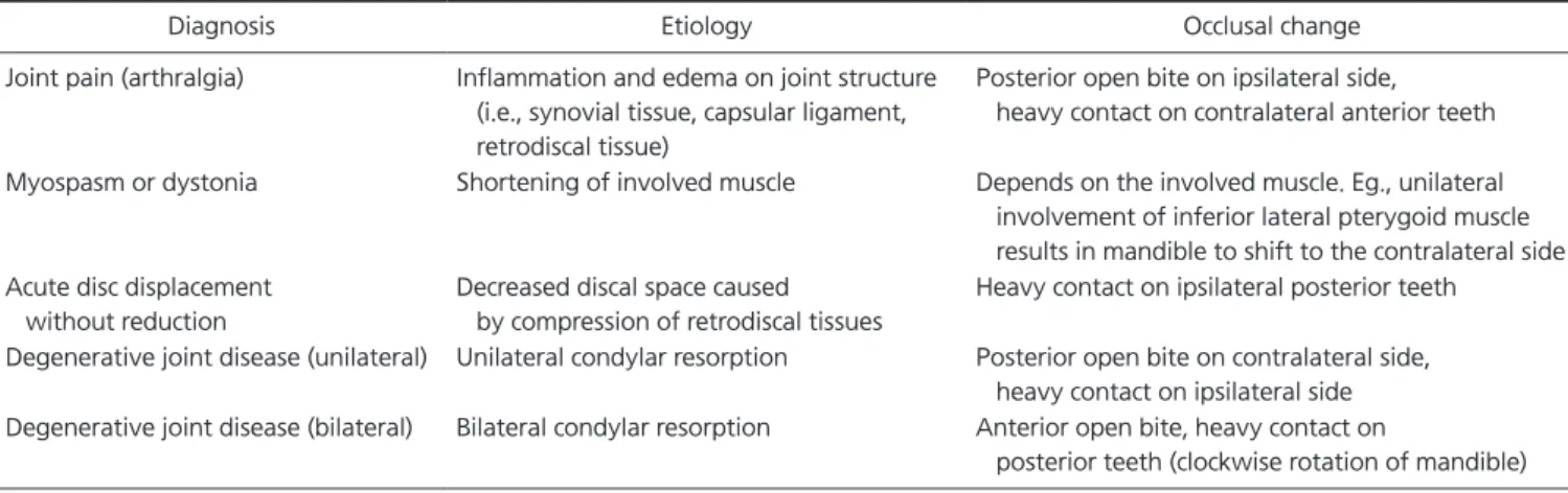
Patients with temporomandibular disorder often present with acute occlusal change and prop- erly managed with conservative treatment. If such change is caused by unusual etiology, dif- ferential diagnosis may be challenged. This article describes the diagnosis of a patient exhibit- ing pain and acute posterior open bite on the ipsilateral side after chewing hard food. After initial conservative treatment failed to resolve the complaint, magnetic resonance imaging was ordered and confirmed partial perforation of articular disc. Disc perforation itself is usually chronic in nature, but sudden macrotrauma may also cause the disorder. However, occlusal discrepancy caused by disc perforation is rare and seldom reported. We present a case of acute malocclusion caused by disc perforation with a review of related literature.

Key Words: Disc perforation; Malocclusion; Posterior disc displacement; Temporomandibular joint

Correspondence to:

Jin-Kyu Kang

Department of Orofacial Pain and Oral Medicine, Wonkwang University Daejeon Dental Hospital, 77 Dunsan- ro, Seo-gu, Daejeon 35233, Korea Tel: +82-42-366-1125 Fax: +82-42-366-1115 E-mail: [email protected] This paper was supported by Wonkwang University in 2018.

JOMP Journal of Oral Medicine and Pain

Copyright Ⓒ 2018 Korean Academy of Orofacial Pain and Oral Medicine. All rights reserved.

CC

This is an open-access article distributed under the terms of the Creative Commons Attribution Non-Commercial License (http://creativecommons.org/licenses/by-nc/4.0/), which permits unrestricted non-commercial use, distribution, and reproduction in any medium, provided the original work is properly cited.

INTRODUCTION

Acute malocclusion is any sudden change of the occlusal condition caused by a disorder. 1) It may be caused by either joint or muscle disorders and is dependent on the respon- sible structure. 2) This change can be a momentary or pro- longed condition. Common disorders by which acute oc- clusal change can be made include joint pain (arthralgia) and myospasm. 3) Each disorder has characteristic patterns of occlusal change. Thus the clinical presentation of acute malocclusion often gives a clue to the diagnosis. 2) In most cases acute malocclusion can be managed with conserva- tive treatment, e.g., physical therapy, pharmacologic thera- py and occlusal appliance, etc. 1)

Perforation of articular disc of temporomandibular joint (TMJ) usually occurs with existing disc disorder (i.e., disc derangement) and/or late stage of degenerative joint

disease. 4,5) It may be associated with a sudden macrotrauma

but usually chronic in nature. 1) Its prevalence is higher in

female and elderly (over 80 years old) with an incidence of

5%-15%. 4,6) Anterior disc displacement is the most common

findings associated with disc perforation. 7) However, on rare

occasion, the perforation may accompany with posterior

disc displacement. 8,9) Clinical symptoms of posterior disc

displacement include pain, clicking, crepitation and poste-

rior open bite on the ipsilateral side. 4,8) Occlusal discrepancy

caused by disc perforation and posterior disc displacement

is rare and seldom reported. We hereby present a rare case

of acute malocclusion caused by perforation of the articular

disc with no previous history of temporomandibular disor-

der (TMD).

(2)

CASE REPORT

A 75-year-old female with noncontributory past medi- cal history presented to the Department of Orofacial Pain and Oral Medicine (Wonkwang University Dental Hospital, Daejeon, Korea), complaining of pain on her left jaw during mastication and mouth opening and reported that her left bite is ‘off’. All of her symptoms had appeared three weeks ago, after chewing hard food. She heard ‘crushing’ noise on her left jaw during mastication, and the symptoms had been developed. She did not have any history of joint noise, pain and dysfunction. Before visiting the hospital, she had vis- ited general dentist and was prescribed with non-steroidal anti-inflammatory drug (NSAID) and muscle relaxant for two weeks. The medicine seemed to have relieved the pain and occlusal discomfort, but her symptoms were relapsed after discontinuation.

On the initial clinical examination, her maximum unas- sisted opening was 45 mm. Pain on the left preauricular

region and click sound was observed during the mouth opening. Her left and right TMJs and masseter muscles showed tenderness to palpation. No dental or periodontal abnormalities were found. Panoramic (Fig. 1A) and TMJ panoramic radiograph (Fig. 1B) revealed no significant findings. Occlusal contacts were initially examined by 8 μ m thickness shim stock (Coltene/Whaledent GmbH, Langenau, Germany). On her right dentition, all teeth except her sec- ond molar were in contact with antagonists. However, only central incisor was in contact on her left dentition, reveal- ing posterior open bite on her left side. Further occlusal as- sessment using 42 μ m thickness occlusal marking film (two sheets of AccuFilm II; Parkell Inc., Edgewood, NY, USA) showed fair contact on her left side. Therefore, tentative di- agnosis of arthralgia on left TMJ and local myalgia on left masseter muscle was made and NSAID and muscle relaxant was prescribed for two weeks.

At two-week-follow-up visit, her maximum unassisted opening was increased to 48 mm without pain. However,

A B

Fig. 1. (A) Panoramic radiograph and (B) temporomandibular joint panoramic radiograph taken on initial clinical visit does not indicate any gross bone pathology including degenerative changes of condyle.

A B C D

Fig. 2. Cone-beam computer tomography image taken on two-week-follow-up check. (A) Right condyle does not show any gross

abnormalities. Sagittal oblique images (B, C) of left temporomandibular joint (TMJ) shows degenerative changes including irregular cortical

margin with erosion and osteophytic growth. (C) Inferior condylar position is also suspected on sagittal oblique image. (D) Coronal oblique

image of left TMJ shows joint space widening on medial aspect and irregular cortical margin with erosion of left condyle.

(3)

tenderness and malocclusion were persisted and crepitation was observed on her left TMJ. Cone-beam computed to- mography (CBCT) was ordered to evaluate bone pathology and joint space. CBCT result showed irregular cortical mar- gin with erosion and osteophytic growth on left condyle (Fig.

2). Inferior condylar position and possibility of space oc- cupying mass were also suspected by CBCT (Fig. 2C). There were no gross abnormalities on right TMJ.

Since unilateral degenerative change of joint results in shifting of mandible toward the ipsilateral side, 10) soft tissue pathology on her left joint space was suspected after the CBCT imaging. Further magnetic resonance imaging (MRI) scan was ordered to rule out any space-occupying lesion on left TMJ. Partially ‘torn’ disc was found on T2-weighted MR image of left TMJ, along with joint effusion and bone irreg- ularities. No anterior disc displacement was found. Posterior segment of this partially perforated disc was posteriorly dis- placed and prohibited left condyle from seating completely in the fossa. Based on MRI findings, the patient was diag- nosed with disc perforation on her left TMJ (Fig. 3).

Based on MRI findings, the patient was diagnosed with disc perforation on her left TMJ (Fig. 3). Occlusal stabilisa- tion splint was suggested, but the patient refused it. Oral meloxicam was prescribed for two weeks with self-manage- ment program including stretch exercise, moist hot pack, and parafunctional habit control. The patient reported some

relief of occlusal symptoms after one month. At six-month follow-up, she adapted to the occlusion and reported no discomfort.

Due to the retrospective nature of this study, it was grant- ed an exemption in IRB of Wonkwang University Daejeon Dental Hospital.

DISCUSSION

It is not uncommon that patients with TMD present with acute occlusal change. The change may present as anterior open bite, unilateral posterior open bite and gross shifting of the mandible. 2) Clinical considerations regarding the dif- ferential diagnosis of occlusal change caused by TMD in- clude 1) onset of occlusal change and 2) resultant occlusal pattern. Characteristic patterns of occlusal change and its etiology is summarised in Table 1.

In this case, diagnosis was complicated because of its acuteness and further confused by its occlusal pattern.

Although CBCT confirmed the degenerative joint disease on her left TMJ, patient’s occlusal manifestation was contra- dictory. Unilateral involvement of degenerative joint disease causes mandible to shift toward the ipsilateral side, making heavier contact on the same side. 10) In this patient, however, open bite was on the ipsilateral side while contralateral side was in contact. This paradoxical manifestation helped to

A B C

D E F

Fig. 3. Magnetic resonance (MR)

images taken on two-week-follow-

up check. (A-C) Images on first row

show T2-weighted MR images of left

temporomandibular joint (TMJ) in close-

mouth position. (A) Inferior condylar

position due to posteriorly displaced disc

can be observed. (B) Disc is separated

as two segments, and an terior segment

is visualised by joint effusion (arrow)

suggests partial disc perforation. (C)

Medial aspect of the disc is seemingly

intact. (D) T2-weighted MR image of

left TMJ in maximum opening shows

no limitation of translation and normal

disc-condyle relationship. (F) On T1-

weighted coronal MR images, widened

joint space of left TMJ is visibile, (E)

compared to no specific widening of

right TMJ, suggestive of swelling and

medial disc displacement.

(4)

make an MRI order, which contributed to the final diagno- sis of disc perforation.

Articular disc perforation usually occurs in patients with chronic TMDs. Disc derangement and/or late stage degener- ative joint disease is commonly associated with the condi- tion. 5,11) Clinical signs of disc perforation include joint noise, restricted mouth opening and pain on jaw movement. 11) In this case patient did not have any history of sound and trismus.

MRI has been considered as gold standard for disc imag- ing, 12) but there are debates on whether MRI has diagnostic value for disc perforation. 7,13) Imaging modality of choice for disc perforation is arthroscopy 12) and demonstration of disc perforation by MRI is difficult in general. Disc perforation may be visualised in T2-weighted MRI when there is suf- ficient joint effusion is present, and it is called ‘arthrogram effect’ image. 10) However, the presence of joint effusion is not frequent and is reported to be only 22.6% of joints with disc perforation. 4) More recent study 7) suggest that MRI has good diagnostic value in diagnosing disc perforation, with a sensitivity of 54.0% and specificity of 97.1%. Still others suggest that MRI with an injection of contrast medium into joint space (magnetic resonance arthrography) is promising imaging modality for more accurate diagnosis of disc per- foration and adhesion. 12,14,15)

Common MRI findings associated with disc perforation include joint effusion, anterior disc displacement, irregu- lar condylar surface and osteophyte formation. 4,7,12) Shen et al. 7) reported that the incidence of joint of perforation was 8.2% (207/2,524 joints) and all of them had anterior disc

displacement. However, there are reports that disc perfora- tion can be present on the normal disc-condyle relation- ship. 4,11,16) In this patient, joint effusion and condylar bone changes were present, but no anterior disc displacement was observed.

In the present case, a portion of disc was slightly poste- riorly displaced and caused posterior open bite on ipsilat- eral side. Posterior disc displacement is a very rare condi- tion, and systematic studies are scarce. 4) According to the previous reports, the disc perforation may cause posterior displacement of disc, and it accounts for 10%-16% of all posterior disc displacement cases. 8,9) Clinical symptoms of posterior disc displacement include pain, clicking, crepita- tion and posterior open bite on the ipsilateral side. 8,9,17) In the present case, the disc was not perforated in the central part; rather, it was ‘torn’ in the lateral aspect of the disc.

Therefore, only slight posterior disc displacement and little occlusal change were observed.

Definitive treatment for articular disc perforation is sur- gery. 1) Traditionally, open surgery (suture of the perforation, discectomy, disc replacement) had been standard choice of treatment. 5,18) More recently Machon et al. 5) reported that ar- throscopic lavage could be effective for reducing pain and increasing joint mobility in disc perforation patients. In this case, however, conservative treatment resulted in good sub- jective outcome, probably because the disc was only par- tially perforated. The subjective symptom of the patient and the degree of structural damage of disc can be prognostic factors in choosing treatment modalities.

Disc perforation and posterior disc displacement is a rare

Table 1. Common temporomandibular disorders and characteristic patterns of occlusal change

Diagnosis Etiology Occlusal change

Joint pain (arthralgia) Inflammation and edema on joint structure (i.e., synovial tissue, capsular ligament, retrodiscal tissue)

Posterior open bite on ipsilateral side, heavy contact on contralateral anterior teeth

Myospasm or dystonia Shortening of involved muscle Depends on the involved muscle. Eg., unilateral involvement of inferior lateral pterygoid muscle results in mandible to shift to the contralateral side Acute disc displacement

without reduction

Decreased discal space caused by compression of retrodiscal tissues

Heavy contact on ipsilateral posterior teeth

Degenerative joint disease (unilateral) Unilateral condylar resorption Posterior open bite on contralateral side, heavy contact on ipsilateral side Degenerative joint disease (bilateral) Bilateral condylar resorption Anterior open bite, heavy contact on

posterior teeth (clockwise rotation of mandible)

(5)

condition. Although it is unusual, it may present as acute form and accompany occlusal changes. Clinicians should maintain a broad perspective when presented with atypical manifestations.

CONFLICT OF INTEREST

No potential conflict of interest relevant to this article was reported.

REFERENCES

1. Okeson JP. Management of temporomandibular disorders and oc- clusion. 7th ed. St. Louis: Elsevier/Mosby; 2013.

2. Caldas W, Conti AC, Janson G, Conti PC. Occlusal changes secondary to temporomandibular joint conditions: a critical review and implications for clinical practice. J Appl Oral Sci 2016;24:411-419.

3. de Leeuw R, Klasser GD. Orofacial pain: guidelines for assess- ment, diagnosis, and management. 5th ed. Chicago: Quintessense;

2013.

4. Kuribayashi A, Okochi K, Kobayashi K, Kurabayashi T. MRI find- ings of temporomandibular joints with disk perforation. Oral Surg Oral Med Oral Pathol Oral Radiol Endod 2008;106:419-425.

5. Machon V, Levorova J, Hirjak D, Drahos M, Foltan R. Temporo- mandibular joint disc perforation: a retrospective study. Int J Oral Maxillofac Surg 2017;46:1411-1416.

6. Widmalm SE, Westesson PL, Kim IK, Pereira FJ Jr, Lundh H, Ta- saki MM. Temporomandibular joint pathosis related to sex, age, and dentition in autopsy material. Oral Surg Oral Med Oral Pathol 1994;78:416-425.

7. Shen P, Huo L, Zhang SY, Yang C, Cai XY, Liu XM. Magnetic resonance imaging applied to the diagnosis of perforation of the temporomandibular joint. J Craniomaxillofac Surg 2014;42:874- 878.

8. Westesson PL, Larheim TA, Tanaka H. Posterior disc displace- ment in the temporomandibular joint. J Oral Maxillofac Surg 1998;56:1266-1273; discussion 1273-1274.

9. Okochi K, Ida M, Honda E, Kobayashi K, Kurabayashi T. MRI and clinical findings of posterior disk displacement in the temporo- mandibular joint. Oral Surg Oral Med Oral Pathol Oral Radiol Endod 2008;105:644-648.

10. Tamimi DF, Hatcher D. Specialty imaging. Temporomandibular joint. Philadelphia: Elsevier; 2016.

11. Cholitgul W, Petersson A, Rohlin M, Akerman S. Clinical and radiological findings in temporomandibular joints with disc per- foration. Int J Oral Maxillofac Surg 1990;19:220-225.

12. Venetis G, Pilavaki M, Triantafyllidou K, Papachristodoulou A, Lazaridis N, Palladas P. The value of magnetic resonance arthrog- raphy of the temporomandibular joint in imaging disc adhesions and perforations. Dentomaxillofacial Radiol 2011;40:84-90.

13. Yura S, Nobata K, Shima T. Diagnostic accuracy of fat-saturated T2-weighted magnetic resonance imaging in the diagnosis of perforation of the articular disc of the temporomandibular joint.

Br J Oral Maxillofac Surg 2012;50:365-368.

14. Toyama M, Kurita K, Koga K, Rivera G. Magnetic resonance ar- thrography of the temporomandibular joint. J Oral Maxillofac Surg 2000;58:978-983; discussion 984.

15. Yang C, Zhang SY, Wang XD, Fan XD. Magnetic resonance arthrography applied to the diagnosis of intraarticular adhe- sions of the temporomandibular joint. Int J Oral Maxillofac Surg 2005;34:733-738.

16. Kondoh T, Westesson PL, Takahashi T, Seto K. Prevalence of morphological changes in the surfaces of the temporomandibular joint disc associated with internal derangement. J Oral Maxillofac Surg 1998;56:339-343; discussion 343-4.

17. Afroz S, Naritani M, Hosoki H, Matsuka Y. Posterior disc dis- placement of the temporomandibular joint: a rare case report.

Cranio 2018. doi: 10.1080/08869634.2018.1509823. [Epub ahead of print]

18. Muñoz-Guerra MF, Rodríguez-Campo FJ, Escorial Hernández V,

Sánchez-Acedo C, Gil-Díez Usandizaga JL. Temporomandibular

joint disc perforation: long-term results after operative arthros-

copy. J Oral Maxillofac Surg 2013;71:667-676.

수치

Fig. 2. Cone-beam computer tomography image taken on two-week-follow-up check. (A) Right condyle does not show any gross  abnormalities
Fig.  3.  Magnetic  resonance  (MR)  images  taken  on   two-week-follow-up check. (A-C) Images on first row  show T2-weighted MR images of left  temporomandibular joint (TMJ) in  close-mouth position
Table 1. Common temporomandibular disorders and characteristic patterns of occlusal change

참조

관련 문서

Purpose: This study was designed to evaluate the clinical characteristics and the treatment of colonoscopic perforation and the availability of

Percutaneous endoscopic cervical discectomy (PECD) is an effective minimally invasivesurgery for soft cervical disc herniation in properly selected cases..

Haemorragic fever with renal syndrome (HFRS) caused by Hantanvirus has been one of the principal acute febrile disease in Korea. Hantaviruses are carried by numerous

A homogeneous disk of mass m and radius r is mounted on the vertical shaft AB as shown Determine the directions of the angular momentum of the disc about the mass

paper disc FDV-3 Staphylococcus aureus, Escherichia coli, Listeria monocytogenes, Pseudomonas aeruginosa, Salmonella typhimurium, Yersinia enterocolitica 와 Lodderomyces

Because the free surface and interface are out of phase, between them a quasi- bottom exists where there is no vertical flow.. The LHS and RHS are imaginary

Recently, for lumbar disc herniation, Trans-Sacral Epiduroscopic Laser Decompression (SELD) has been a minimally invasive procedure before surgery.. Especially,

Just as we achieved good control with a joint-based controller that was based on a linearizing and decoupling model of the arm, we can do the same for the Cartesian case.