pISSN 2288-9272 eISSN 2383-8493 J Oral Med Pain 2015;40(1):28-34 http://dx.doi.org/10.14476/jomp.2015.40.1.28
A Study on the Change of Occlusal Contacts and Lateral Cephalometric Variables after Stabilization Splint Therapy in Temporomandibular Disorders Patients
Hyojung Na, Jeong-Yun Lee
Department of Oral Medicine and Oral Diagnosis, School of Dentistry, Seoul National University, Seoul, Korea
Received January 22, 2015 Revised January 30, 2015 Accepted February 9, 2015
Purpose: The aim of this study is to assess the relationship between possible occlusal change after stabilization splint therapy and the research diagnostic criteria for temporomandibular disorders (RDC/TMD) Axis I diagnoses and lateral cephalometric variables.
Methods: Clinical and radiographic records of 47 TMD patients wearing stablization splint were reviewed. The number of occluding teeth was recorded and lateral cephalogram was taken at pre-treatment and 6-month post-treatment. They were divided into two groups. The control group consists of patients with the unchanged number of occluding teeth throughout 6-month splint therapy (19 females and 4 males), and occlusal-loss group with the number of occluding teeth decreased (19 females and 5 males). The difference of RDC/TMD diagnoses and cephalo- metric variables were compared between two groups.
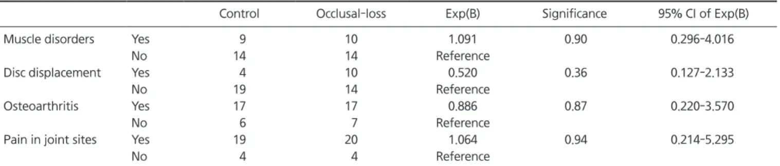
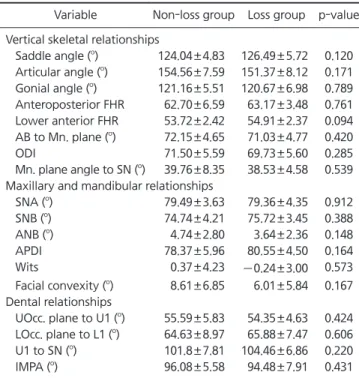
Results: In the control group, RDC group I, muscle disorders, was 39.1% (9/23), group II, disc displacements, was 17.4% (4/23), group III OA, osteoarthritis/osteoarthrosis, was 73.9% (17/23), and group III pain, arthralgia, was 82.6% (19/23). In the occlusal-loss group, group I was 41.7% (10/24), group II was 41.7% (10/24), group III OA was 70.8% (17/24), and group III pain was 83.3% (20/24). The frequency of RDC groups was not different between two groups, ana- lyzed by binomial logistic regression. Pre-treatment cephalometric variables were not different between two groups. However, articular angle, AB to mandibular plane and ODI decreased and gonial angle increased significantly in the occlusal-loss group, implying clockwise rotation of the mandible, between pre-treatment and 6-month post-treatment, while none of cephalomet- ric variables showed any statistical difference in the control group.
Conclusions: Change in the number of occluding teeth was not related to the RDC/TMD diag- noses. Cephalometric values changed only in the occlusal-loss group as a result of mandibular clockwise rotation. None of cephalometric variables before the stabilization splint therapy was statistically different between the control and occlusal loss group.
Key Words: Cephalogram; Occlusal appliance; Occlusion; Temporomandibular joint disorders
Correspondence to:
Jeong-Yun Lee
Department of Oral Medicine and Oral Diagnosis, Dental Research Institute, School of Dentistry, Seoul National University, 101, Daehak-ro, Jongno-gu, Seoul 110-749, Korea
Tel: +82-2-2072-0212 Fax: +82-2-744-9135 E-mail: [email protected]
internal derangement about at 30-35 years and inflam- matory-degenerative disorders about at 50-55 years. 2) And women had higher prevalence rates of TMD than men. 3,4)
Several treatment methods for TMD have been used, in- cluding occlusal splints, behavioral treatment, physical therapy, medications and surgical approaches. 5) de Leeuw et al. 6) concluded that nonsurgical treatment is as effective
INTRODUCTION
Temporomandibular disorders (TMD) are defined as a collective term that embraces a number of clinical prob- lems that involve the masticatory muscles, the temporo- mandibular joint (TMJ), and the associated structures. 1) Epidemiological study shows two distinct age peaks for
JOMP Journal of Oral Medicine and Pain
Copyright Ⓒ 2015 Korean Academy of Orofacial Pain and Oral Medicine. All rights reserved.
CC