Cephalometric Characteristics of the Patients with Developed Anterior Open Bite Following Anterior Disc
Dislocation without Reductions
Yun-Kyung Hur, D.D.S.,M.S.D., Jae-Kap Choi, D.D.S.,M.S.D.,Ph.D.
Department of Oral Medicine, School of Dentistry, Kyungpook National University
Objectives: This article reported three patients developed anterior open bite seemed to be related to TMJ anterior disc dislocation without reduction(ADD WO R), but no evidence of condylar destructive or collapse and analyzed the craniofacial skeletal structure by means of cephalometric analysis.
Results: All patients suddenly developed a centric relation/centric occlusion discrepancy, an increased overjet and an anterior open bite following ADD WO R. All patients had Angle's Class I occlusion and shallow bite, but they had skeletally Class III and Class II pattern and all were vertically significant hyperdivergent type.
Conclusions: These 3 patients had characteristics of common facial morphology including: (1)Angle classification Class I and shallow bite, (2)high mandibular plane angle, (3)high gonial angle. Developed anterior open bite resulted from clockwise rotation of the mandible related TMJ ADD WO R, rather than a result from the eruption of posterior teeth.
We hypothesize rotation may relate to attached direction of masticatory muscle.
Key words: Developed anterior open bite, ADD WO R, Acute CR/CO discrepancy, Clockwise rotation
1)
I. INTRODUCTION
One of the most common temporomandibular disorders is a disc derangement related to anterior disc displacement. Among these patients, we can occasionally find the patients with developed anterior open bite before or during treatment.
Although the anterior open bite is uncommon complication of the temporomandibular disorders, but it often embarrasses clinical practitioners due to its difficulty of management and poor prognosis.
Corresponding author: Prof. Yun-Kyung Hur
Department of Oral Medicine, School of Dentistry, Kyungpook National University 2-188-1, Samduk-Dong, Jung-Ku, Daegu 700-412, Korea
E-mail: [email protected] received: 2006-07-18
accepted: 2006-09-01
Several authors proposed that the ant. open bite
may result from condylar collapse, which was
associated with inflammatory disorders of the TMJ,
such as rheumatoid arthritis.
1-6)However, we have
experienced some cases of anterior open bite, which
developed in the patients with anterior disc
dislocation without reduction(ADD WO R), but no
evidence of condylar destructive or collapse. None
of them show any signs of TMJ arthritis except
occasional tenderness on lateral palpation. In this
article, three clinical cases of acquired anterior open
bite following ADD WO R were analyzed clinically
as well as cephalometrically. The two patients
showed anterior open bite developed during
conservative treatment, such as physical, medical
therapy and they did not taken splint treatment for
a follow-up. The other one patient was clinically
diagnosed as anterior disc displacement with
Yun-Kyung Hur, Jae-Kap Choi
reduction of both TMJs at first visit and did not given any treatment. After 9 months, the patient complained occlusal change accompanied with dull pain of both TMJs. On diagnostic casts, the anterior open bite cann't be seen and pre-existing occlusal facets of upper and lower teeth can make contact precisely.
The aims of this study were to review in the course of time three clinical cases of developed anterior open bite following ADD WO R and to analyze the craniofacial skeletal structure by means of cephalometric analysis.
CASE I
Age : 20
Gender : female(Fig. 1A)
Diagnosis : both TMJs ADD WO R Progress :
2004. 12. 7.
Chief complaint was both TMJ pain during mastication, especially left, and limited opening.
Panoramic and transcranial radiographs showed decreased condylar movement but no condylar resorption(Fig. 1B, 1C).
Medication(NonSteroidal Anti-Inflammatory Drugs) was done.
2004. 12. 21.
Patient complained increased pain.
Medication(NSAIDs) and physical therapy were done.
Clinical diagnosis was ADD WO R of both TMJs.
2004. 12. 29.
On magnetic resonance image(MRI) of TMJ, both TMJs had ADD WO R(Fig. 1D).
2005. 1. 12.
She experienced alteration in occlusion and sound of teeth grinding at reaching centric occlusion.
Impression was taken for study model(Fig. 1H).
2005. 1. 19.
The patient complained suddenly developed ant.
open bite(Fig. 1G).
Cephalometric radiograph were taken(Fig. 1E).
When we compared with occlusal contacts between study model and present occlusion, the discrepancy was found.
CASE II
Age : 32
Gender : female(Fig. 2A)
Diagnosis : Lt TMJ ADD WO R Progress :
2005. 2. 2.
Chief complaint was left TMJ pain during mastication and limited opening.
Panoramic(Fig. 2B) and transcranial radiographs (Fig. 2C) showed decreased condylar movement of Lt. TMJ but no condylar resorption.
2005. 2. 17.
Clinical diagnosis was ADD WO R of left TMJ.
Medication(NSAIDs) and physical therapy were done.
2005. 3. 17.
She suffered from same symptoms.
Patient demanded only medication.
Medication(NSAIDs) and physical therapy were done.
2005. 3. 31.
Patient complained interocclusal sliding at occlusion and occasional headache.
Medication(NSAIDs) was done and impression was taken for study model(Fig. 2G).
2005. 4. 14.
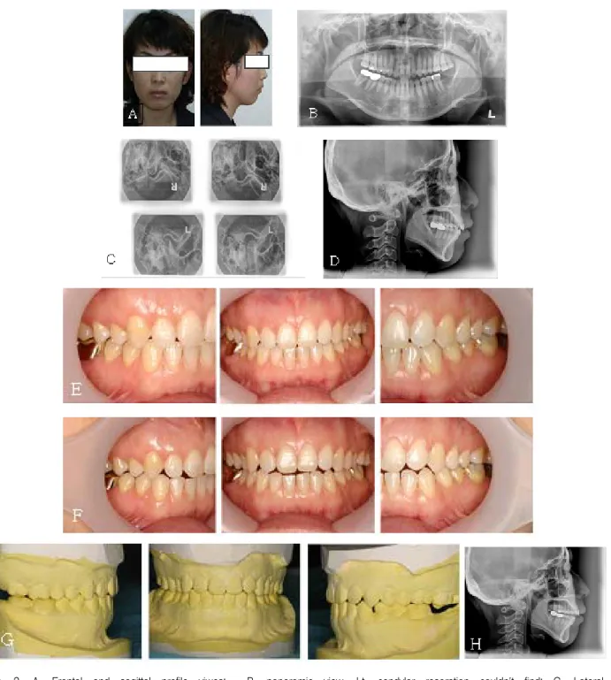
Fig. 1. A, Frontal and sagittal profile viwes; B, panoramic view, no condylar resorption on both sides; C, transcranial view, both condyle movement were decreased; D, MRI, both TMJs were ADD WO R; E, cephalometric radiograph; F, intraoral photograph of centric occlusion at clenching; G, intraoral photograph of centric relation; H, occlusion of study model
Yun-Kyung Hur, Jae-Kap Choi
Fig. 2. A, Frontal and sagittal profile viwes; B, panoramic view, Lt. condylar resorption couldn't find; C, Lateral transcranial view, Lt. condyle movement was decreased; D, cephalometric radiograph; E, intraoral photograph of centric occlusion at clenching; F, intraoral photograph of centric relation at same day; G, occlusion of study model; H, after one month cephalometric radiograph
Fig. 3. A, Frontal and sagittal profile viwes; B, panoramic view, no condylar resorption on both TMJs; C, transcranial view, both condyle movement were not decreased; D, cephalometric radiograph; E, intraoral photograph of present occlusion; F, occlusion of study model
Occlusal contact was only attained between #26 and 36 and ant. open bite was found.
Cephalographic radiograph at CO were taken(Fig.
2D).
When we compared with occlusal contacts between study model and present occlusion, the discrepancy was found.
2005. 5. 18.
Ant. open bite was increased.
Cephalographic radiograph(CR) was taken(Fig. 2H).
CASE III
Age : 21
Gender : female(Fig. 3A)
Diagnosis : both TMJs ADD WO R Progress :
2003. 7. 23.
Clinical diagnosis was anterior disc dislocation with reduction(ADD W R) of both TMJs.
Overbite : 1 mm / Overjet : 2 mm (in previous
chart)
Yun-Kyung Hur, Jae-Kap Choi
Variables case I case II case III Mean
Cranial base relationships
Anterior cranial base length(S-N) (mm) 71.7 63.2 68.7 68.7
Posterior cranial base length(S-Ar) (mm) 34.4 32.8 33.9 36.7
Saddle angle(N-S-Ar) ( ° ) 118.6 130.9 123.5 125.9
Maxillomandibular relationships
SNA angle ( ° ) 80.4 84.5 81.7 81.6
SNB angle ( ° ) 79.7 77.3 77.7 79.2
ANB angle ( ° ) 0.7 7.2 4.0 2.5
Vertical skeletal relationships
FMA ( ° ) 31.2 35.5 33.3 24.3
SN to mandibular plane angle ( ° ) 42.0 45.3 40.9 33.4
FH to palatal plane angle ( ° ) 0.5 0.8 -2.6 0.6
Maxillomandibular plane angle(ANS-PNS/Go-Me) ( ° ) 30.7 34.7 35.9 24.7 Occlusal plane to mandibular plane angle ( ° ) 19.3 17.4 20.1 15.4
Total anterior facial height(N-Me) (mm) 130.7 133.0 130.3 127.4
Total posterior facial height(S-Go) (mm) 76.0 80.4 77.7 85.1
Lower anterior facial height(ANS-Me) (mm) 72.5 80.7 76.8 70.7
Total post. facial height/Total ant. facial height(%) 58.2 60.4 59.6 66.8 Size and form of mandible
Ramus height(Ar-Go) (mm) 44.1 50.9 46.3 51.6
Mandibular body length(Go-Me) (mm) 79.5 72.4 79.6 78.0
Gonial angle (Ar-Go-Me) ( ° ) 133.1 127.6 126.4 118.7
Articular angle(S-Ar-Go) ( ° ) 150.4 146.8 151.1 148.7
Y axis 58.0 68.0 63.5 61.0
mean : mean of Korean adult female with normal occlusion Table 1. Comparison of cephalometric variables of subjects
The patient did not visited since 23. July 2003.
2004. 10. 4.
Patient complained alteration in occlusion and dull pain on both TMJs from several months ago.
Panoramic(Fig. 3B) and transcranial(Fig. 3C)
Overbite : -1.5 mm / Overjet : 3 mm
Clinical diagnosis was ADD WO R of both TMJs.
2004. 11. 3.
Patient complained discomfort of both TMJ
during opening.
2004. 12. 15.
Cephalometric radiograph was taken(Fig. 3D).
When we compared with occlusal contacts between study model and present occlusion, the discrepancy was found(Fig. 3E, 3F).
Ⅱ. RESULTS
All patients had experienced common signs/symptoms of TMJ internal derangement.
All affected TMJs have ADD WO R.
All patients suddenly developed a centric relation/centric occlusion discrepancy, an increased overjet and an open bite following ADD WO R.
All patients had Angle's Class I occlusion and shallow bite, but they had skeletally Class III and Class II pattern and all were vertically significant hyperdivergent type.
The cephalometric findings in the 3 patients are compared with established mean normal values of Korean adult female(Table 1).
Skeletal characteristics of case I patient were as follows: (1)straight facial type with ANB 0.7, (2)high occlusal and high mandibular plane angle, (3)high gonial angle, (4)increased mandibular body length, (5)decreased ramus height. From these results, the patient had skeletal Class III tendency with vertical discrepancy. Case II patient had skeletal Class II pattern with ANB 7 and high mandibular plane angle. Case III patient had also decreased ramus height and increased lower ant.
facial height as hyperdivergent skeletal type.
Ⅲ. DISCUSSION
Cases of developed anterior open bite following severe TMJ destruction have been reported.
1-6)Most of these open bite cases belong to progressive and more rapid destructive disease of the TMJ.
They included rheumatoid arthritis, juvenile RA, psoriatic arthritis, and very rare connective tissue diseases. In severe cases with lost condylar support, an acute malocclusion results in characterized by heavy posterior contacts and an
anterior open bite. However, these 3 patients seemed to be related to TMJs ADD WO R, but no evidence of condylar destructive or collapse and sudden occurrence of a centric relation/centric occlusion discrepancy is a result from the clockwise rotation of the mandible related ADD WO R, not from the eruption of posterior teeth.
Chen and associates reported
7)cases of acquired anterior open bite and all these patients had anteriorly displaced discs. As the etiology of occurrence of open bite, they hypothesized that this bite change might result from self-limiting degenerative joint disease. The etiology of clockwise rotation of the mandible related TMJ ADD WO R has not been studied enough yet.
Takada and associates
8)analyzed the approriate orientation of the superficial masseter and temporalis muscles associated with dentofacial morphology using head films, the origins and insertions being identified by examination of dry skulls for means of anatomic and geometric criteria.
This study suggested that an association exits between an obliquely inclined masseter muscle relative to the occlusal plane and a short posterior face height, a steep mandibular plane and a large gonial angle in long-faced persons. Brachyfacial type with a flat mandibular plane and an acute gonial angle represent a vertically oriented masseter muscle. We hypothesize rotation may relate to attached direction of masticatory muscle.
Further study is needed in the future.
These 3 patients have characteristics of common facial morphology including: (1)Angle’s Class I and shallow bite, (2)high mandibular plane angle, (3)high gonial angle.
Huang et al.
9)and Wolford et al.
10)reported the
skeletal characteristics of the patients with
idiopathic condylar resorption. These were
composed of high mandibular plane, skeletal Class
II morphology. If condyar resorptions of these 3
patients are progressed, vertical height of the
ramus will be decreased and mandible will be
progressively retruded, change to Class II skeletal
pattern will be occurred.
Yun-Kyung Hur, Jae-Kap Choi
After this, the etiology of the developed anterior open bite due to clockwise rotation of the mandible in ADD WO R patients and characteristics of facial morphology appear to be most susceptible to occur anterior open bite should be revealed.
Ⅳ. CONCLUSIONS
This article reported 3 patients developed anterior open bite seemed to be related to TMJ ADD WO R, but no evidence of condylar destructive or collapse and they experienced sudden occurrence of centric relation/centric occlusion discrepancy resulted from clockwise rotation of the mandible related TMJ ADD WO R, rather than a result from the eruption of posterior teeth.
These 3 patients had characteristics of common facial morphology including: (1)Angle classification Class I and shallow bite, (2)high mandibular plane angle, (3)high gonial angle.
REFERENCES
1. Guyuron B. Facial deformity of juvenile rheumatoid arthritis. Plast Reconstr Surg 1988;81:948-951.
2. Chenitz JE. Rheumatoid arthritis and its implications in temporomandibular disorders. Cranio 1992;10:
59-69.
3. Akerman S, Kopp S, Nilner M, Petersson A, Rohlin M. Relationship between clinical and radiologic findings of the temporomandibular joint in rheumatoid arthritis. Oral Surg Oral Med Oral Pathol 1988;66:639-643.
4. Tegelberg A, Kopp., Huddenius K, Forssman L.
Relationship between disorder in the stomatognathic system and general joint involvement in individuals with rheumatoid arthritis. Acta Odontol Scand 1987;45:391-398.
5. Lanigan DT, Myall RW, West RA, McNeill RW.
Condylysis in a patient with a mixed collagen vascular disease. Oral Surg Oral Med Oral Pathol 1979;48:198-204.
6. Haers and Sailer HF. Mandibular resorption due to systemic sclerosis. Case report of surgical correction of a secondary open bite deformity. Int J Oral Maxillofac Surg 1995;24:261-267.
7. Chen YJ, Shih TTF, Wang JS et al. Magnetic resonance images of the temporomandibular joints of patients with acquired open bite. Oral Surg Oral Med Oral Pathol Oral Radiol Endod 2005;99:734-42.
8. Takada K, Lowe AA, Freund VK. Canonical correlations between masticatory muscle orientation and dentoskeletal morphology in children. Am J Orthod 1984;86:331-341.
9. Huang YL, Pogrel MA, Kaban LB. Diagnosis and management of condylar resorption. J Oral Maxillofac Surg 1997;55:114-119.
10. Wolford LM, Cardendas L. Idiopathic condylar resorption. Am J Orthod Dentofacial Orthop 1999;116:667-677.
국문요약
비정복성 관절원판 전위와 연관되어 발생된 전치부 개교합 환자의 측방 두부방사선 계측
경북대학교 치의학전문 대학원 구강내과학 교실