Kim JY, et al: Anterior Anorectocolonic Tubular Duplication in Infant
Journal of Korean Association of Pediatric Surgeons 55
pISSN 2383-5036 eISSN 2383-5508
J Korean Assoc Pediatr Surg Vol. 23, No. 2, December 2017
https://doi.org/10.13029/jkaps.2017.23.2.55
Case Report
Anterior Anorectocolonic Tubular Duplication Presenting as Rectovestibular Fistula in an Infant
Ja-Yeon Kim*,†, Joong Kee Youn*, Soo-Hong Kim, Hyun-Young Kim, Sung-Eun Jung, Kwi-Won Park‡
Department of Pediatric Surgery, Seoul National University Children’s Hospital, Seoul, Korea
Anorectal duplications account for only 5% of gastrointestinal duplications, and cases with involvement of the anal canal are much rarer. Nearly all anorectal duplications are posterior to the rectum; duplications located anterior to the normal rectum are highly un- usual, and only a few cases have been reported. We report the case of an anterior anorectocolonic duplication presenting as a rec- tovaginal fistula in a 2-month-old infant. After diagnosis, the duplication was excised completely without further intestinal complications.
Keywords: Anal canal duplication, Rectal duplication, Colonic duplication, Duplication, Rectovaginal fistula
Received: April 7, 2017, Revised: May 24, 2017, Accepted: June 21, 2017
Correspondence: Hyun-Young Kim, Department of Pediatric Surgery, Seoul National University Children’s Hospital, 101 Daehak-ro, Jongno-gu, Seoul 03080, Korea.
Tel: +82-2-2072-2478, Fax: +82-2-747-5130, E-mail: [email protected]
*These authors contributed equally to this work as first authors.
†Current affiliation: Gastric Cancer Branch, Research Institute & Hospital, National Cancer Center, Goyang, Korea
‡Current affiliation: Department of Surgery, Chung-Ang University Hospital, Seoul, Korea Copyright © 2017 Korean Association of Pediatric Surgeons. All right reserved.
This is an Open Access article distributed under the terms of the Creative Commons Attribution Non-Commercial License (http://creativecommons.org/licenses/by-nc/4.0) which permits unrestricted non-commercial use, distribution, and reproduction in any medium, provided the original work is properly cited.
INTRODUCTION
Gastrointestinal tract duplications are rare conditions, comprising 0.1% to 0.3% of all congenital anomalies.
Duplications can occur throughout the gastrointestinal tract, from the mouth to the anus [1,2]. Anorectal dupli- cations account for only 5% of all duplications and are primarily found in the retro-rectal space. Anterior ano- rectal duplications are extremely rare [3]. Anterior ano- rectal duplications are tubular duplications connected to the colon and open like a distal fistula, but are located anterior to the rectum and behind the bladder and vagina.
It is challenging to diagnose anterior anorectal duplica- tion, both because of its rarity and because it can present in different ways. Perirectal abscess, fistula-in-ano, re- current fistulae due to Crohn’s disease, infected dermoid cysts, sacrococcygeal teratomas, and anterior sacral me- ningoceles must be considered part of the differential di- agnosis [3,4]. Here, we present the case of an anterior anorecto-sigmoid colonic tubular duplication initially presenting as a rectovaginal fistula in a 2-month-old
infant. We also provide a brief literature review.
CASE REPORT
A 2-month-old female infant presented to our hospi- tal with a 1-month history of stool passing through the vagina. She was born at 36 weeks’ gestation weighing 1.875 kg and was the second infant of a twin. She had a ventricular septal defect that closed spontaneously at 1 month of age. Initially, she was diagnosed with a rec- tovaginal fistula. Despite conservative therapy, includ- ing a sitz bath, for 5 months, there was no improvement in her symptoms, and most of the stool still appeared to pass through the vagina. A colon study indicated a rec- tovaginal fistula directly connected to the distal rectum (Fig. 1). Under general anesthesia, a small opening lo- cated posterior to and near the vagina at vestibule was found. The structure that had been misdiagnosed as a fis- tula based on the colon study was not present. The patient had a normally sited anus, an anterior ectopic anus, and a double vagina (Fig. 2). A catheter was inserted through
J Korean Assoc Pediatr Surg 2017;23(2):55-58
56 Journal of Korean Association of Pediatric Surgeons
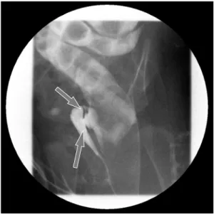
Fig. 1. A preoperative colon study appeared to show that the contrast had passed to the vagina through a rectovaginal fistula (arrows).
Fig. 3. Intraoperative findings. (A) The intraoperative contrast study revealed that the contrast injected via the an- terior anorectal duplication filled the normal colon and rectum and that there was a connection between the dupli- cation and the normal colon at the level of the proximal sigmoid colon. (B) Intraoperative sigmoidoscopy at the merging point revealed that a catheter inserted via the anterior anorectal duplication could be observed by a scope inserted via the normal anus.
Fig. 2. Examination under general anesthesia revealed a normal anus, an anterior anorectal duplication (catheter-inserted state), and dupli- cate vaginal openings.
the ectopic anus, and an intraoperative contrast study and sigmoidoscopy were performed. A parallel tubular du- plication with a common wall was found. The common wall extended 5 cm from the anal verge to the sigmoid colon and then 20 cm to the level of the proximal sigmoid colon. Proximally, the anterior duplication and normal colon merged and formed a normal colon. Because most of the stool moved through the anterior duplication and because the diameter of the anterior duplication was larger than that of the normally located colon (Fig. 3), complete resection of both the anterior duplicated bowel and posterior native bowel up to the merging point with colo-anal anastomosis was performed (Fig. 4). Fig. 5
shows a schematic diagram of surgical findings. Pathologic examination confirmed a duplicated colonic segment with a common proper muscle layer. The patient was dis- charged on the 8th postoperative day without incident.
During a 3-year follow-up, she defecated normally without complications.
DISCUSSION
Gastrointestinal tract duplications occur with an in- cidence of 1 in 4,000 to 5,000 live births. The incidence of anorectal and colonic duplications has been reported as 3% to 8% and 4% to 18% of all duplications, re-
Kim JY, et al: Anterior Anorectocolonic Tubular Duplication in Infant
Journal of Korean Association of Pediatric Surgeons 57
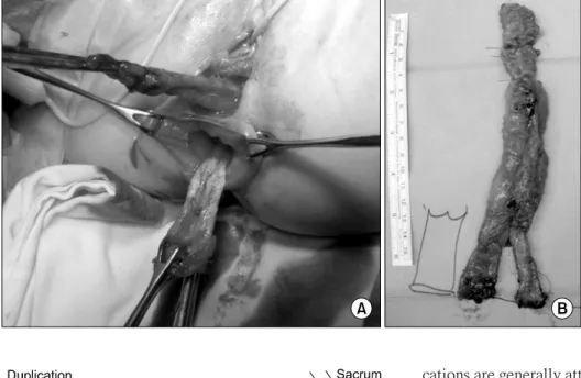
Fig. 4. Operative findings. (A) The tu- bular anorectocolonic duplication and the normal anorectum were dissected from neighboring structures. (B) Re- sected specimen.
Fig. 5. Schematic diagram of surgical findings.
spectively [3,5,6]. Anorectal duplications comprise a small proportion of enteric duplications, with only 70 cases reported to date in the literature. Based on their embryogenesis, anorectal duplications would be ex- pected to be located posterior to the rectum. In fact, most anorectal duplications are located posterior to the anus;
anterior rectal duplications are very rare, with fewer than 10 cases reported [7].
Although the causes of alimentary tract duplication are uncertain, many theories have been proposed to ex- plain their pathophysiology, including the abortive twinning theory, persistent embryonic diverticular theo- ry, split notochord theory, and aberrant luminal recanal- ization theory. However, no single theory can explain all forms of intestinal duplication [2,6,8]. Anorectal dupli-
cations are generally attributed to a simple “pinching off”
of the diverticulum in the 8th to 9th week of gestation or to more complex “caudal twinning” occurring when the embryo is 10 mm in length during the 5th week of ges- tation [3].
Symptoms and findings depend on the location of the duplication [2]. Most patients with anorectal duplica- tions are asymptomatic. The most frequent symptoms in- clude progressive constipation, prolapse of the anorectal mucosa, and mucosal discharge. More severe symptoms have also been reported, including recurrent fistulae, pain, or infection in the duplicated cyst, rectal bleeding from ulceration, or ectopic gastric mucosa. The severity of these symptoms increases with the age at presentation.
Malignant degeneration of duplications has also been re- ported [3,9,10]. The symptoms reported for anterior ano- rectal duplications are similar to those for posterior ano- rectal duplications, including recurrent gastroenteritis and failure to thrive, isolated rectal bleeding, chronic constipation with soiling, and rectal prolapse [7]. Due to the differing presentations for anterior anorectal dupli- cations, various diseases must be considered part of the differential diagnosis, including sacrococcygeal ter- atoma, anal fistula due to Crohn’s disease, rectal pro- lapse, dermoid cysts, hydrocolpos, chordoma, leiomyo- sarcoma, and hydrometrocolpos [3,4,7]. Because it is challenging to distinguish anterior anorectal duplica- tions from other diseases, delayed diagnosis can occur [4].
J Korean Assoc Pediatr Surg 2017;23(2):55-58
58 Journal of Korean Association of Pediatric Surgeons
Duplications of the hindgut, such as anorectal dupli- cations, are frequently accompanied by genitourinary tract anomalies and have complicated structures; thus, they tend to be challenging and complex. When encoun- tering such duplications, genitourinary tract involve- ment should be considered [2].
Various diagnostic modalities can assist in diagnosis, such as abdominal ultrasonography, contrast studies, computed tomography, and MRI [7]. In particular, MRI can provide multiplanar views, allowing identification of pelvic structures and ruling out other diagnoses [6,7]. To evaluate communication between duplications and nor- mal bowels, contrast studies can be helpful in accurately delineating the anatomy of the duplicated rectum [6]. In the present case, we performed a preoperative contrast study, but we initially misinterpreted the contrast spill as a fistula. We reached a final diagnosis intraoperatively after re-performing the contrast study. The second con- trast study clearly revealed the anterior anorectocolonic tubular duplication.
Total surgical excision of all duplications is recom- mended because of the potential for long-term compli- cations. If an anorectal duplication is not treated, secre- tions accumulate in the lumen of the duplication, leading to infection with age [1]. Malignant changes in anorectal duplications have also been reported in adult patients, although not in pediatric populations [1,10]. When there was no evidence of fistula formation between the dupli- cation cyst and vagina, cyst unroofing and residual mu- cosa ablation was applied [7]. In the other case of ante- rior rectal duplication without rectovaginal fistula, sep- tum division via surgical staplers was performed [3].
The location of the duplication determines the direc- tion of approach. Perineal or posterior sagittal appro- aches in the prone position have primarily been used in cases of posterior rectal duplication and anal canal du- plication [1], whereas abdominoperineal approaches have been chosen for anterior rectal duplications [3,7].
One reported case of posterior anorectal duplication ex- tending to the colon was initially treated with a posterior sagittal approach, but as the dissection proceeded cepha-
lad, it became difficult to manage, and laparotomy was also performed [6]. In our case, a perineal approach was first attempted because the lesion was misdiagnosed as a rectovaginal fistula; however, when the anterior ano- rectosigmoid duplication was found, the operation pro- ceeded using an abdominal approach.
In summary, for infants who present with stool dis- charging from the vagina, anterior anorectal duplication should be considered as a differential diagnosis, despite its rarity. Because it is challenging to diagnose, careful evaluation is required.
CONFLICTS OF INTEREST
No potential conflict of interest relevant to this article was reported.
REFERENCES
1. Narci A, Dilek FH, Cetinkurşun S. Anal canal duplication. Eur J Pediatr 2010;169:633-5.
2. Stern LE, Warner BW. Gastrointestinal duplications. Semin Pediatr Surg 2000;9:135-40.
3. Prasil P, Nguyen LT, Laberge JM. Delayed presentation of a con- genital recto-vaginal fistula associated with a recto-sigmoid tubu- lar duplication and spinal cord and vertebral anomalies. J Pediatr Surg 2000;35:733-5.
4. Knudtson J, Jackson R, Grewal H. Rectal duplication. J Pediatr Surg 2003;38:1119-20.
5. Hernandez Troya AC, Gebara S, Bloom DA, Chan W. Occult colon- ic duplication. Clin Pediatr (Phila) 2011;50:550-2.
6. Zhang Z, Huang Y, Wang D, Su P. Rectosigmoid tubular duplica- tion presenting as perineal sepsis in a neonate. J Pediatr Surg 2010;45:627-9.
7. Amjadi K, Poenaru D, Soboleski D, Hurlbut D, Kamal I. Anterior rectal duplication: a diagnostic challenge. J Pediatr Surg 2000;35:
613-4.
8. Macpherson RI. Gastrointestinal tract duplications: clinical, pathologic, etiologic, and radiologic considerations. Radiographics 1993;13:1063-80.
9. Kratz JR, Deshpande V, Ryan DP, Goldstein AM. Anal canal dupli- cation associated with presacral cyst. J Pediatr Surg 2008;43:
1749-52.
10. Parvaiz A, Stevens RJ, Lamparelli MJ, Jeffery PJ. A rare case of ad- enocarcinoma arising within a duplication cyst of the rectum: cu- rative excision with 9-year follow-up. Ann R Coll Surg Engl 2005;
87:W8-10.