Cobalt Chloride Attenuates Oxidative Stress and Inflammation through NF-κB Inhibition in Human Renal Proximal Tubular Epithelial Cells
We evaluated the effect of cobalt chloride (CoCl2) on TNF-α and IFN-γ-induced-
inflammation and reactive oxygen species (ROS) in renal tubular epithelial cells (HK-2 cells).
We treated HK-2 cells with CoCl2 before the administration of TNF-α/IFN-γ. To regulate hemeoxygenase-1 (HO-1) expression, the cells were treated CoCl2 or HO-1 siRNA. CoCl2
reduced the generation of ROS induced by TNF-α/IFN-γ. TNF-α/IFN-γ-treated-cells showed an increase in the nuclear translocation of phosphorylated NF-κBp65 protein, the DNA- binding activity of NF-κBp50 and NF-κB transcriptional activity and a decrease in IκBα protein expression. These changes were restored by CoCl2. We noted an intense increase in monocyte chemoattractant protein-1 (MCP-1) and regulated on activation normal T cell expressed and secreted (RANTES) production in TNF-α/IFN-γ-treated cells. We
demonstrated that this effect was mediated through NF-κB signaling because an NF-κB inhibitor significantly reduced MCP-1 and RANTES production. CoCl2 effectively reduced MCP-1 and RANTES production. The expression of HO-1 was increased by CoCl2 and decreased by HO-1 siRNA. However, knockdown of HO-1 by RNA interference did not affect MCP-1 or RANTES production. We suggest that CoCl2 has a protective effect on TNF-α/IFN-γ-induced inflammation through the inhibition of NF-κB and ROS in HK-2 cells. However, CoCl2 appears to act in an HO-1-independent manner.
Keywords: Cobalt Chloride; Hemeoxygenase-1; Inflammation; Nuclear Factor-kappa B;
Renal Tubular Epithelial Cells Se Won Oh,1 Yun-Mi Lee,2
Sejoong Kim,2,3 Ho Jun Chin,2,3 Dong-Wan Chae,2,3 and Ki Young Na2,3
1Department of Internal Medicine, Inje University College of Medicine, Ilsan Paik Hospital, Goyang;
2Department of Internal Medicine, Seoul National University Bundang Hospital, Seongnam;
3Department of Internal Medicine, Seoul National University College of Medicine, Seoul, Korea Received: 10 May 2014
Accepted: 3 July 2014 Address for Correspondence:
Ki Young Na, MD
Division of Nephrology, Department of Internal Medicine, Seoul National University Bundang Hospital, 82 Gumi-ro 173beon-gil, Bundang-gu, Seongnam 463-707, Korea Tel: +82.31-787-7014, Fax: +82.-31-787-4051 E-mail: [email protected]
http://dx.doi.org/10.3346/jkms.2014.29.S2.S139 • J Korean Med Sci 2014; 29: S139-145
INTRODUCTION
Inflammation is an inevitable phenomenon in all forms of acute and chronic kidney diseases, regardless of the underlying cause. Renal proximal tubular epithelial cells are an important source of cytokines/chemokines and play a central role in the regulation of the local inflammatory response (1). Tumor ne- crosis factor-α (TNF-α) is a pleiotropic cytokine that has been implicated in the inflammatory cascade leading to renal injury.
TNF-α is not typically detected in normal kidneys. However, re- nal tubular epithelial cells express TNF-α after noxious stimula- tion (2). TNF-α increases vascular permeability, the expression of adhesion molecules on endothelial cells, cytokine and che- mokine release, and the production of reactive oxygen species (ROS) (3-5). TNF-α has been shown to increase ROS produc- tion through the NADPH oxidase-like system or the cytosolic phospholipase A2-linked cascade in various cells (6, 7). The in- flammatory response to TNF-α is mediated through the nuclear factor-kappa B (NF-κB) pathway, and NF-κB activation leads to cytokine and chemokine production in renal tubular epithelial cells, including monocyte chemoattractant protein-1 (MCP-1)
and regulated on activation normal T cell expressed and secret- ed (RANTES) (2,8).
The effect of cobalt chloride (CoCl2) on inflammation has rarely been reported. CoCl2 has been used to induce inflamma- tion in in vitro experiments (9, 10). CoCl2 was the first metal ion identified with a hemeoxygenase-1 (HO-1)-inducing property (11). HO-1 is the rate-limiting enzyme in the degradation of heme, and its products, including biliverdin and carbon mon- oxide (CO), have anti-oxidant and anti-inflammatory effects (12, 13). The administration of heme protein to HO-1-/- mice has been shown to result in severe renal interstitial inflamma- tion accompanying a drastic upregulation of MCP-1 and NF-κB expression (12). The constitutive expression of HO-1 has been shown to reduce MCP-1 production induced by albumin in re- nal proximal tubular epithelial cells (14). Collectively, CoCl2 has shown mixed results, with both pro-inflammatory and anti-in- flammatory effects. Therefore, we investigated the role of CoCl2
in an experiment of TNF-α- and interferon gamma (IFN-γ)- induced NF-κB activation, cytokine production, and ROS pro- duction in renal tubular epithelial cells. We also attempted to determine the role of HO-1 in the generation of inflammatory
cytokine production induced by TNF-α and IFN-γ.
MATERIALS AND METHODS Cell culture
HK-2 cells (ATCC CRL-2190), which are proximal tubular epi- thelial cells derived from normal human kidney tissue, were cultured using Renal Epithelial Basal Medium (Lonza Walkers- ville Inc., Walkersville, MD, USA) with the recommended sup- plements included in the EGM® SingleQuot Bullet Kit. Cells were fed two to three times weekly and subcultivated via tryp- sinization when near confluence. HK-2 cells between passages 10 and 25 were used for these experiments.
Cell treatment
Cells were grown to 80% confluence before treatment for all ex- periments. TNF-α (R&D Systems Inc., Minneapolis, MN, USA) and IFN-γ (PeproTech Inc., Rocky Hill, NJ, USA) were dissolved in phosphate-buffered saline (PBS) containing 0.1% bovine se- rum albumin. CoCl2 (Sigma, St. Louis, MO, USA) was dissolved in PBS.
The cells were divided into four groups: 1) control cells (C), 2) TNF-α/IFN-γ-treated cells (TI), 3) TNF-α/IFN-γ-treated cells with CoCl2 pre-treatment (TIC), and 4) CoCl2-pre-treated cells (Co). For the inhibition of HO-1, two additional groups were added: 5) TNF-α/IFN-γ-treated cells with CoCl2 pre-treatment and negative control small interfering RNA (siRNA) transfection (TICN) and (6) TNF-α/IFN-γ-treated cells with CoCl2 pre-treat- ment and HO-1 siRNA transfection (TIC-HO-1). Before the ex- periments, the cells were incubated in basal medium in the ab- sence of supplements for 24 hr. On the day of the experiment, the cells were pre-treated with 150 μM CoCl2 for 24 hr to activate HO-1. The cells were then treated with 5 ng/mL TNF-α and 50 ng/mL IFN-γ for an additional 24 hr. To evaluate the relation- ship between NF-κB activation and TNF-α/IFN-γ-induced RANTES or MCP-1 production, HK-2 cells were pre-treated in the absence or presence of the NF-κB inhibitor pyrrolidinedi- thiocarbamate (PDTC, 10, 15, 20, or 25 μM) (Sigma, St. Louis, MO, USA) for two hr before exposure to TNF-α/IFN-γ.
Western blotting analysis
Western blotting was performed as previously described (15).
Nuclear extracts were prepared from the HK-2 cell lysates using a nuclear extraction kit (Sigma, St Louis, MO, USA). Protein concentrations were measured using a bicinchoninic acid pro- tein assay kit (Thermo Fisher Scientific, Rockford, IL, USA). The samples were run on SDS-polyacrylamide mini-gels (Bio-Rad Mini-Protean III), and the proteins were transferred to nitrocel- lulose membranes by electroelution. Antibodies to phosphory- lated NF-κB p65 (p-NF-κB p65) (Ser276) (Cell Signaling Tech- nology, Danvers, MA), NF-κB p65 (Santa Cruz Biotech, Santa
Cruz, CA, USA), inhibitor of kappa B-α (IκBα) (Santa Cruz Bio- tech, Santa Cruz, CA, USA), HO-1 (Assay Designs, Inc., Ann Ar- bor, MI, USA), β-actin (Santa Cruz Biotech, Santa Cruz, CA), and GAPDH (Santa Cruz Biotech, Santa Cruz, CA) were used for this study. Incubation with horseradish peroxidase-conju- gated secondary antibodies (Santa Cruz Biotech, Santa Cruz, CA, USA) was followed by band visualization using an en- hanced chemiluminescence substrate (Thermo Fisher Scientif- ic, Rockford, IL, USA). The band densities were quantified by densitometry (GS-700 Imaging Densitometry, Bio-Rad, Hercu- les, CA, USA). To facilitate comparisons, the densitometry val- ues were normalized to a control, thereby defining the mean for the control group as one.
ELISA for RANTES and MCP-1
HK-2 cells were reseeded in 24-well plates and treated as de- scribed above. Supernatants were collected, and ELISAs for RANTES (R&D Systems Inc., Minneapolis, MN, USA) and MCP- 1 (R&D Systems Inc., Minneapolis, MN, USA) were performed using Quantikine kits following the manufacturer’s instructions.
The samples and known standard RANTES and MCP-1 protein preparations were incubated in 96-well plates pre-coated with monoclonal anti-RANTES and anti-MCP-1 antibodies, respec- tively. The reaction was read in a microplate reader (Bio-Rad Laboratories, Inc., CA, USA) and measured at 450 nm with a correction wavelength set at 560 nm. The mean values of the measurements for three independent wells were used for sta- tistical analysis.
NF-κB binding assay
NF-κB p50 DNA binding was detected by ELISA using a Trans- Factor p50 Colorimetric Kit (Clontech Laboratories, Palo Alto, CA). A total of 30 μg of nuclear extracts from HK-2 cell lysates was bound to wells coated with their consensus DNA-binding sequence. Anti-p50 was then added to the wells and incubated, followed by anti-rabbit HRP-conjugated IgG and detection by measuring the color development of tetramethylbenzidine at 655 nm using a microplate reader (Bio-Rad Laboratories, Inc., CA, USA).
Detection of intracellular reactive oxygen species (ROS) Oxidation-sensitive 2’,7’-dichlorofluorescein diacetate (DCFH- DA) (Sigma, St. Louis, MO, USA) was used to evaluate the intra- cellular production of ROS. The cells were loaded with DCFH- DA at a final concentration of 10 μM and incubated at 37oC for 30 min. Next, the cells were washed with PBS, removed from the dishes by scraping, and measured for fluorescence intensi- ty. Fluorescence was measured with a fluorescence spectrome- ter at 490 nm excitation and 526 nm emission.
Transfection and luciferase assay
The day before transfection, HK-2 cells were seeded at 90%-95%
confluence in 24-well plates. NF-κB luciferase reporter plasmids (luc2P/NF-κB-RE/Hygro) and the internal control reporter plas- mid (pRL-TK) were purchased (Promega Corp., Madison, WI, USA). For the analysis of reporter gene expression, 0.76 μg of NF- κB Photinus luciferase reporter plasmid was transfected into cells in each well using Lipofectamine 2000 (Invitrogen Corp., Carlsbad, CA, USA). Each construct was transfected with 0.04 μg of pRL-TK, where the thymidine kinase promoter drove Renilla luciferase expression. After transfection, the cells were main- tained in serum-free medium for 4 hr and then switched to fresh serum-free medium for an additional 20 hr. Photinus and Renil- la luciferase activities in extracts of the transfected cells were de- termined using the commercial Dual-Luciferase Reporter Assay System kit (Promega Corp., Madison, WI, USA). For each sam- ple, the Photinus luciferase activity was divided by the Renilla luciferase activity to correct for transfection efficiency.
RNA interference
In experiments using HO-1 siRNA (25 nM, 5’-AUGCUGAGUU- CAUGAGGAA-3’, 5’-ACACUCAGCUUUCUGGUGG-3’, 5’-CAGUUGCUGUAGGGCUUUA-3’, 5’-AGAUUGAGCGCAA- CAAGGA-3’) (ON-TARGETplus SMARTpool Human HMOX1, Dharmacon Inc.), the transfection of HK-2 cells was performed using DharmaFECT Transfection Reagent (Thermo Fisher Sci- entific) according to the manufacturer’s recommendations 24 hr before other experimental procedures. A nonspecific siRNA (ON-TARGETplus Non-Targeting siRNA #1, Dharmacon Inc.) was used as a negative control.
Statistical analysis
All of the data are presented as the mean ± standard deviation of the mean. The statistical analyses were performed using SPSS (version 18.0. for Windows; SPSS Inc., Chicago, IL, USA).
Comparisons between groups were performed using analysis of variance followed by Tukey’s test. The level of statistical sig- nificance was set at P < 0.05.
RESULTS
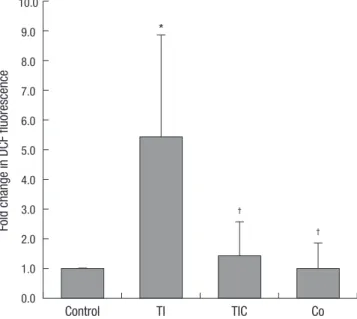
CoCl2 ameliorated TNF-α/IFN-γ-induced oxidative stress We examined the effect of CoCl2 on the generation of ROS (Fig.
1). Treatment with TNF-α and IFN-γ significantly increased the generation of intracellular ROS in HK-2 cells. CoCl2 pre-treat- ment significantly reduced the generation of ROS induced by TNF-α and IFN-γ. Cell treatment with CoCl2 alone did not affect the generation of intracellular ROS.
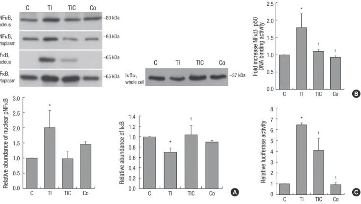
CoCl2 restored the activation of NF-κB induced by TNF-α and IFN-γ
NF-κB p65 protein phosphorylation was significantly increased in TNF-α/IFN-γ-treated cells (P = 0.049). This phosphorylation was significantly decreased by CoCl2 pre-treatment (P = 0.039).
The abundance of IκBα protein, which inactivates NF-κB by masking its nuclear localization signal, was significantly de- creased in TNF-α/IFN-γ-treated HK-2 cells (P = 0.001). Howev- er, this change was restored by CoCl2 (P < 0.001) (Fig. 2A). Next, we investigated the DNA-binding activity of NF-κB p50. Treat- ment with TNF-α and IFN-γ significantly increased the DNA- binding activity of NF-κB p50, but pre-treatment with CoCl2 re- stored its activity to the level of control cells (Fig. 2B). NF-κB- driven transcriptional activity was also evaluated. NF-κB-driven luciferase expression increased approximately six-fold in re- sponse to TNF-α and IFN-γ treatment (P < 0.001). As expected, pre-treatment with CoCl2 significantly decreased luciferase ex- pression (P = 0.007) (Fig. 2C). Collectively, CoCl2 restored TNF- α/IFN-γ-induced NF-κB activation.
CoCl2 increased the expression of HO-1
We investigated whether CoCl2 induced HO-1 protein expres- sion (Fig. 3A). The amount of HO-1 protein did not differ be- tween control cells and TNF-α/IFN-γ-treated cells. However, CoCl2-pre-treated cells showed markedly increased HO-1 ex- pression, and treatment with CoCl2 alone also increased its ex-
Fold change in DCF fluorescence
Control TI TIC Co
10.0 9.0 8.0 7.0 6.0 5.0 4.0 3.0 2.0 1.0 0.0
Fig. 1. Effects of CoCl2 pre-treatment on TNF-α/IFN-γ-induced ROS generation.
TNF-α/IFN-γ-induced increases in intracellular ROS were revealed by the fluores- cence intensity of 2’,7’-dichlorofluorescein. The fluorescence intensities of the cells are shown as the relative intensities of the experimental groups compared with control cells. A representative set of three independent experiments is shown. The results are expressed as the mean±s.d. *P<0.05, compared with control cells. †P<0.05, com- pared with TNF-α-+INF-γ-treated cells. C, control cells; TI, TNF-α/IFN-γ-treated cells;
TIC, TNF-α/IFN-γ-treated cells with CoCl2 pre-treatment; Co, CoCl2-pre-treated cells.
*
†
†
Fig. 2. Effects of CoCl2 pre-treatment on TNF-α/IFN-γ-induced NF-κB activation. (A) Western blotting of NF-κB, phosphorylated NF-κB, and IκBα in nuclear and cytoplasmic ex- tracts of HK-2 cell lysates. The intensities of the bands corresponding to phosphorylated NF-κB in nuclear extracts and IκBα in whole cell lysates are shown as the relative inten- sities of experimental groups compared with control cells. (B) The DNA-binding activity of NF-κBp50 was analyzed in the nuclear extracts of HK-2 cells using ELISA. (C) NF-κB- driven transcription is expressed as relative luciferase activity in HK-2 cells. A representative set of three independent experiments is shown. The results are expressed as the mean±s.d. *P<0.05, compared with control cells. †P<0.05, compared with TNF-α-+IFN-γ-treated cells. C, control cells; TI, TNF-α/INF-γ-treated cells; TIC, TNF-α/IFN-γ-treated cells with CoCl2 pre-treatment; Co, CoCl2-pre-treated cells.
Relative abundance of nuclear pNFκB
C TI TIC Co
3.0 2.5 2.0 1.5 1.0 0.5 0.0
*
Relative abundance of IκB
C TI TIC Co
1.4 1.2 1.0 0.8 0.6 0.4 0.2 0.0
*
†
A
C TI TIC Co
pNFκB, nucleus
–80 kDa pNFκB,
cytoplasm
–80 kDa
NFκB,
nucleus –65 kDa
NFκB,
cytoplasm –65 kDa
C TI TIC Co
IκBα, whole cell
–37 kDa
B Fold increase NFκB p50 DNA binding activity
C TI TIC Co
2.5
2.0 1.5
1.0
0.5
0.0
*
†
†
C
Relative luciferase activity
C TI TIC Co
8 7 6 5 4 3 2 1 0
*
†
†
C TI TIC Co
HO-1 HO-1
β-actin GAPDH
–32 kDa –32 kDa
–43 kDa –37 kDa
Relative abundance of HO-1 Relative abundance of HO-1
Control TI TIC Co
Control 2.5
2.0
1.5
1.0
0.5
0.0
2.5 2.0 1.5 1.0 0.5 0.0
Fig. 3. The expression of hemeoxygenase-1 (HO-1). (A) Western blotting of HO-1 in HK-2 cells. HK-2 cells were pre-treated with 150 μM CoCl2 for 24 hr and then treated with 5 ng/mL TNF-α and 50 ng/mL IFN-γ for an additional 24 hr. Beta-actin was used as a loading control. A representative set of three independent experiments is shown. The densi- tometric ratios of HO-1 and β-actin in cells are shown as the relative ratios of experimental groups compared with control cells. The results are expressed as the mean ± s.d.
*P<0.01, compared with control cells. C, control cells; TI, TNF-α/IFN-γ-treated cells; TIC, TNF-α/IFN-γ-treated cells with CoCl2 pre-treatment; Co, CoCl2-pre-treated cells. (B) Western blotting of HO-1 in HK-2 cells. GAPDH was used as a loading control. A representative set of three independent experiments is shown. The densitometric ratios of HO-1 and GAPDH in cells are shown as the relative ratios of experimental groups compared with control cells. The results are expressed as the mean±s.d. *P<0.001, compared with control cells. †P<0.005, compared with CoCl2-pre-treated cells.
* * *
†
*
A B
CoCl2 – – – + + +
HO-1 siRNA – – + – – +
Control siRNA – + – – + –
pression. HO-1 protein expression was markedly decreased af- ter transfection with a siRNA specific for human HO-1 in CoCl2- pre-treated cells, demonstrating very efficient siRNA-mediated knockdown in HK-2 cells (Fig. 3B).
CoCl2 reduced TNF-α/IFN-γ induced cytokine production in an HO-1-independent manner
Treatment with TNF-α and IFN-γ remarkably increased RAN- TES production by 239-fold (981.0 ± 253.6 vs. 4.1 ± 1.7 pg/mL, TI vs. C, P < 0.001). Pre-treatment with CoCl2 significantly decreased its production (226.4 ± 70.2 pg/mL, P < 0.001). However, RAN- TES production was not affected by HO-1 inhibition (TICN, 216.7 ± 43.1 pg/mL; TIC-HO-1, 255.0 ± 76.7 pg/mL, P > 0.05).
Similarly, MCP-1 production was markedly increased in
TNF-α/IFN-γ-treated HK-2 cells (308.6 ± 86.7 vs. 11.2 ± 6.7 pg/
mL, TI vs. C, P = 0.001). CoCl2 pre-treatment significantly re- duced MCP-1 production (45.6 ± 8.3 pg/mL, P < 0.005). How- ever, MCP-1 production was not affected by HO-1 siRNA treat- ment (TIC-HO1; 52.2 ± 13.0 pg/mL, P > 0.05) (Fig. 4A). Collec- tively, the results indicated that CoCl2 reduced TNF-α/IFN-γ- induced RANTES and MCP-1 production in an HO-1-indepen- dent manner.
Dose-dependent inhibition of TNF-α- and IFN-γ-induced cytokine production by an NF-κB inhibitor
To investigate whether NF-κB is involved in the TNF-α and IFN- γ-induced production of RANTES and MCP-1, we pre-treated HK-2 cells with an NF-κB inhibitor before exposure to TNF-α and
† † †
†
*
RANTES (pg/mL)MCP-1 (pg/mL) RANTES (pg/mL)MCP-1 (pg/mL)
C
C
Free media
Free media TI
TI
TI+PDTC 5 mM
TI+PDTC 5 mM
TI+PDTC 10 mM
TI+PDTC 10 mM
TI+PDTC 15 mM
TI+PDTC 15 mM
TI+PDTC 20 mM
TI+PDTC 20 mM TI
TI TIC
TIC TICN
TICN
TIC-HO-1
TIC-HO-1 Co
Co 1,400
1,200
1,000
800
600
400
200
0
500
400
300
200
100
0
1,000 900 800 700 600 500 400 300 200 100 0
500
400
300
200
100
0
Fig. 4. Effects of CoCl2, an NF-κB inhibitor, and HO-1 RNA interference on TNF-α/IFN-γ-induced cytokine production. (A) The production of RANTES and MCP-1 was analyzed in the supernatants of HK-2 cells using ELISA. A representative set of three independent experiments is shown. The results are expressed as the mean ± s.d. *P<0.05, compared with control cells. †P<0.05, compared with TNF-α-+IFN-γ-treated cells. C, control cells; TI, TNF-α/IFN-γ-treated cells; TIC, TNF-α/IFN-γ-treated cells with CoCl2 pre-treatment;
TICN, TNF-α/IFN-γ-treated cells with CoCl2 pre-treatment and negative control siRNA transfection; TIC-HO1, TNF-α/IFN-γ-treated cells with CoCl2 pre-treatment and HO-1 siRNA transfection; Co, CoCl2-pre-treated cells. (B) HK-2 cells were pre-treated with different doses of the NF-κB inhibitor PDTC for 2 hr before exposure to TNF-α and IFN-γ. TI, TNF-α/
IFN-γ-treated cells; PDTC, pyrrolidinedithiocarbamate.
*
* † * † * †
†
A B
IFN-γ. Pre-treatment with PDTC for 2 hr reduced RANTES and MCP-1 production in a dose-dependent manner (Fig. 4B).
DISCUSSION
In this study, we evaluated the effect of CoCl2 on TNF-α/IFN-γ- induced inflammation and oxidative stress in renal tubular epi- thelial cells. We found that CoCl2 effectively reduced the pro- duction of inflammatory mediators and the generation of ROS by inhibiting the NF-κB pathway in renal tubular injury. Howev- er, this beneficial effect of CoCl2 was not related to HO-1 activity.
NF-κB is a transcription factor that regulates many genes re- lated to immunity, apoptosis, cell proliferation, differentiation, and inflammation (16-18). NF-κB induces or represses numer- ous genes by binding to specific DNA sequences in the enhanc- er elements and promoters of target genes (8). NF-κB has a Rel- homology domain that permits DNA binding and dimerization and includes a nuclear localization signal. Inducers of TNF-α activate NF-κB through the classical/canonical pathway (19).
The activated IKK complex triggers the phosphorylation and proteasomal degradation of IκBα, allowing p65/p50 complexes to migrate to the nucleus and bind DNA (20). Classical NF-κB activation by TNF-α influences the transcription of a variety of genes. TNF-α activates the transcription of either early genes, including inflammatory cytokines (MCP-1/CCL-2, IL-6, IL-8, and IP-10 [interferon-γ introducing protein-10]), and negative regulators of NF-κB activity (IκB-α, IκB-ε, and A20) or late genes that are transcribed when NF-κB activation lasts for at least 1 hr including some chemokines (RANTES/CCL5) (8). We noted in- tense increases in MCP-1 and RANTES in TNF-α/IFN-γ-treated renal tubular epithelial cells. We stimulated renal tubular epi- thelial cells with TNF-α and INF-γ in combination because MCP-1 and RANTES production has been shown to signifi- cantly increase after their combined application but not after stimulation with only TNF-α (data not shown) (1). This in- creased production of MCP-1 and RANTES was mediated through NF-κB signaling because an NF-κB inhibitor signifi- cantly reduced their production. Additionally, we confirmed that TNF-α/IFN-γ-treated renal tubular epithelial cells exhibited increased nuclear translocation of phosphorylated NF-κB p65 protein, NF-κB p50 DNA-binding activity, and NF-κB transcrip- tional activity, as well as reduced IκBα protein expression.
NF-κB activation has been reported in renal cells such as tu- bular and mesangial cells after exposure to inflammatory stim- uli (8, 21, 22). In human renal disease, histologic studies have shown that NF-κB signaling is activated in glomerulonephritis (23, 24). NF-κB is primarily detected in the renal tubular epithe- lium in patients with proteinuric glomerulonephritis and the expression of NF-κB, MCP-1, and RANTES has been correlated with disease severity (23). In crescentic glomerulonephritis, NF-κB-positive cells are also primarily detected in crescentic le-
sions, the interstitium, and tubular epithelial cells (24). These data suggest that NF-κB activation is important in the promo- tion of inflammation in human renal disease. Therefore, the in- hibition of NF-κB activity can be used to reduce renal inflam- mation. Repression of IκBα ameliorates proteinuria-induced tubulointerstitial injury (25). The inhibition of NF-κB p50 atten- uates renal inflammation in sepsis-induced acute kidney injury (AKI) (26). In our in vitro study, CoCl2 reduced inflammation through NF-κB inhibition. Therefore, CoCl2 might be useful as an anti-inflammatory agent in the treatment of renal disease.
HO exists in two isoforms. In a healthy state, HO-1 is weakly expressed in the outer medulla and the cortex of the kidney.
When the kidneys are injured, HO-1 is primarily induced in tu- bular epithelial cells (27). HO-1 expression in proximal tubular epithelial cells has been correlated with proteinuria, hematuria, and tubulointerstitial disease (28). However, preconditioning with HO-1 can prevent AKI induced by repeated exposure to endotoxin (29). The beneficial effects of HO-1 have been docu- mented in toxic, ischemic, and diabetic nephropathy and in- clude vasorelaxant, anti-apoptotic, and anti-inflammatory ef- fects (30). Based on the beneficial effect of HO-1 and the poten- tial of CoCl2 to induce HO-1, we hypothesized that HO-1 over- expression induced by CoCl2 could attenuate TNF-α/IFN-γ- induced inflammation and oxidative stress in renal tubular epi- thelial cells. We showed that CoCl2 effectively reduced the pro- duction of RANTES and MCP-1 and that CoCl2 led to IκBα deg- radation and the subsequent restoration of NF-κB activity in re- nal inflammation. These observations might have resulted from the reduction of oxidative stress. Although CoCl2 induced HO-1 overexpression as expected, knockdown of HO-1 by RNA inter- ference did not affect cytokine production in activated HK-2 cells. Therefore, it does not appear that the anti-inflammatory effect of CoCl2 is related to HO-1 expression.
Our study has some limitations. First, although CoCl2 is known to be a potent inducer of HO-1, chemical effects other than HO-1 activation by CoCl2 may bring about these results.
Further evaluation of the CoCl2 anti-inflammatory signaling pathway in renal tubular epithelial cells should be performed in future studies. Second, an electrophoretic mobility shift as- say (EMSA) of NF-κB could provide more direct evidence indi- cating CoCl2 is involved in the NF-κB pathway.
In summary, this study demonstrated that CoCl2 effectively inhibited the production of ROS and cytokines in activated HK-2 cells via an NF-κB-mediated mechanism. CoCl2 might therefore have potential therapeutic value in renal inflammation, and fur- ther investigations are needed to clarify the mechanism.
DISCLOSURE
The authors have no potential conflicts of interest relevant to this article to disclose.
ORCID
Se Won Oh http://orcid.org/0000-0003-3795-9322 Ho Jun Chin http://orcid.org/0000-0003-1185-2631 Ki Young Na http://orcid.org/0000-0002-8872-8236 REFERENCES
1. Deckers JG, Van Der Woude FJ, Van Der Kooij SW, Daha MR. Synergis- tic effect of IL-1alpha, IFN-gamma, and TNF-alpha on RANTES pro- duction by human renal tubular epithelial cells in vitro. J Am Soc Nephrol 1998; 9:194-202.
2. Ernandez T, Mayadas TN. Immunoregulatory role of TNF alpha in in- flammatory kidney diseases. Kidney Int 2009; 76: 262-76.
3. Pfeffer K. Biological functions of tumor necrosis factor cytokines and their receptors. Cytokine Growth Factor Rev 2003; 14: 185-91.
4. Lee BH, Lee TJ, Jung JW, Oh DJ, Choi JC, Shin JW, Park IW, Choi BW, Kim JY. The role of keratinocyte-derived chemokine in hemorrhage-in- duced acute lung injury in mice. J Korean Med Sci 2009; 24: 775-81.
5. Gibbs LS, Lai L, Malik AB. Tumor necrosis factor enhances the neutro- phil-dependent increase in endothelial permeability. J Cell Physiol 1990;145: 496-500.
6. Lee IT, Luo SF, Lee CW, Wang SW, Lin CC, Chang CC, Chen YL, Chau LY, Yang CM. Overexpression of HO-1 protects against TNF-alpha-medi- ated airway inflammation by down-regulation of TNFR1-dependent oxidative stress. Am J Pathol 2009;175: 519-32.
7. Woo CH, Eom YW, Yoo MH, You HJ, Han HJ, Song WK, Yoo YJ, Chun JS, Kim JH. Tumor necrosis factor-alpha generates reactive oxygen spe- cies via a cytosolic phospholipase A2-linked cascade. J Biol Chem 2000;
275: 32357-62.
8. Sanz AB, Sanchez-Niño MD, Ramos AM, Moreno JA, Santamaria B, Ruiz-Ortega M, Egido J, Ortiz A. NF-kappaB in renal inflammation. J Am Soc Nephrol 2010; 21: 1254-62.
9. Kim KS, Rajagopal V, Gonsalves C, Johnson C, Kalra VK. A novel role of hypoxia-inducible factor in cobalt chloride- and hypoxia-mediated ex- pression of IL-8 chemokine in human endothelial cells. J Immunol 2006;
177: 7211-24.
10. Wan X, Yang J, Xing L, Fan L, Hu B, Chen X, Cao C. Inhibition of IκB Ki- nase β attenuates hypoxia-induced inflammatory mediators in rat renal tubular cells. Transplant Proc 2011; 43: 1503-10.
11. Maines MD, Kappas A. Studies on the mechanism of induction of haem oxygenase by cobalt and other metal ions. Biochem J 1976; 154: 125-31.
12. Nath KA. Heme oxygenase-1: a redoubtable response that limits reperfu- sion injury in the transplanted adipose liver. J Clin Invest 1999; 104: 1485-6.
13. Choi AM, Alam J. Heme oxygenase-1: function, regulation, and implica- tion of a novel stress-inducible protein in oxidant-induced lung injury.
Am J Respir Cell Mol Biol 1996; 15: 9-19.
14. Murali NS, Ackerman AW, Croatt AJ, Cheng J, Grande JP, Sutor SL,
Bram RJ, Bren GD, Badley AD, Alam J, et al. Renal upregulation of HO-1 reduces albumin-driven MCP-1 production: implications for chronic kidney disease. Am J Physiol Renal Physiol 2007; 292: F837-44.
15. Ahn JM, You SJ, Lee YM, Oh SW, Ahn SY, Kim S, Chin HJ, Chae DW, Na KY. Hypoxia-inducible factor activation protects the kidney from genta- micin-induced acute injury. PLoS One 2012; 7: e48952.
16. Basak S, Hoffmann A. Crosstalk via the NF kappaB signaling system.
Cytokine Growth Factor Rev 2008; 19: 187–97.
17. Hayden MS, Ghosh S. Shared principles in NF-kappaB signaling. Cell 2008; 132: 344–62.
18. Perkins ND. Integrating cell-signalling pathways with NF-kappaB and IKK function. Nat Rev Mol Cell Biol 2007; 8: 49–62.
19. Wan F, Lenardo MJ. Specification of DNA binding activity of NF-kappaB proteins. Cold Spring Harb Perspect Biol 2009; 1: a000067.
20. Haas AL. Linear polyubiquitylation: the missing link in NF-kappaB sig- nalling. Nat Cell Biol 2009; 11: 116–8.
21. Häussler U, von Wichert G, Schmid RM, Keller F, Schneider G. Epider- mal growth factor activates nuclear factor-kappaB in human proximal tubule cells. Am J Physiol Renal Physiol 2005; 289: F808–15.
22. Zhang Z, Yuan W, Sun L, Szeto FL, Wong KE, Li X, Kong J, Li YC.
1,25-Dihydroxyvitamin D3 targeting of NF-kappaB suppresses high glu- cose-induced MCP-1 expression in mesangial cells. Kidney Int 2007; 72:
193–201.
23. Mezzano SA, Barría M, Droguett MA, Burgos ME, Ardiles LG, Flores C, Egido J. Tubular NF-kappaB and AP-1 activation in human proteinuric renal disease. Kidney Int 2001; 60: 1366–77.
24. Sakai N, Wada T, Furuichi K, Iwata Y, Yoshimoto K, Kitagawa K, Kokubo S, Kobayashi M, Takeda S, Kida H, et al. p38 MAPK phosphorylation and NF-kappa B activation in human crescentic glomerulonephritis.
Nephrol Dial Transplant 2002; 17: 998–1004.
25. Takase O, Hirahashi J, Takayanagi A, Chikaraishi A, Marumo T, Ozawa Y, Hayashi M, Shimizu N, Saruta T. Gene transfer of truncated IkappaB al- pha prevents tubulointerstitial injury. Kidney Int 2003; 63: 501–13.
26. Höcherl K, Schmidt C, Kurt B, Bucher M. Inhibition of NF-kappaB ameliorates sepsis-induced downregulation of aquaporin-2/V2 receptor expression and acute renal failure in vivo. Am J Physiol Renal Physiol 2010; 298: F196–204.
27. da Silva JL, Zand BA, Yang LM, Sabaawy HE, Lianos E, Abraham NG.
Heme oxygenase isoform-specific expression and distribution in the rat kidney. Kidney Int 2001; 59: 1448-57.
28. Morimoto K, Ohta K, Yachie A, Yang Y, Shimizu M, Goto C, Toma T, Kasahara Y, Yokoyama H, Miyata T, et al. Cytoprotective role of heme oxygenase(HO)-1 in human kidney with various renal diseases. Kidney Int 2001; 60: 1858-66.
29. Nath KA. Renal response to repeated exposure to endotoxin: implica- tions for acute kidney injury. Kidney Int 2007; 71: 477–9.
30. Nath KA. Heme oxygenase-1: a provenance for cytoprotective pathways in the kidney and other tissues. Kidney Int 2006; 70: 432-43.