ⓒ 2017 Korean Association of Physical Anthropologists
This is an Open Access article distributed under the terms of the Creative Commons Attribution Non-Commercial License(http://creativecommons.org/ licenses/by-nc/3.0) which permits unrestricted non-commercial use, distribution, and reproduction in any medium, provided the original work is properly cited.
ISSN 2287-626X (Online)·ISSN 1225-150X (Print) Korean J Phys Anthropol Vol. 30, No. 1(2017) pp.29~38
https://doi.org/10.11637/kjpa.2017.30.1.29 Original Article
Introduction
CD44 is a transmembrane glycoprotein which acts as a receptor for adhesion molecule and as a cell surface
receptor for hyaluronic acid[1,2]. The hyaluronan is pro- duced by reactive astrocytes, microglia and oligodendro- cytes both accumulate in the extracellular matrix of the damaged central nervous system(CNS)[3]. CD44 was expressed early from 5 somite stage in chick embryo, which was found in neural crest cells and later at 18 to 20 som- ite stages CD44 was expressed in some hematopoietic cells[4], which suggested CD44 might be required for the migration of cells and for the interaction with appro- priate extracellular environment[4]. CD44 was firstly used as an early marker for astrocytic differentiation in chicken spinal cord during development[5]. CD44 was
CD44 Expression in Microglia of the Retina and Cerebellum of Developing and Adult Chicken
Ji Young Kim, Hyun Joon Sohn, Je Hoon Seo, Eun Young Lee
Department of Anatomy, College of Medicine, Chungbuk National University (Received 14 February 2017, revised 20 March 2017, accepted 24 March 2017)
Abstract : CD44 is a transmembrane protein that acts as a receptor for an adhesion molecule, hyaluronic acid.
The type of cells expressing CD44 and roles of CD44 are still controversial and need to be elucidated. The aim of the present study was to examine the type of cells expressing CD44 and the changes in their distribution in the retina and the cerebellum of the developing and adult chicken. Embryonic day 14(E14) and post-hatch day 90 (P90) chickens were used in this study. CD44-immunoreactive(ir) cells were observed both in the retina and the cerebellum of the two developmental stages examined. In the retina of E14, CD44-ir cells were mainly located in the nerve fiber layer. In adults, most of the CD44-ir cells were in the nerve fiber layer and some were dispersed in other layers of the retina. In the cerebellum of E14, CD44-ir cells were distributed throughout the cerebellar cortex, including the external and internal granular layers. CD44-ir cells were more frequently found in the cerebellum of P90 adult chickens than in that of E14 embryos. At higher magnification, CD44-ir cells showed ramified cytoplasmic processes irradiating from their cell bodies. In the retina and in the cerebellum of all ages examined, double staining showed that most of the CD44-ir cells also expressed RCA-1, a marker of microglia. In contrast to that, at the same locations, GFAP and CD44 were not co-expressed in cells. When the adult retina was stimulated by LPS, CD44 immunoreactivity increased, and CD44-ir cells were also RCA-1-positive. The present results indicated that CD44 was expressed in microglia of the retina and the cerebellum of the developing and adult chicken even in normal conditions, and microglial CD44 expression was increased upon LPS stimulation.
Keywords : CD44, Microglia, Chicken, Retina, Cerebellum
*This work was supported by the research grant of the Chungbuk National University in 2014.
The author(s) agree to abide by the good publication practice guideline for medical journals.
The author(s) declare that there are no conflicts of interest.
Correspondence to : Eun Young Lee(Department of Anatomy, College of Medicine, Chungbuk National University, Cheongju, 28644, Korea) E-mail : [email protected]
Je Hoon Seo(Department of Anatomy, College of Medicine, Chungbuk National University, Cheongju, 28644, Korea)
E-mail : [email protected]
30 Ji Young Kim, Hyun Joon Sohn, Je Hoon Seo, Eun Young Lee localized in astrocyte lineage cells in developing CNS of the chick embryo[4], and developing CNS of the rat and mouse[6,7]. In the development of murine cerebellum, hyaluronic acid, a ligand for CD44 was suggested a role in astrocyte differentiation[8]. Besides the CNS, CD44 expression was also reported in the retina. In mature mice retina, CD44 expressed on Müller glial cells[9]. In devel- oping retina of the mice, CD44 was not detected at E15, transiently expressed in cells at around P1 day which will differentiate into Müller glial cells, and after P15 day it was increased to adult level[9].
On the other hand, CD44 expression in microglia has been also reported. Several studies have showed that up- regulation of CD44 expression after brain injury such as ischemia[10,11] and brain traumas[12]. In inflammatory conditions CD44 have been suggested to play a role in endothelial cell recognition, migration of inflammatory cells, cell-matrix interactions, and regulation of cytokine expression[11,13]. After ischemic brain injury of the rat, CD44 was induced in microglia and a subset of mac- rophage while basal expression of CD44 was localized primarily in the area of microvessel[10]. In a study using SOD1G93A mice[13], CD44 was expressed in most ac- tivated astrocytes and in a portion of activated microglia during ALS progression whereas CD44 was hardly de- tected in wild type mice. When primary cultured microg- lia from WT mice were exposed to LPS and IFN-gamma, CD44 was induced in microglia, whereas cultured astro- cytes from WT mice produced CD44 even without stim- ulation, but they produced more CD44 with stimulation [13]. These suggest that interaction between CD44 and its ligand promotes glial cell migration and their accumula- tion in lesion sites. Increased CD44 induced by α-synu- clein made microglial cells to migrate into the substantia nigra of Parkinson mice model[14]. Increased microglial migration in relation to the CD44 was also reported in mice microglial cell culture[14]. CD44 expression in nonmyelinating Schwann cells has been suggested in re- lation to the neurodegeneration-induced glial plasticity in rat model[15].
In the present study, we examined the distribution of CD44 and the type of cells that express it, in the retina and cerebellum of developing and adult chicken. The use of the retina for this study has several advantages, includ- ing the fact that the spectrum of glial cells in the retina is similar to that in the CNS and the retina consists of
clearly defined layers. Among brain regions, the cerebel- lum has been reported as a preferred site for bone-marrow derived microglia[16,17]. We observed that CD44 was expressed in microglia and its expression increased upon LPS stimulation.
Materials and Methods
1. Experimental animals
We used embryonic day 14(E14) embryos(n=3) and post-hatch day 90(P90) chickens(n=3). Fertilized eggs (Pulmuwon, Seoul, Korea) were incubated at 38℃ in a hu- mid atmosphere until they reached the appropriate embry- onic stage according to Hamburger and Hamilton method [18].
2. Tissue preparation
Eyes were quickly obtained from decapitated adult chic kens and chick embryos, and the cornea, lens, and vitreous body were sequentially removed. Cerebellums were obtained from adult chickens. Tissue was fixed in 4% paraformaldehyde(PFA) for 3~6 hours. The tissue was cryoprotected by 20% sucrose infiltration, embedded in OCT compound, and frozen rapidly. Tissue section were cut into 10~20μm horizontal sections on a cryostat (CM3050S, Leica, Wetzlar, Germany).
3. Immunofluorescence
Tissue were incubated in 1% normal goat serum in PBS for 30 minutes for blocking of nonspecific staining. The tissue were incubated overnight at 4℃ with a monoclonal CD44(1D10, 1:5, Developmental Study of Hybridoma Bank, Iowa, USA), RCA-I(Ricinus communis agglutinin-1, 1:5000, Vector Laboratories, CA, USA), GFAP(1:100, Biogenex, CA, USA). Secondary antibodies were used with biotinylated goat anti-m IgG(1:500, Jackson Immu- noResarch Laboratories, West Grove, USA), Cy2-labeled streptavidin(1:1000, Jackson ImmunoResarch Labora- tories, West Grove, USA), Cy3-labeled anti-mouse IgG (1:500, Jackson ImmunoResarch Laboratories, West Grove, USA) and they were incubated for 2 hours at room temperature. In contrast to staining of cell nuclei stained with 0.05% 4′,6-diamidino-2-phenylindole(DAPI, Sig- ma, Saint Louis, USA) for 3 minutes. Between each step,
tissues were washed with PBS three times for 5 minutes and stained cells were observed under a multipurpose microscope with an epifluorescence attachment(DMLB, Leica, Wetzlar, Germany). Lectin RCA-1 has been used as a marker of microglia in normal human nervous tissues [19] and developing and adult avian retina[20].
4. Lipopolysaccaride(LPS) injection
ThreeμL of LPS solution(2μg/μL, Sigma, Saint Louis, USA) was injected into adult chicken eyeball. After 24 hours of LPS injection we took eyes with the same man- ner of tissue preparation described above including fixa- tion in 4% PFA.
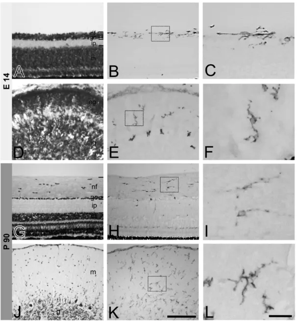
Fig. 1. CD44 expression in the retina(A-C, G-I) and cerebellum(D-F, J-L) of E14 chick embryo and adult chicken. Cresyl violet staining shows layers of the retina(A, G) and cerebellum(D, J). Images in small boxes of B, E, H, and K are magnified to C, F, I, and L, respective- ly. At E14 day, CD44-ir cells in the retina are mainly located in the nerve fiber layer(B). At P90 CD44-ir cells are also observed mainly in the nerve fiber layer(H). In the cerebellum, CD44-ir cells are distributed throughout the cerebellar cortex of E14 embryo(E) and P90 adult (K). CD44-ir cells show ramified cytoplasmic processes from their cell bodies in higher magnified images(C, F, I, L). nf, nerve fiber layer;
gc, ganglion cell layer; ip, inner plexiform layer; in, inner nuclear layer; eg, external granular layer; ig, internal granular layer; m, molecular layer; g, granule cell layer. Bars represent 100μm(A, B, D, E, G, H, J, K) and 30μm(C, F, I, L).
32 Ji Young Kim, Hyun Joon Sohn, Je Hoon Seo, Eun Young Lee
Results
1. Distribution of CD44 expressing cells in the retina and cerebellum of chick embryo(E14) and adult chicken(P90)
Even at embryonic stage(E14), CD44-immunoreactive (ir) cells were observed in both, the retina and the cer- ebellum(Fig. 1A-F). In E14 embryonic retina, CD44-ir cells were mainly located in the nerve fiber layer(Fig. 1B,
C). In E14 embryonic cerebellum, CD44-ir cells were dis- tributed throughout the cerebellar cortex including the ex- ternal and internal granular layer(Fig. 1D-F). In P90 adult chicken retina, most of CD44-ir cells were also found mainly in the nerve fiber layer(Fig. 1H) and some cells were dispersed through other layers. In the cerebellum at this age, CD44-ir cells were distributed in overall layers of the cerebellar cortex including the molecular, Purkinje cell, and granular layers(Fig. 1K). At higher magnified Fig. 2. Double immunofluorescent staining for CD44 and RCA-1 in the retina(A, C) and cerebellum(B, D) of E14 chick embryo and adult chicken. All merged images(A3, B3, C3, D3) represent that CD44-ir cells are colocalized with RCA-1-immunoreactivity, a marker of mi- croglia. nf, nerve fiber layer; gc, ganglion cell layer; ip, inner plexiform layer. Bar indicates 50μm.
images morphological features of CD44-ir cells showed ramified cytoplasmic processes from their cell bodies(Fig.
1C, F, I, L).
2. Type of cells expressing CD44 in the retina and the cerebellum
As indicated by double immunofluorescent staining for CD44 and RCA-1, a marker of microglia, almost all the
CD44-ir cells were also immunoreactive to RCA-1, both in the retina and cerebellum at E14 and P90(Fig. 2). In the retina from E14 embryos, CD44-ir cells were observed mainly in nerve fiber layer(Fig. 2A1), which also showed RCA-1-immunoreactivity(Fig. 2A2, A3). Similarly, most of the CD44-ir cells in the cerebellum from E14 embryos (Fig. 2B1) were also RCA-1-immunoreactivity(Fig. 2B2, B3). The co-localization of CD44 and RCA-1 was also Fig. 3. Double immunofluorescent staining for CD44 and GFAP in the retina(A, C) and cerebellum(B, D) of E14 chick embryo and adult chicken. GFAP-immunoreactiviry is observed only in adult cerebellum(D2), not in E14(A2) and P90 retina(C2), and E14 embryonic cer- ebellum(B2). All merged images(A3, B3, C3, D3) represent that CD44-ir cells are not labeled with GFAP, a marker of astrocytes.(A) E14 embryonic retina,(B) E14 embryonic cerebellum,(C) P90 adult retina,(D) P90 adult cerebellum. nf, nerve fiber layer; gc, ganglion cell lay- er; ip, inner plexiform layer. Bar indicates 50μm for all figures.
34 Ji Young Kim, Hyun Joon Sohn, Je Hoon Seo, Eun Young Lee
found in cells from the retina(Fig. 2C1-C3) and the cere- bellum(Fig. 2D1-D3) of P90 adult chicken. These results indicate that almost all CD44-ir cells in the developing and adult chicken retina and cerebellum also express RCA-1, a marker of microglia.
On the other hand, double immunofluorescent stain- ing for GFAP and CD44 did not show co-localization of these two proteins either in the retina and the cerebellum of E14 and P90(Fig. 3). GFAP, a marker of astrocytes was not detected in the chick embryonic(Fig. 3A2) and adult chicken retina(Fig. 3C2). Thus, CD44-ir cells in the retina did not show any GFAP-immunoreactivity(Fig. 3A3, C3) with double immunofluorescent staining. To understand which cell types expressed CD44, LPS was injected in the eyeball of P90 adult chicken: the retina was subsequently examined. After LPS stimulation, the number of CD44- ir cells did not seem to be increased. Rather, the intensity of CD44-immunoreactivity was increased in cells of the nerve fiber layer of P90 adult chicken retina(Fig. 4A1, B1).
These CD44-ir cells also showed RCA-1 immunoreactiv- ity(Fig. 4A2, A3). Even after LPS stimulation, GFAP-im- munoreactivity was not detected(Fig. 4B2) and thus,
co-localization of CD44 and GFAP was not observed(Fig.
4B3).
Discussion
The present study showed that CD44 is expressed in the developing and adult chicken retina and cerebellum. The CD44-ir cells in these tissues are microglia, not astrocytes.
Many other studies have reported the CD44 expression in microglia; canine retinal microglia in normal condition [21], microglia after ischemic brain injury of the rat[10,11], and microglia in Parkinson mice model by treatment with α-synuclein[14]. However, some studies have showed CD44 expression in astrocytes during development and adult; astrocyte precursor cells in developing chick[4,5], mice and rat[6,7,22], Müller glial cells in mice retina[9].
In some human disease, CD44 expression was reported in astrocyte-restricted precursor cells or reactive astrocytes [3,23]. On the other hand, CD44 expression both in the microglia and astrocytes has been also reported[13]. The discrepancy between the results obtained here and those Fig. 4. CD44 expression in P90 adult chicken retina after LPS stimulation and double immunofluorescent staining for RCA-1 and GFAP, respectively. At 24 hours after LPS injection, CD44 is overexpressed and its immunoreactivity is observed mainly in the nerve fiber lay- er of the retina(A1, B1). CD44-immunoreactivity is colocalized with RCA-1(A3) but not with GFAP(B3). Despite of LPS stimulation, GFAP-immunoreactivity is not observed(B2). These results represent that CD44 is expressed both in resting and activated microglia of adult retina. Bar indicates 50μm for all figures.
in previous studies regarding the type of cells expressing CD44 might be related to the presence of many isoforms of the CD44 protein. In a review of Ponta et al.[24], CD44 pre-mRNA is encoded by 20 exons, 10 of which can be regulated by alternative splicing. This alternative splicing is regulated by tissue-specific factors[24]. CD44 antibod- ies used in previous studies might have been generated against different epitopes of different CD44 isoforms. The monoclonal antibody for CD44 used in the present study was developed by the Developmental Studies Hybridoma Bank of University of Iowa. According to the data sheet provided by the Institute, immunogen substance is proteo- glycans from chick embryo and it labels macrophages in chick and quail of optic nerve. One of the possible ex- planations of the finding that CD44 is observed to be re- stricted to microglia might be due to the use, in this study, of a monoclonal antibody which is probably specific for microglial CD44.
Transmembrane hyaluronan receptor CD44 has been suggested to regulate cellular response to hyaluronan and assembly of hyaluronan-rich extracellular matrix[25]. Fur- ther, the degradation of hyaluronan begins when high- molecular weight hyaluronan molecule bind to the trans- membrane receptor, CD44, before internalization and ly- sosomal degradation[3]. Within the cytoplasm, the cyto- plasmic domain of CD44 interacts with actin cytoskeleton by which CD44 can influence cell signaling[2], by which CD44 participates in many cellular processes including cell migration, survival, and differentiation[2].
During development of the mouse, CD44 expressed in chiasmatic neurons was suggested to play a role in mid- line crossing of retinal axons in optic chiasm[26]. In the retina, most of CD44-ir cells were observed in nerve fiber layer, a layer forming the optic nerve. Although the CD44-ir cells were microglia, not neurons in our study using chick, the results obtained here might implicate that CD44 could influence the behavior of retinal axons in the nerve fiber layer during development and in its adult stage. When the retina was stimulated with LPS, CD44 immunoreactivity increased in the microglia, but not in astrocytes in the present study. The possible reason for the absence of GFAP expression, even upon LPS stimulation may be related to the fact that in avians, unlike in mam- mals, most of the retina does not contain definite astrocytes, with the exception of the vascularized optic nerve head region[27,28]. The results of this study support these pre-
vious avian-specific retinal findings.
Regarding the cerebellum, CD44-ir microglia was not concentrated to one specific layer. Rather, CD44-ir cells were scattered throughout the whole cerebellar cortex. In addition, the CD44-ir microglial cells were more frequent- ly observed in adult chicken cerebellum than in the E14 embryonic stage. These findings are different from other studies reporting changes in CD44 expression patterns through life span including from glial progenitor cells in the developing stage to mature cells in the adult stage; in the mouse cerebellum, CD44 was expressed in develop- ing astrocyte precursor cells but not in the mature astro- cyte[7]. Additionally, in the study of Naruse[22], CD44 in the mouse cerebellum of embryonic period was widely expressed in astrocyte precursor cells, neural stem cells, and oligodendrocyte precursor cells, however, over the course of development, CD44 became restricted to gran- ule neurons in the adult. Although in the present study we did not perform any double staining of CD44 and markers of neurons or oligodendrocytes, the CD44-ir cells in the retina were morphologically different from neurons or oligodendrocytes, based on our previous findings[20].
Oligodendrocytes exhibit small round cytoplasm located in the nerve fiber layer, while neurons present relatively large cell bodies located in the ganglion cell layer of the retina[20].
There are no reports about CD44 expressing microglia in the developing retina or cerebellum. Unlike astrocytes, microglia are of hematopoietic origin. Hematopoietic cells were also previously reported to express CD44[4]. The appearance of microglia in the CNS during the develop- ment and the adult life is the cause of great controversies.
Recent study using rat showed that in early embryonic day, microglia/macrophage cells were located in ventri- cles, thereafter until neonate, cells were dispersed in brain matrix[29]. From young adult microglia/macrophage were found mostly in capillaries and vascular margines in the CNS parenchyme[29]. Greter and Merad[30] suggest- ed that bone marrow-derived myeloid cells were constant- ly recruited to the brain in the steady state. Interestingly, these bone marrow derived microglia show brain regional preference; they are detected in the olfactory bulb, cere- bellum, and hippocampus rather than the cerebral cortex [16]. In injured brain, bone marrow derived cells were found only in the cerebellum and in injury sites[17].
The surface molecules of microglia can be rapidly upreg-
36 Ji Young Kim, Hyun Joon Sohn, Je Hoon Seo, Eun Young Lee ulated in response to inflammatory stimuli, e.g., IFN-gam- ma or LPS[30]. In the present study, CD44 expression was increased by LPS stimulation. Microglia is generally known to participate in immunological function as an anti gen presenting cells and subsequent responses in the CNS. Thus, increased expression of CD44 in activated microglia could play a certain role in immunological res- ponses in the CNS. In relation to immunologic roles, the CD44 was suggested to have role in monocyte and/or lymphocyte adhesion and subsequent migration into the brain across the blood-brain barrier[20]. In experimental autoimmune uveitis mice model, macrophage migration inhibitory factor(MIF), an inflammatory cytokine was increased with simultaneous increase of CD44[31]. One of the immune regulator MIF signal transduction pathway is mediated by CD44, a transmembrane receptor in both normal physiological conditions and uveitis[31]. Several studies implicated that CD44 might mediate signal trans- duction necessary for endocytosis of bacteria, which lead to events of linking between CD44 and actin cytoskeleton [20]. The microglia monitor microenvironmal changes even in the resting state. They are capable of morphologi- cal and functional rapid adaptation to dynamic microenvi- ronmental conditions[32]. The microglial transmembrane protein CD44, a receptor for an adhesion molecule, seems to be an essential component for these general microgli- al functions. In summary, the present study showed that CD44 is expressed in the retina and the cerebellum of the developing and adult chicken. The CD44-ir cells are mi- croglia. Microglia activated by LPS stimulation showed increased expression of CD44. Those results suggested that, in microglia, the transmembrane protein CD44 may play important roles in development, in inflammatory condition, and even in the normal resting state of the ret- ina and the CNS. The specific functions of the microglial CD44 need to be clarified by further studies.
REFERENCES
1. Aruffo A, Stamenkovic I, Melnick M, Underhill CB, Seed B.
CD44 is the principal cell surface receptor for hyaluronate.
Cell. 1990; 61:1303-13.
2. Ponta H, Sherman I, Herrlich PA. CD44: from adhesion molecules to signaling regulators. Nat Rev Mol Cell Biol.
2003; 4:33-45.
3. Sherman LS, Back SA. A ‘GAG’ reflex prevents repair of the damaged CNS. Trends Neurosci. 2008; 31:44-52.
4. Corbel C, Lehmann A, Davison F. Expression CD44 during early development of the chick embryo. Mech Dev. 2000;
96:111-4.
5. Alfei L, Aita M, Caronti B, Vita RD, Margotta V, Albani LM, et al. Hyaluronate receptor CD44 is expressed by as- trocytes in the adult chicken and in astrocyte cell precursors in early development of the chick spinal cord. Eur J Histo- chem. 1999; 43:29-38.
6. Liu Y, Han SSW, Wu Y, Tuohy TMF, Xue H, Cai J, et al.
CD44 expression identifies astrocyte-restricted precursor cells. Dev Biol. 2001; 276: 31-46.
7. Cai N, Kurachi M, Shibasaki K, Okano-Uchida T, Ishizaki Y. CD44-positive cells are candidates for astrocytes pre- cursor cells in developing mouse cerebellum. Cerebellum.
2011; 11:181-93.
8. Baier C, Baader SL, Jankowski J, Gieselmann V, Schilling K, Rauch U, et al. Hyaluronan is organized into fiber-like structures along migratory pathways in the developing mouse cerebellum. Matrix Biol. 2007; 26:348-58.
9. Shinoe T, Kuribayashi H, Saya H, Seiki M, Aburatani H, Watanabe S. Identification of CD44 as a cell surface marker for Müller glia precursor cells. J Neurochem. 2010;
115:1633-42.
10. Wang H, Zhan Y, Xu L, Feuerstein GZ, Wang X. Use of suppression subtractive hybridization for differential gene expression in stroke: Discovery of CD44 gene expression and localization in permanent focal stroke in rats. Stroke.
2001; 32:1020-27.
11. Wang X, Xu L, Wang H, Zhan Y, Puré E, Feuerstein GZ.
CD44 deficiency in mice protects brain from cerebral isch- emia injury. J Neurochem. 2001; 83:1172-79.
12. Jones LL, Liu Z, Shen J, Werner A, Kreutzberg GW, Raiv- ich G. Regulation of the cell adhesion molecule CD44 after nerve transaction and direct trauma to the mouse brain. J Comp Neurol. 2000; 23:468-92.
13. Matsumoto T, Imagama S, Hirano K, Ohgomori T, Natori T, Kobayashi K, et al. CD44 expression in astrocytes and microglia is associated with ALS progression in a mouse model. Neurosci Lett. 2012; 520:115-20.
14. Kim S, Cho SH, Kim KY, Shin KY, Kim HS, Park CH, et al. Α-synuclein induces migration of BV-2 microglial cells by up-regulation of CD44 and MT1-MMP. J Neurochem.
2009; 109:1483-96.
15. Gorlewicz A, Wlodarczyk J, Wilczek E, Gawlak M, Cabaj A, Majczynski H, et al. CD44 is expressed in non-myelin- ating Schwann cells of the adult rat, and may play a role in neurodegeneration-induced glial plasticity at the neuromus- cular junction. Neurobiol Dis. 2009; 34:245-58.
16. Priller J, Flugel A, Wehner T, Boentert M, Haas CA, Prinz M, et al. Targeting gene-modified hematopoietic cells to the central nervous system: use of green fluorescent protein uncovers microglial engrafment. Nat Med. 2001; 7:1356- 17. Vallieres L, Sawchenko PE. Bone marrow-derived cells 61.
that populate the adult mouse brain preserve their hemato- poietic identity. J Neurosci. 2003; 23:5197-207.
18. Hamburger V, Hamilton HL. A series of normal stages in the development of the chick embryo. Dev Dyn. 1992;
195:231-72.
19. Mannoji H, Yeger H, Becker LE. A specific histochemical marker(lectin Ricinus communis agglutinin-1) for normal human microglia, and application to routine histopathology.
Acta Neuropathol. 1986; 71:341-3.
20. Kim JY, Song SH, Kim HN, Kim DW, Sohn HJ, Lee EY, et al. αB-crystallin is expressed in myelinating oligodendro- cytes of the developing and adult avian retina. Neurochem Res. 2012; 37:2135-42.
21. Genini S, Beltran WA, Stein VM, Aguirre GD. Isolation and ex vivo characterizarion of the immunophenotype and function of microglia/macrophage populations in normal dog retina. Adv Exp Med Biol. 2014; 801:339-45.
22. Alfei L, Aita M, Caronti B, Vita RD, Margotta V, Albani LM, et al. Hyaluronate receptor CD44 is expressed by as- trocytes in the adult chicken and in astrocyte cell precursors in early development of the chick spinal cord. Eur J Histo- chem. 1999; 43:29-38.
23. Naruse M, Shibasaki K, Yokoyama S, Kurachi M, Ishizaki Y. Dynamic changes of CD44 expression from progenitors to subpopulations of astrocytes and neurons in developing cerebellum. PLoS One. 2013; 8:1-12.
24. Buiani M, Postma N, Polder E, Dieleman N, Scheffer PG, Sim FJ, et al. Hyaluronan accumulation and arrested oligo- dendrocyte progenitor maturation in vanishing white matter disease. Brain. 2013; 136:209-22.
25. Sherman LS, Struve JN, Rangwala R, Wallingford NM, Tu- ohy TMF, Iv CK. Hyaluronate-based extracellular matrix:
keeping glia in their place. Glia. 2002; 38:93-102.
26. Lin L, Chan SO. Pertubation of CD44 function affects chi- asmatic routing of retinal axons in brain slice preparations of the mouse retinofugal pathway. Eur J Neurosci. 2003;
17:2299-312.
27. Fischer AJ, Zelinka C, Scott MA. Heterogeneity of glia in the retina and optic nerve of birds and mammals. PLoS One. 2010; 5:1-15.
28. Kim JY, Sohn HJ, Lee EY, Goo YS, Kim DW, Seo JH. Ex- pression of αB-crystallin in the peripapillary glial cells of the developing chick retina. Neurochem Res. 2011; 36:76- 29. Ghosh P, Mukherjee N, Ghosh K, Mallick S, Pal C, Las-82.
kar A, et al. Prospective microglia and brain macrophage distribution pattern in normal rat brain shows age sensitive dispersal and stabilization with development. Indian J Exp Biol. 2015; 53:561-7.
30. Greter M, Merad M. Regulation of microglia development and homeostasis. Glia. 2013; 61:121-7.
31. Yang H, Zheng S, Mao Y, Chen Z, Zheng C, Li H, et al.
Modulating of ocular inflammation with macrophage mi- gration inhibitory factor is associated with notch signaling in experimental autoimmune uveitis. Clin Exp Immunol.
2015; 183:280-93.
32. Nayak D, Roth TL, McGavern DB. Microglia development and function. Annu Rev Immunol. 2014; 32:367-402.
38 Ji Young Kim, Hyun Joon Sohn, Je Hoon Seo, Eun Young Lee
발생 및 성체 닭의 망막과 소뇌에서 미세아교세포에서의 CD44 발현
김지영, 손현준, 서제훈, 이은영
충북대학교 의과대학 해부학교실
간추림 : CD44는 막관통단백질로서 히알루론산에 대한 막 표면 수용체로 작용한다고 알려져 있다. CD44가 발현되
는 세포 종류 및 세포 내에서의 역할에 대하여 많은 연구가 시행되고 있으나 모든 결과들이 일치하지는 않는다. 본
연구에서는 발생 중 및 성체 조류의 망막과 소뇌에서 CD44가 발현되는지 확인하고 CD44가 발현되는 세포의 종류
와 그 분포를 알아보고자 하였다.
본 연구에서는 발생 14일과 부화 후 90일(성체) 된 닭의 망막과 소뇌를 사용하였다. 면역화학염색을 위해
CD44, RCA-1 그리고 GFAP 항체를 사용하였다. CD44 면역반응 세포들은 모든 시기의 망막과 소뇌에서 관찰
되었다. 발생 14일 망막에서 CD44 면역반응 세포들은 주로 신경섬유층에서 관찰되었다. 성체에서도 대부분의
CD44 면역반응 세포들이 신경섬유층에서 관찰되었고 몇몇은 망막의 다른 층에 흩어져서 보였다. 발생 14일 소
뇌의 CD44 면역반응 세포들은 바깥 및 속과립층을 포함한 소뇌 겉질에서 관찰되었다. 성체 소뇌의 CD44 면역
반응 세포들은 소뇌 전체 층에 흩어져 존재하였으며, 발생 14일 때보다 더 많은 세포들이 관찰되었다. 고배율에
서 관찰한 결과, CD44 면역반응 세포들은 세포체로부터 분지한 세포질돌기를 나타내었다. 미세아교세포 표지자
인 RCA-1과 이중형광 염색을 한 결과, 발생 중과 성체의 망막과 소뇌에서, 대부분의 CD44 면역반응물은 RCA-1
에 면역 염색성을 보이는 세포에 함께 존재하였다. 그러나 GFAP와 CD44 면역반응은 동일한 세포 내에서 관찰
되지 않았다. 성체 망막에 LPS로 자극을 주었을 때 CD44 면역반응은 증가하였으며, 이 세포는 RCA-1에도 면역
염색성을 나타내었다. 따라서, 결과를 종합하면, 정상 환경에서도 CD44는 발생 중 및 성체 조류의 망막과 소뇌
의 미세아교세포에서 발현되었으며, 이는 LPS 자극에 의해 발현이 증가하였음을 보여줌으로써, 미세아교세포의
역할에 CD44가 중요하리라는 가능성을 시사하였다.
찾아보기 낱말 : CD44, 미세아교세포, 닭, 망막, 소뇌
교신저자 : 이은영, 서제훈(충북대학교 의과대학 해부학교실)
전자우편 : [email protected], [email protected]