서 론
법의인류학적 검사는 개인식별항목(biological profile) 즉, 인구집단 (ethnicity), 성별 (sex), 신장 (height), 사망당 시의 나이 (age) 등을 추정하여 신원확인을 하는 데 도 움을 준다 [1]. 기본적인 네 가지 개인식별항목 중에서 도 성별의 추정은 나머지 세 가지 개인식별항목을 추
정하는 데 영향을 미칠 수 있으므로 중요하다 [2]. 체질 인류학적 연구를 통해 밝혀진 형태학적 특징은 인구집 단에 따라 다르기 때문에, 어떤 뼈대의 형태학적 특징 을 이미 연구된 체질인류학적 결과와 비교하면 개인식 별항목을 추정할 수 있다 [3]. 그런데 한국인의 개인식 별항목에 대한 연구는 뼈표본이 부족하기 때문에 외국 의 연구에 비해 미진한 상황이다[3].
한국과학기술정보연구원 (KISTI)은 2000년부터 한국 인의 고유한 인체 특징을 연구할 수 있는 기반 마련을 위하여 디지털 코리언 (Digital Korean) 데이터베이스라 는 인체정보 구축사업을 시작하였다 [4]. 디지털 코리언 데이터베이스는 가톨릭대학교 의과대학 해부학교실과 순천향대학교 의과대학 해부학교실에 기증된 약 100여
한국인 3차원 인체형상모델을 이용한 고리뼈의 성별판별 검증
홍종근
1, 정재광
2, 이우영
3, 김덕수
1, 박대균
1, 박경호
11순천향대학교 의과대학 해부학교실, 2경희대학교 의학계열 실습지원센터,
3가톨릭대학교 의과대학 해부학교실∙가톨릭응용해부연구소
(2014년 11월 6일 접수, 2014년 12월 10일 수정접수, 2014년 12월 15일 게재승인, Published Online 30 December 2014)
간추림 : 사망 당시의 정보를 알고 있는 마른뼈들을 통계학적으로 분석한 연구가 있다면 마른뼈를 법의인류학 적으로 분석하여 개인식별항목(biological profile)을 추정하는 것이 가능하다. 한국인의 경우 개인식별항목을 추 정하는 데 필요한 뼈표본이 거의 없기 때문에 개인식별항목을 추정하기 위한 통계학적 자료가 부족하다. 그러 므로 뼈대에 붙어 있는 물렁조직을 모두 제거하여 뼈표본을 제작하는 방법의 대안으로 방사선으로 촬영된 진단 영상을 활용하여 뼈대를 3차원으로 재구성하는 연구가 제안되고 있다. 이 연구는 해부용 시신을 컴퓨터단층촬 영 (computed tomography)하여 구축한 3차원 인체형상모델에서 뼈대 정보를 재구성하여 계측하였고, 통계학적 분석을 통해 성별을 추정하는 데 도움이 되는지를 조사하였다.
가톨릭대학교 가톨릭응용해부연구소에서 관리하고 있는 디지털 코리언 (Digital Korean) 데이터베이스에서 고 리뼈 (atlas) 100개 (남성 51개, 여성 49개)를 계측하였다. 3차원 컴퓨터 프로그램인 미믹스 (Mimics, Simulation module, Version 13.0, Materialise NV, Belgium)를 이용하여 계측항목들을 계측하였고, IBM® SPSS® (version 20.0, Armonk, New York)를 이용하여 통계학적 분석을 하였다.
계측 결과를 실제 마른뼈의 연구에서 얻은 판별함수를 적용하였을 때 정확도가 떨어졌기 때문에 새롭게 판별 함수를 구하였고, 19개 계측항목들 중에서 고리뼈너비(Width of Atlas)가 성별을 추정하는 데 가장 유용한 계측 항목이었다. 본 연구 결과 고리뼈너비가 76.7 mm보다 큰 값일 경우 남성으로 판별할 수 있었다.
뼈표본이 부족한 현실에서 한국인 뼈대의 체질인류학적 특징을 연구하는 것은 한계가 있다. 그러므로 방사선 진단영상에서 3차원으로 재구성한 뼈대를 활용하여 형태학적 특징을 규명하려고 시도된 이번 연구의 결과는 디지털 코리언 데이터베이스를 활용하여 체질인류학적 연구를 할 수 있는 가능성을 보여주었다고 생각되고 현 장에서도 계측값을 통해 성별을 추정하는 데 도움이 될 것으로 판단된다.
찾아보기 낱말 : 고리뼈, 한국인, 디지털 코리언, 계측적 연구, 컴퓨터단층사진 http://dx.doi.org/10.11637/kjpa.2014.27.4.225
*이 논문은 2008년 정부 (교육과학기술부)의 재원으로 한국연구재단 (한 국학술진흥재단)의 지원을 받아 수행된 연구임(NRF-2008-313-E00022).
저자(들)는 ‘의학논문 출판윤리 가이드라인’을 준수합니다.
저자(들)는 이 연구와 관련하여 이해관계가 없음을 밝힙니다.
교신저자 : 박경호(순천향대학교 의과대학 해부학교실) 전자우편 : [email protected]
Original Article
명의 한국인 해부용 시신을 컴퓨터단층촬영 (computed tomography)한 영상이며, 이 영상에서 개별 뼈를 구역 화 (segmentation)하면 3차원으로 재구성된 뼈 정보를 획득할 수 있다 [4]. 한국인 맞춤형 무릎인공관절을 개 발하기 위한 연구 [5]와 열두째등뼈를 활용한 성별판별 연구 [6]에서 디지털 코리언 데이터베이스가 사용되었 다고 보고된 바 있다.
체질인류학에서는 뼈대에 붙어 있는 물렁조직을 모 두 제거하여 마른뼈 상태로 변형시켜 뼈대를 계측는 것이 일반적이다. 그런데 발굴된 인체를 그대로 컴퓨터 단층촬영하여 3차원으로 재구성된 뼈를 계측한 연구에 의하면 실제 뼈와 계측값이 통계학적으로 거의 동일하 였다 [7]. 이 연구에서 연구자들은 현장에서 발굴된 인 체 또는 뼈대를 3차원으로 재구성한 뒤 그 정보를 실 험실로 전송하는 방식으로도 체질인류학적 검사가 가 능할 것이라고 하였다 [7]. Robinson 등 (2008) [7]의 연 구에서는 인체를 1.25 mm 간격으로 단층촬영하였지만 본 연구에 이용된 디지털 코리언[4] 데이터베이스는 이 보다 세밀한 1.0 mm 간격으로 단층촬영하였기 때문에 디지털 코리언 데이터베이스에서 3차원으로 재구성된 뼈를 계측하는 것은 더욱 정밀하다.
이 연구는 해부용 시신을 컴퓨터단층촬영하여 구축 한 3차원 인체형상모델인 디지털 코리언 데이터베이스 에서 고리뼈 (atlas)를 구역화하여 계측한 것으로, 실제 고리뼈를 직접 계측했던 연구결과 [3]를 적용해 봄으로 써 디지털 코리언 데이터베이스가 체질인류학적 연구 에 도움이 될 수 있는지를 확인하는 것과 현장에서 활 용 가능한 결과가 도출될 수 있는지를 알아보는 것이 연구의 목적이다.
재료 및 방법
가톨릭대학교 가톨릭응용해부연구소에서 관리하고 있 는 디지털 코리언 데이터베이스 [4-6]에서 구역화한 고 리뼈 100개(남성 51개, 여성 49개)를 연구 재료로 이용 하였으며, 남성의 평균 나이는 50.7세 (20세부터 60세), 여성의 평균나이는 53.7세 (27세부터 60세)였다. 컴퓨터 단층촬영에서 얻은 진단영상들은 DICOM이란 확장자로 표현된 2차원 영상인데, 미믹스 (Mimics, Version 13.0, Materialise NV, Belgium)라는 컴퓨터 프로그램에서 2차 원 영상을 3차원으로 재구성하여 재구성된 3차원 영상 에서 고리뼈에 대한 정보만을 구역화하였다.
본 연구에서는 Simulation module을 갖춘 미믹스를 이용하여 고리뼈를 계측하였고, 계측항목은 이전 연구 자가 마른뼈를 계측한 항목을 참고로 하였다 [8]. 실제
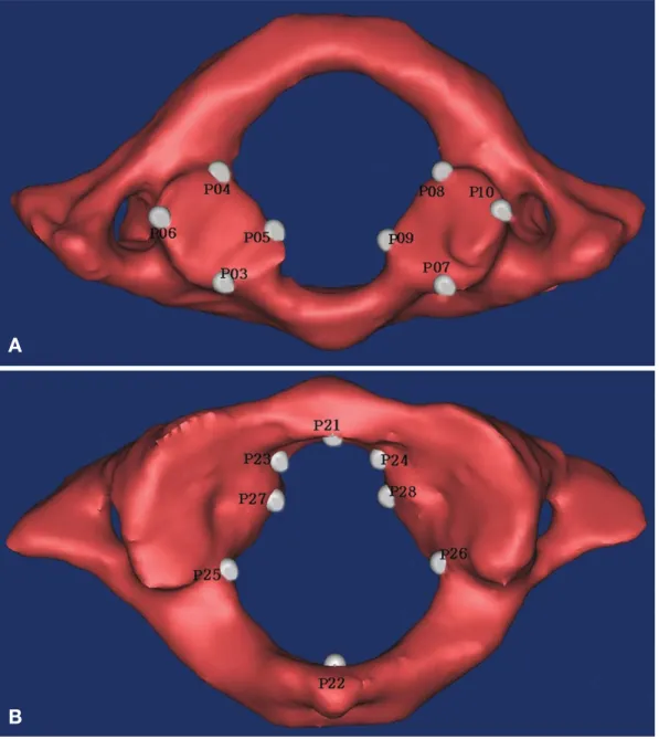
마른뼈에서는 잘 구별되지만 3차원으로 재구성된 영상 에서는 정확한 경계를 알 수 없는 관절면에 대한 계측 항목은 제외하였다. Simulation module에는 연구자가 임 의로 계측점과 계측항목을 정의하여 추가할 수 있는 기 능이 있다. 연구자는 미믹스 프로그램에서 재구성된 3차 원 영상을 여러 각도로 움직여 계측항목들의 시작점과 끝점 찾아 3차원 영상 위에 표시하였고, 시작점과 끝점 이 표시되면 자동으로 계측값이 측정된다. 미믹스에서 정의한 계측점은 38개였고, 두 개의 계측점 사이인 19개 항목을 계측하였으며, 각 38개의 계측점(Fig. 1)과 19개 의 계측항목(Fig. 2)은 다음과 같다.
1. 계측점 (Point on 3D data)
P01) 위관절돌기 (왼쪽)의 가쪽점 (Lateral point of supe- rior articular process, Left)
P02) 위관절돌기(오른쪽)의 가쪽점(Lateral point of supe- rior articular process, Right)
P03) 아래관절면(왼쪽)의 앞점(Anterior point of inferior articular surface, Left)
P04) 아래관절면(왼쪽)의 뒤점(Posterior point of inferior articular surface, Left)
P05) 아래관절면(왼쪽)의 안쪽점(Medial point of inferior articular surface, Left)
P06) 아래관절면(왼쪽)의 가쪽점(Lateral point of inferior articular surface, Left)
P07) 아래관절면 (오른쪽)의 앞점 (Anterior point of infe- rior articular surface, Right)
P08) 아래관절면(오른쪽)의 뒤점(Posterior point of infe- rior articular surface, Right)
P09) 아래관절면(오른쪽)의 안쪽점(Medial point of infe- rior articular surface, Right)
P10) 아래관절면(오른쪽)의 가쪽점(Lateral point of infe- rior articular surface, Right)
P11) 아래관절돌기(왼쪽)의 가쪽점(Lateral point of infe- rior articular process, Left)
P12) 아래관절돌기 (오른쪽)의 가쪽점 (Lateral point of inferior articular process, Right)
P13) 앞결절의 앞점(Anterior point of anterior tubercle) P14) 뒤결절의 뒤점(Posterior point of posterior tubercle) P15) 가로돌기(왼쪽)의 가쪽점(Lateral point of transver-
se process, Left)
P16) 가로돌기(오른쪽)의 가쪽점(Lateral point of transv- erse process, Right)
P17) 앞고리의 위점(Superior point of anterior arch) P18) 앞고리의 아래점(Inferior point of anterior arch)
P19) 뒤고리의 위점(Superior point of posterior arch) P20) 뒤고리의 아래점(Inferior point of posterior arch) P21) 앞고리의 뒤점(Posterior point of anterior arch) P22) 뒤고리의 앞점(Anterior point of posterior arch) P23) 앞고리 (왼쪽)의 안쪽점 (Medial point of anterior
arch, Left)
P24) 앞고리 (오른쪽)의 안쪽점 (Medial point of anterior arch, Right)
P25) 가쪽덩이(왼쪽)의 가장속점(Innermost point of late-
ral mass, Left)
P26) 가쪽덩이 (오른쪽)의 가장속점 (Innermost point of lateral mass, Right)
P27) 가쪽덩이 (왼쪽)의 돌출점 (Projected point of lateral mass, Left)
P28) 가쪽덩이(오른쪽)의 돌출점(Projected point of late- ral mass, Right)
P29) 가로구멍(왼쪽)의 위점(Superior point of transverse foramen, Left)
Fig. 1.Captured images from Mimics simulation module. In these images, only a few examples of points were demonstrated. After the points were marked by researcher, measurements were performed automatically by the Mimics stimulation module. Each numbered P was explained at methods and materials. A. Inferior view of the Atlas, B. Superior view of the Atlas
A
B
P30) 가로구멍(왼쪽)의 아래점(Inferior point of transver- se foramen, Left)
P31) 가로구멍(오른쪽)의 위점(Superior point of transver- se foramen, Right)
P32) 가로구멍(오른쪽)의 아래점(Inferior point of transver- se foramen, Right)
P33) 치아돌기오목의 왼쪽점(lateral point of fovea dentis, Left)
P34) 치아돌기오목의 오른쪽점(lateral point of fovea den- tis, Right)
P35) 가쪽덩이 (왼쪽)의 위점 (Superior point of lateral mass, Left)
P36) 가쪽덩이 (왼쪽)의 아래점 (Inferior point of lateral mass, Left)
P37) 가쪽덩이 (오른쪽)의 위점 (Superior point of lateral mass, Right)
P38) 가쪽덩이 (오른쪽)의 아래점 (Inferior point of lateral
mass, Right)
2. 계측항목 (Measurements)
1) 위관절면사이최대거리(Maximum distance between superior articular surface)
�정의 : 오른쪽 위관절면과 왼쪽 위관절면 사이의 최 대거리
�계측방법 : P01번 계측점인 위관절돌기(왼쪽)의 가 쪽점(Lateral point of superior articular process, Left) 과 P02번 계측점인 위관절돌기 (오른쪽)의 가쪽점 (Lateral point of superior articular process, Right)의 거리
2) 아래관절면길이(왼쪽) (Length of inferior articular surface, Lt.)
�정의 : 왼쪽 아래관절면의 최대길이
�계측방법 : P03번 계측점인 아래관절면(왼쪽)의 앞 Fig. 2.Captured images from Photoshop and Mimics simulation module. In these images, only a few examples of measurements were demonstrated. Definition for each number was explained at table 1. A. Superior view of the Atlas B. Inferior view of the Atlas, C. Anterior view of the Atlas D. Posterior view of the Atlas
A B
C
D
점(Anterior point of inferior articular surface, Left)과 P04 번 계측점인 아래관절면 (왼쪽)의 뒤점 (Posterior point of inferior articular surface, Left)의 거리
3) 아래관절면너비(왼쪽) (Width of inferior articular surface, Lt.)
�정의 : 왼쪽 아래관절면의 최대너비
�계측방법 : P05번 계측점인 아래관절면(왼쪽)의 안 쪽점 (Medial point of inferior articular surface, Left) 과 P06번 계측점인 아래관절면(왼쪽)의 가쪽점(La- teral point of inferior articular surface, Left)의 거리
4) 아래관절면길이(오른쪽) (Length of inferior articular surface, Rt.)
�정의 : 오른쪽 아래관절면의 최대길이
�계측방법 : P07번 계측점인 아래관절면 (오른쪽)의 앞점(Anterior point of inferior articular surface, Right) 과 P08번 계측점인 아래관절면(오른쪽)의 뒤점(Po- sterior point of inferior articular surface, Right)의 거 리
5) 아래관절면너비(오른쪽) (Width of inferior articular surface, Rt.)
�정의 : 오른쪽 아래관절면의 최대너비
�계측방법 : P09번 계측점인 아래관절면 (오른쪽)의 안쪽점 (Medial point of inferior articular surface, Ri- ght)과 P10번 계측점인 아래관절면(오른쪽)의 가쪽 점(Lateral point of inferior articular surface, Right)의 거리
6) 아래관절면사이최대거리(Maximum distance between inferior articular surface)
�정의 : 오른쪽 아래관절면과 왼쪽 아래관절면 사이 의 최대거리
�계측방법 : P11번 계측점인 아래관절돌기 (왼쪽)의 가쪽점(Lateral point of inferior articular process, Left) 과 P12번 계측점인 아래관절돌기 (오른쪽)의 가쪽 점 (Lateral point of inferior articular process, Right) 의 거리
7) 고리뼈길이(Length of Atlas)
�정의 : 고리뼈의 최대길이
�계측방법 : P13번 계측점인 앞결절의 앞점(Anterior point of anterior tubercle)과 P14번 계측점인 뒤결절 의 뒤점(Posterior point of posterior tubercle)의 거리
8) 고리뼈너비(Width of Atlas)
�정의 : 고리뼈의 최대너비
�계측방법 : P15번 계측점인 가로돌기(왼쪽)의 가쪽
점(Lateral point of transverse process, Left)과 P16번 계측점인 가로돌기 (오른쪽)의 가쪽점 (Lateral point of transverse process, Right)의 거리
9) 앞고리높이(Height of anterior arch)
�정의 : 고리뼈의 앞고리부분의 높이
�계측방법 : P17번 계측점인 앞고리의 위점(Superior point of anterior arch)과 P18번 계측점인 앞고리의 아래점(Inferior point of anterior arch)의 거리
10) 뒤고리높이(Height of posterior arch)
�정의 : 고리뼈의 뒤고리부분의 높이
�계측방법 : P19번 계측점인 뒤고리의 위점(Superior point of posterior arch)과 P20번 계측점인 뒤고리의 아래점(Inferior point of posterior arch)의 거리
11) 시상지름(Sagittal diameter of vertebral foramen)
�정의 : 고리뼈 척추뼈구멍에서 앞-뒤방향의 최대지름
�계측방법 : P21번 계측점인 앞고리의 뒤점(Posterior point of anterior arch)과 P22번 계측점인 뒤고리의 앞점(Anterior point of posterior arch)의 거리
12) 앞고리지름(Diameter of anterior arch)
�정의 : 고리뼈 척추뼈구멍에서 앞고리부분의 최대 지름
�계측방법 : P23번 계측점인 앞고리(왼쪽)의 안쪽점 (Medial point of anterior arch, Left)과 P24번 계측점 인 앞고리(오른쪽)의 안쪽점(Medial point of anteri- or arch, Right)의 거리
13) 가로최대지름(Maximum transverse diameter of vertebral foramen)
�정의 : 고리뼈 척추뼈구멍에서 가로방향의 최대지름
�계측방법 : P25번 계측점인 가쪽덩이(왼쪽)의 가장 속점 (Innermost point of lateral mass, Left)과 P26번 계측점인 가쪽덩이 (오른쪽)의 가장속점 (Innermost point of lateral mass, Right)의 거리
14) 가로최소지름(Minimum transverse diameter of vertebral foramen)
�정의 : 고리뼈 척추뼈구멍에서 가로방향의 최소지름
�계측방법 : P27번 계측점인 가쪽덩이(왼쪽)의 돌출 점(Projected point of lateral mass, Left)과 P28번 계 측점인 가쪽덩이 (오른쪽)의 돌출점 (Projected point of lateral mass, Right)의 거리
15) 가로구멍길이(Length of transverse foramen, Lt.)
�정의 : 왼쪽 가로구멍의 길이
�계측방법 : P29번 계측점인 가로구멍(왼쪽)의 위점
(Superior point of transverse foramen, Left)과 P30번 계 측점인 가로구멍 (왼쪽)의 아래점 (Inferior point of trans- verse foramen, Left)의 거리
16) 가로구멍길이(Length of transverse foramen, Rt.)
�정의 : 오른쪽 가로구멍의 길이
�계측방법 : P31번 계측점인 가로구멍(오른쪽)의 위 점 (Superior point of transverse foramen, Right)과 P32번 계측점인 가로구멍(오른쪽)의 아래점(Inferior point of transverse foramen, Right)의 거리
17) 치아돌기오목너비(Width of surface of dens)
�정의 : 고리뼈 척추뼈구멍에서 치아돌기오목의 최대 너비
�계측방법 : P33번 계측점인 치아돌기오목의 왼쪽점 (lateral point of fovea dentis, Left)과 P34번 계측점 인 치아돌기오목의 오른쪽점 (lateral point of fovea dentis, Right)의 거리
18) 위-아래관절면사이 높이(왼쪽) (Height of superior- inferior articular surface, Lt.)
�정의 : 왼쪽위관절면과 왼쪽아래관절면사이의 최대 높이
�계측방법 : P35번 계측점인 가쪽덩이(왼쪽)의 위점 (Superior point of lateral mass, Left)과 P36번 계측점 인 가쪽덩이(왼쪽)의 아래점(Inferior point of lateral mass, Left)의 거리
19) 위-아래관절면사이 높이(오른쪽) (Height of superior- inferior articular surface, Rt.)
�정의 : 오른쪽위관절면과 오른쪽아래관절면사이의 최대높이
�계측방법 : P37번 계측점인 가쪽덩이(오른쪽)의 위 점(Superior point of lateral mass, Right)과 P38번 계 측점인 가쪽덩이(오른쪽)의 아래점(Inferior point of lateral mass, Right)의 거리
계측값은 이전 연구결과 [3]에서 고리뼈너비 (width of atlas)항목에서 생성된 성별판별함수 (0.268×고리뼈너 비-20.316)에 적용해 보았고, 또한 IBM® SPSS® (ver- sion 20.0, Armonk, New York)를 이용하여 통계학적으 로 분석하였다. 성별판별 함수를 계산할 때에는 19개 계 측항목을 활용하여 성별판별함수(discriminant functions) 를 계산하였다.
결 과
3차원으로 재구성된 고리뼈를 컴퓨터 프로그램으로 형태 분석하여 얻은 19개의 계측값들에 대한 평균값을 정리하여 제시하였고 (Table 1), 이전 연구결과 [3]에서 고리뼈너비 (width of atlas)항목에서 생성된 성별판별함 수 (0.268×고리뼈너비-20.316)에 본 연구결과를 대입 하여 판별력을 계산하였다.
이전 연구결과의 성별판별함수에서 고리뼈너비의 기
Table 1.Mean of metric value of measurement for atlas in Korean
Measurements Male Female
(mean±SD) (mean±SD)
1 Maximum distance between superior articular surface 48.87±2.96 48.80±2.86
2 Length of inferior articular surface, Lt. 16.31±1.27 15.12±1.34
3 Width of inferior articular surface, Lt. 16.06±1.44 14.33±1.45
4 Length of inferior articular surface, Rt. 16.11±1.31 15.25±1.30
5 Width of inferior articular surface, Rt. 16.30±1.14 14.47±1.39
6 Maximum distance between inferior articular surface 47.14±2.31 44.15±2.47
7 Length of Atlas 46.69±2.89 43.35±2.53
8 Width of Atlas 80.99±3.35 72.63±4.02
9 Height of anterior arch 13.02±0.29 12.31±1.36
10 Height of posterior arch 13.15±2.30 12.12±1.69
11 Sagittal diameter of vertebral foramen 29.65±1.70 28.35±1.66
12 Diameter of anterior arch 14.89±1.58 13.89±1.59
13 Maximum transverse diameter of vertebral foramen 28.28±2.00 26.88±1.82
14 Minimum transverse diameter of vertebral foramen 16.67±1.59 15.34±1.57
15 Length of transverse foramen, Lt. 6.71±1.07 5.95±1.17
16 Length of transverse foramen, Rt. 6.73±1.02 5.95±1.61
17 Width of surface of dens 9.49±1.10 8.72±0.98
18 Height of superior-inferior articular surface, Lt. 22.58±2.38 21.60±2.22
19 Height of superior-inferior articular surface, Rt. 23.08±2.46 21.00±2.25
SD denotes the standard deviation.
All unit is millimeter.
준 점수(cut-off score)는 74.1로 계산되었기에 성별판별 함수에서 계산된 값이 74.1 mm보다 크면 남성, 작으면 여성으로 판별할 수 있었다. 본 연구의 고리뼈너비 값 을 이전 연구의 함수에 대입해 본 결과, 51개의 남성 고리뼈 중에서 50개는 74.1 mm보다 큰 값을 나타냈다.
그러나 49개의 여성 고리뼈 중에서 31개는 74.1 mm보 다 작은 값을 나타냈다. 정리하면 남성은 98%, 여성은 63%가 제대로 판별된 결과를 얻었다. 반별의 정확도에 문제가 있다고 판단하여 본 연구의 계측값을 활용하여 새롭게 성별판별함수를 구하였다.
총 19가지 계측항목 중에서 이전 연구 [3]와 마찬가 지로 고리뼈너비 항목을 활용한 성별판별함수의 정확 도가 88%로 가장 높은 것으로 나타났으며, 성별판별함 수는 0.271×고리뼈너비-20.823, 기준 점수는 76.7로 계산되었다. 새롭게 얻은 성별판별함수에 본 연구의 고 리뼈너비 값을 대입해 본 결과, 51개의 남성 고리뼈 중 에서 45개는 76.7 mm보다 큰 값을 나타냈기에 남성 판 별의 정확도는 88%였고, 49개의 여성 고리뼈 중에서 43개는 76.7 mm보다 작은 값을 나타냈기에 여성 판별 의 정확도는 88%였다. 따라서 현장에서 고리뼈가 발견 된다면 고리뼈너비를 계측하여 7.7 cm보다 큰 경우 약 90%의 정확도로 남성임을 추정할 수 있다고 판단된다.
고 찰
체질인류학 (physical anthropology)은 인류학의 한 분 야로서 생물학적 진화, 유전정보의 전달 및 사람의 형 태학적 특징을 연구하는 분야이며, 그 중에서도 연구가 많이 진행된 분야는 정해진 인구집단, 즉 한국인 또는 미국인을 대상으로 인체의 형태학적 특징을 계측적 및 비계측적 방법으로 연구하는 것이다. 어떤 인구집단의 체질인류학적 연구결과를 다른 인구집단의 결과들과 통계학적으로 분석하면 각 인구집단의 형태학적 특징 을 알아낼 수 있다.
1949년 창립된 미국법과학회 (American Academy of Forensic Sciences)는 11개 분과로 이루어져 있으며, 체 질인류학 분과(physical anthropology section)는 1972년 에 새로운 분과로 인정을 받았다[9,10]. 그 이후에 체질 인류학자들은 자신들의 전문분야인 사람뼈를 분석함으 로써 법의학 체계 (medicolegal system)를 돈독히 하는 데 도움을 주기 시작하였고 새로운 학문분야인 법의인 류학 (forensic anthropology)을 탄생시키게 된다 [11]. 법 의인류학자들은 주로 신원을 알 수 없는 마른뼈(skele- tal remains)를 분석하여 개인식별항목 즉, 인구집단(eth- nicity), 성별 (sex), 신장 (height), 사망당시의 나이 (age)
등의 추정 가능성에 대하여 기록해야 한다 [1]. 미국의 체질인류학자들이 개인식별항목(biological profile)을 추 정하는 연구를 수행하는 과정에서 가장 많이 활용한 뼈 대가 Terry Collection (Smithsonian Institute의 자연사박 물관 소장)과 Hamann-Todd Collection (Cleveland 자연 사박물관 소장)이다[11].
그 중에서도 Terry Collection은 Robert J. Terry가 1898 년부터 연구와 교육을 목적으로 수집한, 살아생전의 기 록이 있는 사람뼈 표본이다 [12]. 그가 은퇴 (1941년)한 이후에도 1967년까지 표본의 수집은 계속되었으며, 1967 년에 Smithsonian Institute의 자연사박물관으로 옮겨져서 현재까지 보관되어 다양한 연구에 활용되고 있다 [12].
Terry Collection의 구성은 모두 1,728 개체이고 출생 연 도는 1928년부터 1943년이며, 인구집단 및 성별에 따른 구성을 보면 백인 남성은 461구, 흑인 남성은 546구, 백 인 여성은 323구 및 흑인 여성 392구로 되어 있고, 나 이를 정확히 알고 있는 표본은 1,608 개체라고 한다[12].
살아생전의 기록이 있는 뼈대를 바탕으로 한 체질인류 학적 연구에 따라 법의인류학이 한층 더 발전하게 되 었으며, 미국의 법의인류학자들이 제시한 기준에 따라 법의인류학적 연구들이 검증되고 있다 [13]. 유럽의 법 의인류학자들은 이런 상황에 대응하기 위해 다양한 방 법을 고려하고 있는 것으로 알려져 있다[14].
우리나라의 경우 1990년대에 한국인의 체질인류학적 특징을 밝히려는 연구가 활발하게 진행되기 시작하였 지만, 살아생전의 기록이 있는 뼈대가 부족하기 때문에 뼈대를 대상으로 연구한 결과가 부족한 형편이다. 또한, 우리나라에서 법의인류학이 학문분야로 각인된 것은 대구지하철참사 이후라고 할 수 있다[15]. 한국인의 개 인식별항목을 추정하기 위한 연구를 활발하게 진행하 기 위해서 뼈표본을 확보하는 것이 필수적이지만 해부 용 시신을 기증받기도 어려운 현실에서 뼈표본까지를 확보하는 것은 거의 불가능한 상황이다. 따라서 뼈표본 을 제작하는 것 이외의 다른 방법을 통해 한국인의 뼈 정보를 확보하려는 노력이 필요하다.
우리나라에서 백골시신으로 발견되어 국립과학수사 연구원에 부검이 의뢰된 뼈대 중에서 고리뼈가 포함되 어 있는 비율이 약 44.4%이라고 보고된 바 있다 [16].
현장에서 비교적 흔하게 발견되는 뼈인 고리뼈에 대한 이번 연구결과를 현장에서 활용한다면 한국인의 신원 을 확인하는 과정에 도움을 줄 수 있다고 판단된다. 게 다가 고리뼈너비의 경우 비전문가라 할지라도 간단한 교육을 통해 쉽게 계측할 수 있는 항목이다.
사람의 머리뼈는 환경의 영향보다 유전적 영향을 많 이 받는 부위로 알려져 있고 [17,18], 남성 성인의 머리 뼈는 여성의 머리뼈와 비교했을 때 크기가 크고 더 거
칠고 튼튼한 모습을 나타낸다고 한다[18,19]. 따라서 머 리뼈는 성별을 추정하는 연구에서 매우 중요한 부분을 차지하고 있다. 한국인의 머리뼈를 대상으로 한 성별추 정연구는 머리뼈[20]와 아래턱뼈[21-23]에 대한 연구가 보고되어 있고, 머리이후뼈대의 경우 볼기뼈[24]에 대한 연구도 있다. 또한, 부검에 의뢰된 시신에서 얻은 목뿔 뼈 [25]와 방패연골 [26]의 형태분석을 통한 성별판별함 수도 보고된 바 있다. 그러나 사망이전 자료가 있는 한 국인 뼈표본이 매우 부족한 현실에서 한국인의 개인식 별항목을 추정하기 위한 연구는 제한적일 수밖에 없는 상황이다.
Robinson 등 (2008) [7]은 다리부위를 컴퓨터 단층촬 영하여 3차원으로 재구성한 뼈의 계측값과 이 부위의 물렁조직을 제거하여 제작한 마른뼈의 계측값을 비교 하여 두 값 사이에 통계학적으로 의미 있는 차이가 없 다는 것을 증명함으로써 컴퓨터진단영상을 활용한 뼈 계측 연구의 유용성을 주장하였고, 이들 연구를 토대로 하여 한국인의 진단영상을 체질인류학 및 법의인류학 연구에 활용할 수 있을 것이다. 이 밖에도 뼈표본으로 제작하는데 어려움이 많은 신생아 머리뼈를 진단영상 으로 촬영한 뒤, 3차원으로 재구성하여 인류학적 연구 를 시도한 연구 [27,28]가 보고되어 있으나, 표본수가 적고 컴퓨터 단층촬영기기의 해상도가 최근의 연구결 과와 비교하기에는 어려움이 있었다. 일반적으로 성별 판별의 기준은 인구집단에 따라 다르게 적용되어야 하 는데, 이번 연구에서 고리뼈너비가 한국인 성별판별에 가장 유용한 항목이란 사실은 실제 뼈대를 계측한 연 구 결과 [3]와 같았으며, 정확도에 차이가 다소 존재하 였지만 이전 연구 [3]의 성별판별함수의 가치를 확인할 수 있었다. 이와 같은 결과는 한국인 뼈표본이 부족한 현실에서 디지털 코리언 데이터베이스의 활용 가능성 을 제시하기에 충분하다고 판단된다.
참 고 문 헌
1. Pickering RB, Bachman DC. The use of forensic anthropo- logy. 2nd Ed., Boca Raton: CRC Press; 2008. pp. 15-26.
2. Blau S, Ubelaker DH. Handbook of forensic anthropology and archaeology (World Archaeological Congress Research Handbooks in Archaeology). Walnut, California: Left Coast Press; 2008. pp. 201-7.
3. Park DK, Ra JJ, Park KH, Ko JS, Kim DI, Kim YS, et al.
Determination of sex in Korean using atlas. Korean J Phys Anthropol. 2009; 22:205-12. Korean.
4. Lee SH, Lee SB. Production and Usage of Korean Human Information in KISTI. J Korea Contents Associ. 2010; 10:
416-21.
5. Kwak DS, Surendran S, Pengatteeri YH, Park SE, Choi KN, Gopinathan P, et al. Morphometry of the proximal tibia to design the tibial component of total knee arthroplasty for the Korean population. Knee. 2007; 14: 295-300.
6. Yu SB, Lee UY, Kwak DS, Ahn YW, Jin CZ, Zhao J, et al.
Determination of sex for the 12th thoracic vertebra by mor- phometry of three-dimensional reconstructed vertebral mo- dels. J Forensic Sci. 2008; 53:620-5.
7. Robinson C, Eisma R, Morgan B, Jeffery A, Graham EA, Black S, et al. Anthropological measurement of lower limb and foot bones using multi-detector computed tomography.
J Forensic Sci. 2008; 53:1289-95.
8. Kim BS. Morphometric analysis of the atlas in Korean. Ma- ster degree dissertation, Soonchunhyang University Gradu- ate School, 2008.
9. Kerley ER. Recent development in forensic anthropology.
YEARB PHYS ANTHROPOL. 1978; 21:160-73.
10. Snow CC. Forensic Anthropology. Annu Rev Anthopol.
1982; 11:97-131.
11. I•s¸can MY. Rise of forensic anthropology. YEARB PHYS ANTHROPOL. 1988; 31:203-30.
12. Hunt DR, Albanese J. History and demographic composi- tion of the Robert J. Terry anatomical collection. Am J Phys Anthropol. 2005; 127:406-17.
13. Reichs KJ. Forensic osteology, Advances in the identifica- tion of human remains. 2nd Ed., Springfield, Illinois: Char- les C Thomas Publisher; 2005. pp. 13-33.
14. Cattaneo C, Baccino E. A call for forensic anthropology in Europe. Int J Legal Med. 2002; 116:N1-N2.
15. Park DK, Park KH, Ko JS, Kim YS, Chung NE, Ahn YW, et al. The Role of Forensic Anthropology in the Examina- tion of the Daegu Subway Disaster (2003, Korea). J For- ensic Sci. 2009; 54:513-8.
16. Kim DI, Lee SS, Kim YS. Statistical analysis of bone ele- ments excavated from the forensic context. Korean J Phys Anthropol. 2010; 23:1-8. Korean.
17. Krogman WM,I•s¸can MY. The human skeleton in forensic medicine. 1st Ed., Springfield, Illinois: Charles C Thomas Publisher; 1986.
18. Stewart TD. Essentials of forensic anthropology; Especially as developed in the United State. 1st Ed., Springfield, Illin- ois: Charles C Thomas Publisher; 1979. pp. 85-127.
19. White TD, Folkens PA. The human bone manual. 1st Ed., San Diego, California: Elsevier Academic Press; 2005. pp.
385-98.
20. Han SH, Kim IB, Kim YH, Park DK, Kim DW. Anthropo- logical analysis of the Korean skull by Moiré contourogra- phy. Korean J Phys Anthropol. 1998; 11:223-36. Korean.
21. Hu KS, Koh KS, Jeong HS, Kang MK, Choi BY, Kim HJ.
Physical anthropological characteristics and sex determina- tive analysis by the metric traits of Korean mandibles. Kor- ean J Phy Anthropol. 2000; 13:369-82. Korean.
22. Hu KS, Koh KS, Han SH, Shin KJ, Kim HJ. Sex determi- nation using nonmetric characteristics of the mandible in Koreans. J Forensic Sci. 2006; 51:1376-82.
23. Kim YR, Lee JY, Song WC, Koh KS. Sex Determination of the Mandible Focusing on the Ramus. Korean J Phys Anthropol. 2009; 22:269-77. Korean.
24. Choi BY, Chung IH. Sex discrimination with the metric measurements of the Korean dried pelvic bones by discri- minant function analysis. Korean J Phys Anthropol. 1999;
12:151-57. Korean.
25. Kim DI, Lee UY, Park DK, Kim YS, Han KH, Kim, KH,
et al. Morphometrics of the hyoid bone for human sex deter- mination from digital photographs. J Forensic Sci. 2006; 51:
979-84.
26. Park DK, Kim DI, Lee UY, Han KH, Kim KH, Han SH.
Morphometric analysis of the Korean thyroid cartilage for identification of sex: metric study. Korean J Phys Anthropol.
2003; 16:129-36. Korean.
27. Hildebolt CF, Vannier MW. Three-dimensional measure- ment accuracy of skull surface landmarks. Am J Phys An- thropol. 1988; 76:497-503.
28. Hildebolt CF, Vannier MW, Knapp RH. Validation study of skull three-dimensional computerized tomography mea- surements. Am J Phys Anthropol. 1990; 82:283-94.
Validation Study of Discriminating Sex using the Atlas from the Digital Korean 3D Human Body Modeling
Chong-Kun Hong
1, Jae-Kwang Chung
2, U-Young Lee
3, Duk-Soo Kim
1, Dae-Kyoon Park
1, Kyung-Ho Park
11Department of Anatomy, College of Medicine, Soonchunhyang University
2Kyunghee Medical Practice Support Center, Kyunghee University
3Department of Anatomy, Catholic Institute for Applied Anatomy, College of Medicine, The Catholic University of Korea
Abstract: Estimation of biological profile for skeletal remains by forensic anthropological examination would be possible based on physical anthropological studies which were statistically analyzed. However, physical anthropological studies for Korean population are not enough to establish biological profile for Korean because there is lack of documented osteological specimens in Korea. Recent study suggested that physical anthro- pological examination could be possible on the three dimensional data which were reconstructed from computed tomography, instead of preparing skeletal specimen by defleshing bone. One of the aim of this study is to evaluate Digital Korean database which were composed of computed tomographic images of 100 cadavers. The other is to perform statistical analysis on morphometrics for segmented atlas whether the results could be helpful to estimate sex in Korean.
We measured 100 segmentation atlases (male 51, female 49) from the Digital Korean database which were under the control of Catholic Institute for Applied Anatomy, The Catholic University of Korea. Measurements of segmented atlas were carried out by using Mimics software (Simulation module, Version 13.0, Materialise NV, Belgium), and values of measurement were performed statistical analysis by IBM® SPSS® (version 20.0, Armonk, New York).
Among 19 measurements, the width of atlas showed most crucial element for estimating sex, which was consistent with the result using dried bones of atlas and hit ratio of discriminant functions was 88.0%. 76.7 was the cut-off score of discriminant functions.
This results showed that morphometrics for segmented atlas could be helpful in estimating sex using the Digital Korean database. In the future, we expect that the other researchers could more actively use the Digital Korean database as a good specimen for physical and forensic anthropological study.
Keywords: Atlas, Korean, Digital Korean, Metric data, Computed tomography
Correspondence to : Kyung-Ho Park (Department of Anatomy, College of Medicine, Soonchunhyang University of Korea) E-mail : [email protected]