MR Imaging of Endometrial Cancer that Occurs After Radiation Therapy for Cervix Cancer
1Youn Jeong Kim, M.D., Yong Yeon Jeong, M.D.2, Nam Yeol Lim, M.D.2, Seok Wan Ko, M.D.3, Bo Hyun Kim, M.D.4
1Department of Diagnostic Radiology, Inha University Hospital, Korea
2Department of Diagnostic Radiology, Chonnam National University Medical School, Chonnam National University Hwasun Hospital
3Department of Diagnostic Radiology, Kwangju Christian Hospital, Korea
4Department of Diagnostic Radiology, Mayo Clinic College of Medicine, Rochester, Minnesota, USA.
Received December 19, 2006 ; Accepted February 28, 2007
Address reprint requests to : Yong Yeon Jeong, M.D., Department of Radiology, Chonnam National University Medical School, Chonnam National University Hwasun Hospital, 160, Ilshim-li Hwasun-eup, Hwasun-gun, Jeollanam-Do, Korea.
Tel. 82-61-379-7102 Fax. 82-61-379-7133 E-mail: yjeong@chonnam.ac.kr
Purpose: We wanted to describe the MR imaging findings of endometrial cancer in pa- tients with a history of prior radiation therapy for cervical cancer (ECRT) and we com- pare them to the MR imaging findings of patients with spontaneously occurring en- dometrial cancer (SEC).
Materials and Methods: Twenty-two patients with endometrial cancer that was diag- nosed by operation or endometrial biopsy were included in the study. The patients were divided into two groups according to the presence of past RT for cervical cancer:
ECRT (n = 4) and SEC (n = 18). The MR images were retrospectively analyzed by con- sensus of two experienced radiologists. The MR imaging findings were analyzed by the size, shape and signal intensity of the mass, distension of the uterine cavity, the presence of cervical stenosis and the nature of the fluid collection.
Results: For the mass shape, all the ECRT lesions were polypoid masses. However, the SEC patients had 5 polypoid masses and 13 wall thickenings. The maximal diameter, signal intensity and enhancement pattern of the masses were not different between the ECRT and SEC patients. The width of the endometrial cavity varied between 3.9 cm in the ECRT patients and 0.4 cm in the SEC patients (p =0.002). All the ECRT pa- tients had cervical stenosis. However, none of the SEC patients had cervical stenosis.
Conclusion: MR imaging of ECRT patients demonstrated prominent distension of their uterine cavity and cervical stenosis, which may be the result of radiation fibrosis in the uterus.
Index words :Uterine neoplasms, MR Uterus, endometrium Radiations
Injurious effects
Complications of therapeutic radiology
Endometrial cancer is the seventh most common ma- lignant disorder worldwide, but its incidence varies be- tween different geographic regions (1). Clinically, pa- tients who have endometrial cancer generally present with abnormal uterine bleeding. The findings, when performing endocervical curettage and suction endome- trial biopsy, confirm the diagnosis of endometrial cancer and provide the tumor grade and histologic type (2, 3).
MR imaging has only a small role in detecting endome- trial cancer. However, MR imaging is considered the most accurate imaging technique for the preoperative assessment of endometrial cancer due to its excellent soft-tissue contrast (4-7).
Radiation therapy (RT) is the standard treatment for most patients with stages IIB-IVA cervical cancer (8).
The patients who have survived after RT for cervical cancer have frequently developed secondary malignan- cies such as uterine sarcoma, bladder cancer, vaginal cancer and cancer of the overlying integument because RT treatment is relatively successful and many patients survive long enough to be at risk for the late complica- tions from radiotherapy. The incidence of endometrial cancer, as a second malignancy following radiation, is very low. The endometrial carcinomas arising after RT for treating cervical cancers are poorly differentiated and they are usually diagnosed at an advanced stage (9- 11). We hypothesized that the MR imaging findings of endometrial cancer in patients with a history of prior ra- diation therapy for cervical cancer (ECRT) can be differ- entiated from the MR imaging findings of spontaneously occurring endometrial cancer (SEC). SEC patients usual- ly undergo MRI and they have a known histological di- agnosis via endometrial biopsy. In contrast, ECRT pa- tients, because of their cervical stenosis, cannot undergo endometrial biopsy and they are being imaged when cancer is suspected. Thus, for ECRT patients, the radiol- ogist must make the initial diagnosis of cancer. The pur- pose of this study was to describe the MR imaging find- ings of ECRT patients and we compared these findings with the MR findings of SEC patients.
Materials and Methods
The records in the radiology-pathology databases from October 2002 to September 2004 at two institutions in- cluded four ECRT patients. From October 2002 to September 2004, 18 consecutive patients with SEC with- out RT were retrospectively evaluated at one institution.
All the patients were referred to the radiology depart-
ment for MR imaging. Their mean age was 55.4±12.8 years. All the patients had their diagnosis confirmed by endometrial biopsy (n=3) or operation (n=19). The pa- tients were divided into two groups according to the past performance of RT. The mean age of the ECRT pa- tients was 53.5±4.2 and the mean age of the SEC pa- tients was 55.8±14.0. The mean latency period of the ECRT was 10.3 years. The stage was determined by ret- rospectively applying the FIGO staging criteria via the operative and pathologic findings at surgery for the ECRT patients and via the imaging and biopsy patholog- ic findings in the other SEC patients (12).
All the patients underwent MR imaging with a 1.5T MR scanner (GE Signa Horizon; GE Healthcare, Milwaukee, WI, U.S.A.) and with using a body coil. The T1-weighted spine-echo images with a repetition time of 500 ms and an echo time of 8 ms were obtained in the axial plane. The T2-weighted spine-echo images with a repetition time of 3,500-3,800 ms and an echo time of 80-90 ms were obtained in the axial and sagittal planes.
The Imaging parameters were a 5 mm section thickness, a 1 mm intersection gap and a 512×224 matrix. The con- trast-enhanced, fat-suppressed, gradient echo T1-weight- ed images (TR/TE = 120/1.7 ms, Flip angle = 90°, 5 mm section thickness) were obtained after the administration of 0.1 mmol gadolinium per kilogram of body weight.
The shape of the mass, the maximal diameter of the mass, the signal intensity of the mass and the enhance- ment pattern of the mass, the width of the endometrial cavity and the signal intensity of the fluid on the MR im- ages were all analyzed by two experienced radiologists working in consensus. The shape of mass was classified as an irregular polypoid mass and/or wall thickening.
When a mass was pedunculated, which resulted in an acute angle between the mass and the uterine wall, then the shape of the mass was considered as a polypoid mass. The maximal diameter of the mass was classified according to the thickest diameter of the thick walled le- sion and the maximal size of the polypoid mass. The sig- nal intensity of the mass was classified into hyperin- tense, hypointense or isointense, relative to the signal in- tensity of the myometrium.
The size of the mass and the width of the uterine body between the ECRTs and SECs were analyzed with using the Mann-Whitney U test.
Results
The distribution of the tumor stages for the ECRT was
stage I: 1, stage II: 1, stage III: 2 and stage IV: 1. All the SEC patients were stage I. The histologic types of ECRT were as follows; subtypes of papillary serous carcinoma:
2, clear cell carcinoma: 1 and endometroid carcinoma:
1. In contrast, all the SEC patients had endometroid car- cinoma.
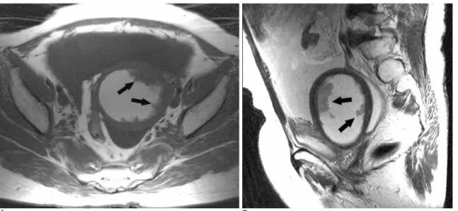
Regarding the mass shape, all lesions of the ECRT pa- tients were polypoid masses (Fig. 1). However, the SEC
lesions showed five polypoid masses and 13 wall thick- enings (Fig. 2). The maximal diameter of the masses var- ied between 1 cm and 2.6 cm (mean±SD: 1.53±0.75 cm) for the ECRT patients and between 0.5 cm and 3.5 cm (mean±SD: 2.25±1.07 cm) for the SEC patients (p
= 0.157).
For the SEC patients, all the lesions of the 22 patients demonstrated iso-signal intensity lesion on the T1-
A B
Fig. 1. A 48-year-old woman with endometrial cancer 14 years after radiation therapy.
A. The axial T1-weighted MR image shows multiple, polypoid isointense masses (arrows) within the distended endometrial cavity.
The signal intensity of the fluid within the endometrial cavity is hyperintense, which is suggestive of hematoma.
B. The sagittal T2-weighted MR image reveals polypoid, hyperintense masses (arrows) in the markedly distended endometrial cavi- ty.
A B
Fig. 2. A 48-year-old woman with spontaneous endometrial cancer.
A. The sagittal T2-weighted image shows the hyperintense thickened lesion of the endometrium (arrows).
B. The contrast-enhanced gradient echo sagittal T1-weighted image demonstrates the homogenous enhancing endometrial cancer (arrows) with an intact junction zone.
weighted images and high signal intensity lesion on the T2-weighted images. For the ECRT patients, three of four patients (75%) showed heterogeneous enhance- ment and one patient showed homogeneous enhance- ment on the contrast enhanced T1-weighted images.
However, for the SEC patients, 13 of 18 patients (72.2%) showed heterogeneous enhancement.
The width of the endometrial cavity varied between 1.6 cm and 5.2 cm (mean±SD: 3.9±1.7 cm) in the ECRT patients, whereas for the SEC patients, the width of the endometrial cavity varied between 0.1 cm and 2.7 cm (mean±SD: 0.6±0.7 cm). The width of the endome- trial cavity in the ECRT patients was wider than that of the SEC patients (p=0.002) (Fig. 1). In all patients, the signal intensity of the fluid in the endometrial cavity ap- peared cystic as hypointensity on the T1-weighed im- ages and as hyperintensity on the T2-weighted images, except for 2 ECRT patients. These 2 patients had hematoma in the endometrial cavity.
All ECRT patients had cervical stenosis and this was established via pelvic exams. However, there was no cervical stenosis in all the SEC patients.
Discussion
Endometrial tissue can persist after administering RT for cervical carcinoma, and so the risk of developing en- dometrial carcinoma must be considered. ECRT may develop from the remnant endometrial tissue; however, theses cases are very rare. Most ECRT patients are asymptomatic, but the advanced cases complain of low- er abdominal distension or pain. The MRI findings of ECRT are a polypoid mass with prominent distension of the endometrial cavity or a distended endometrial cavi- ty with a large tumor filling it. The prognosis of ECRT is poorer than that for SEC.
Several studies have reported that aggressive adeno- carcinomas developed after administering RT for cervi- cal carcinoma, implicating radiation as a carcinogenic factor for the development of this aggressive histological subtype (10, 13). In our study, 75% of the patients (3/4) had aggressive subtypes of adenocarcinoma such as papillary serous carcinoma or clear cell carcinoma.
Papillary serous carcinoma and clear cell carcinoma are significantly more common in black women. Patients with papillary serous carcinoma and clear cell carcino- ma are more likely to have pelvic and periaortic tumor involvement (14).
The significance of ionizing radiation as a risk factor
for the development of endometrial cancer is currently unresolved. However, given the age of diagnosis, the la- tent period and the preponderance of high-risk histolog- ic types, radiation is likely to be the cause. Therefore, it is important to continue careful annular surveillance even if the patient has been free of disease for many years, as the mean latency period for developing en- dometrial cancer is 10-14 years (9, 13). In our study, endometrial cancers were diagnosed at a mean of 10.3 years after administering radiation for cervical cancer, supporting that this should be sufficient time to exclude the presence of this tumor at the time of irradiation (9- 11).
Radiation-induced cervical stenosis may prevent the patient from displaying early symptoms and this compli- cates efforts to obtain a tissue sample for diagnosis.
Unlike the SEC patients, whose first symptoms are typi- cally vaginal bleeding, the patients with ECRT usually presented with the signs and symptoms of an enlarge uterus and pelvic pain, indicating relatively advanced disease (13). In our study, three ECRT patients (75%) had their disease detected at advanced stages (stages II- IV), whereas all the cases of SEC were stage I.
The MR images of endometrial carcinoma have shown various abnormal endometrial findings. The en- dometrium may be thickened focally or diffusely, and this seen as being irregular in thickness and configura- tion, or as widened via polypoid tumor (5, 7). Polypoid masses were revealed in all ECRT patients (100%), whereas polypoid masses were seen in five SEC patients (27.8%) in our study. It seems to us that a polypoid mass was seen only in the ECRT patients who have hydrome- tra. On the other hand, the endometrial thickening was more commonly seen in SEC patients whose cavities were collapsed.
The signal intensity of the tumor shows various pat- terns on the T1-weighted and T2-weighted images (7). In our study, although the signal intensity and enhance- ment pattern on the MR imaging were not different be- tween the ECRT and SEC patients, prominent widening of the endometrial cavity was revealed in the ECRT pa- tients compared with the SEC patients. The cause of this may be radiation fibrosis with cervical stenosis.
The limitation of this study is the small number of ECRT patients. Because the development of ECRT is quite rare, only 4 patients belonging to this group were included in this study. A future study is needed to de- scribe the MR imaging in a large number of ECRT pa- tients.
In summary, it is important to recognize that ECRT patients may show a high-risk histological subtype and a history of pelvic radiation for treating cervical cancer.
The MR imaging findings of ECRT patients showed a polypoid mass with prominent distension of the en- dometrial cavity, as compared with SEC patients.
References
1. Hoffman M, Roberts WS, Cavanagh D. Second pelvic malignan- cies following radiation therapy for cervical cancer. Obstet Gynecol Surv 1985;40:611-617
2. Ben-Shachar I, Pavelka J, Cohn DE, Copeland LJ, Ramirez N, Manolitsas T, et al. Surgical staging for patients presenting with grade 1 endometrial carcinoma. Obstet Gyencol 2005;105:487-493 3. Daniel AG, Peters WA 3rd. Accuracy of office and operating room
curettage in the grading of endometrial carcinoma. Obstet Gynecol 1988;71:612-614
4. Yamashita Y, Harada M, Sawada T, Takahashi M, Miyazaki K, Okamura H. Normal uterus and FIGO stage I endometrial carcino- ma: dynamic gadolinium-enhanced MR imaging. Radiology 1993;
186:495-501
5. Sironi S, Colombo E, Villa G, Taccagni G, Belloni C, Garancini P, et al. Myometrial invasion by endometrial carcinoma: assessment with plain and gadolinium-enhanced MR imaging. Radiology 1992;
185:207-212
6. Ito K, Matsumoto T, Nakada T, Nakanishi T, Fujita N, Yamashita H. Assessing myometrial invasion by endometrial carcinoma with dynamic MRI. J Comput Assist Tomogr 1994;18:77-86
7. Manfredi R, Gui B, Maresca G, Fanfani F, Bonomo L. Endometrial cancer: magnetic resonance imaging. Abdom Imaging 2005;30:626- 636
8. Morris M, Eifel PJ, Lu J, Grigsby PW, Levenback C, Stevens RE, et al. Pelvic radiation with concurrent chemotherapy compared with pelvic and para-aortic radiation for high-risk cervical cancer. N Engl J Med 1999;340:1137-1143
9. Fehr PE, Prem KA. Malignancy of the uterine corpus following ir- radiation therapy for squamous cell carcinoma of the cervix, Am J Obstet Gynecol 1974;119:685-692
10. Gallion HH, van Nagell JR Jr, Donaldson ES, Powell DE.
Endometrial cancer following radiation therapy for cervical carci- noma. Gynecol Oncol 1987;27:76-83
11. Rodriguez J, Hart WR. Endometrial cancers occurring 10 or more years after pelvic irradiation for carcinoma. Int J Gyencol Pathol 1982;1:135-144
12. Creasman WT. Malignant tumors of the uterine corpus. In: Rock JA, Jones HW, ed. Te Lind’s Operative gynecology, 9th ed. Philadelphia:
Lippincott Williams & Wilkins, 2003;1445-1486
13. Pothuri B, Ramondetta L, Martino M, Alektiar K, Eifel PJ, Deavers MT, et al. Development of endometrial cancer after radiation treat- ment for cervical carcinoma. Obstet Gynecol 2003; 101:941-945 14. Matthews RP, Hutchinson-Colas J, Maiman M, Fruchter RG,
Gates EJ, Gibbon D, et al. Papilary serous and clear cell type lead to poor prognosis of endometrial carcinoma in black women.
Gynecol Oncol 1997;65:206-212
대한영상의학회지 2007;56:491-495
자궁 경부암으로 방사선 치료 후 발생한 자궁 내막암종의 자기공명영상소견
11인하대병원 방사선과, 2전남대 화순병원 방사선과,
3광주기독병원 방사선과, 4Myo대학병원 김윤정・정용연2・임남열2・고석완3・김보현4
목적: 자궁 경부암으로 방사선치료를 받았던 환자들에서 발생한 자궁 내막암종의 자기공명영상소견을 기술하고 자 발적으로 발생하는 자궁 내막암종과의 차이를 알아보고자 한다.
대상과 방법: 수술 혹은 자궁내막 조직검사에 의해 발견된 22명의 자궁 내막암 환자를 대상으로 하였다. 환자들을 자궁 경부암으로 방사선치료를 받았던 과거력의 유무에 따라 나누었을 때 각각 4명(방사선 치료 후), 18명(자연발 생)이었다. 자기공명영상 소견은 종괴의 크기, 모양, 신호강도와 자궁강 확장, 자궁협부협착, 자궁내 저류된 액체성 상을 분석하였다.
결과: 방사선 치료 후는 모든 종괴 모양은 폴립 양인 데 비해 자연발생은 5명만 폴립 양이고 나머지 13명은 두꺼 운 내벽으로 보였다. 최대 크기, 신호강도, 종괴의 조영증강은 두 그룹 간에 차이가 없었다. 자궁 강이 확장된 너비 는 방사선 치료 후에서는 3.9 cm이고 자연발생에서는 0.4 cm으로 통계학적으로 유의하였다. 모든 방사선 치료 후 의 환자들은 자궁협부가 협착되었고 반면, 자연발생의 환자들은 자궁협부협착을 볼 수 없었다.
결론: 자궁 경부암으로 방사선치료를 받았던 환자에서 발생한 자궁 내막암종은 자발적으로 생긴 자궁 내막암종에 비해 자궁강 확장과 자궁협부협착의 자기공명영상소견을 보였다. 자궁협부협착은 방사선 치료에 따른 섬유화 변화 에 의한 소견일 것이다.
