Effects of Environmental Reinforcement Through Motivation on Motor and Cognitive Function in Rats With Focal Ischemic Brain Injury
Myoung Heo, Ph.D., P.T.
Dept. of Occupational Therapy, College of Economics & Welfare, Gwangju University
Abstract
1)It is known that individual factors as cognitive, perception, emotion, and motivation may greatly in- fluence on recovery from neurologic region. This study was to investigate the effects of environmental reinforcement through motivation to perform the tasks voluntarily on motor and cognition function in rats with focal ischemic brain injury. Focal ischemic brain injury was induced in Sprague-Dawley rats (15 rats, 250±50 g) through middle cerebral artery occlusion (MCAo). And then, experiment groups were ran- domly divided into three groups; The control group: MCAo induction (n1=5), the environmental reinforce- ment (ER) group: the application for ER after MCAo induction (n2=5), the environmental reinforcement through motivation (ERM) group: the application for ERM after MCAo induction (n3=5). The climbing test (CT) and the modified limb placing tests (MLPTs) to measure the motor function and the Morris water maze acquisition test (MWMAT) and the Morris water maze retention test (MWMRT) to measure the cognitive function were performed. For the CT, the ERM group was significantly larger than the ER group. For the MLPTs, the ERM group was significantly decreased compared to other groups. For the MWMAT, the time to find the circular platform in the ERM group significantly decreased compared to other groups. For the MWMRT, the time to dwell on the quadrant circular platform in the ERM group was significantly increased compared to other groups. These results suggested that the ERM could im- prove the motor and cognitive functions in the rats with focal ischemic brain injury.
Key Words: Cognitive function; Environmental reinforcement through motivation; Focal ischemic brain injury; Motor function.
Introduction
Ischemic brain injury is one of the most frequent etiological factors of death and impairment in the world and causes multiple disability such as motor impairment, sensory impairment, perception impair- ment, and cognition impairment (Kotila et al, 1984;
Pedersen et al, 1996; Sims and Anderson, 2002).
The physical therapy approach for stroke patients include the muscle re-education (Knapp, 1955), the reflex theory of the neurofacilitation approach such as the Bobath, Brunnstrom, PNF (Proprioceptive neuro- muscular facilitation), and Rood (Perry, 1967; Semans, 1967; Stockmeyer, 1967; Voss, 1967), and the system
theory, a new field starting which appeared because of the limitation of existing therapy approaches where the neurophysiological approach did not have. The system theory argued that the central nerve system was not a vertical hierarchy structure but consisted of horizontal interaction of many systems and move- ment or behavior originated from the interaction among individual factors, task factors, and environ- mental factors (Shumway-Cook and Woollacott, 2006).
Recently the importance of environmental factors has been perceived in physical therapy for stroke.
Environmental reinforcement provides an opportunity of social interaction and physical activity, increases sensory-motor functions in experimental animals Corresponding author: Myoung [email protected]
This study was conducted by research funds from Gwangju University.
with brain injury (Belayev et al, 2003; Biernaskie and Corbett, 2001; Dahlqvist et al, 2004), and rises the number of neurons and synapses (morphological changes). As a result, learning and memory becomes improved (behavioral changes) (Briones et al, 2004;
Johansson, 2004). However, some researches reported that reinforced environment alone had no a great ef- fect on skilled reaching and grasping of affected forelimb (Biernaskie and Corbett, 2001).
Tang et al (2007) indicated that early willed move- ment treatment can increase the expression level of AMPA receptor subunits and thus might increase synaptic transmission and enhance brain plasticity af- ter ischemia at the subacute stage. Motivation is the motive power that induces behaviors for some goals and needs satisfaction by an individual and has a function of maintaining and reinforcing behaviors (You and Ann, 2009). Shontz (1978) indicated that patient's lack of motive for his treatment was an im- portant problem in the process of rehabilitation and Hafen et al (2001) indicated that patient's motive for his treatment or active attitude toward his treatment would significantly act on the process or outcomes of treatment. Although some researches have observed the effects of environmental reinforcement, there are few researches on animal experiments in environ- mental reinforcement through motivation.
Accordingly, this study was intended to examine the effects on motor and cognitive functions in the rats with focal ischemic brain injury and provide clinical basic materials by conducting environmental reinforcement through motivation using food for rats with focal ischemic brain injury to perform willed target tasks.
Methods Experimental Animals
Fifteen adult male Sprague-Dawley rats of eight weeks weighting 250±50g were used for the experiment. For individual selection, neurological
evaluation developed by Bederson et al (1986) was conducted. Only rats that had a stroke symptom of grade 2 or 3 in neurological evaluation were selected for the test (Table 1). The climbing test and modi- fied limb placing tests (1st, 3rd, 7th, and 14th day) were conducted to test motor functions, and the Morris water maze acquisition test (10th, 11th, 12th and 13th day) and retention test (14th day) were conducted to test cognitive functions. A one-week environmental adaption period was given for task training before the experiment to reduce stress caused by task training. All rats were randomly div- ided into three groups 24h after middle cerebral ar- tery occlusion (MCAo) induction; The control group:
MCAo induction (n1=5), the environmental reinforce- ment (ER) group: the application for ER after MCAo induction (n2=5), the environmental reinforcement through motivation (ERM) group: the application for ERM after MCAo induction (n3=5).
Instruments
Rats in the control group were housed in standard cage (40 ㎝ × 30 ㎝ × 18 ㎝ high). Rats in the ER were put into a ER cage. The cage was covered with wire netting of square iron frame of 50 ㎝ × 60
㎝ × 40 ㎝ using modified Tang et al. method (2007) for ER training. The floor was laid with acrylite covered with straw and a rope with 20 ㎜ thick was hung. A T-shaped tunnel of 50 ㎜ in diameter and 100 ㎜ long was installed in the floor and a ladder of 8 ㎝ × 10 ㎝ × 22 ㎝ and the frame of a sieve of 22 ㎝ in diameter × 8 ㎝ in width were installed in the side. While in the ER group, food and water was installed 18 ㎝ high as the general cage, in the ERM group, food and water was installed 35 ㎝ high to eat by climbing up the ladder (Figure 1).
Interventions
Rats in the ERM group went through four stag- gered phases: (1) MCAo induction, (2) post-MCAo induction recovery, (3) climbing ladder induction in ERM cage and (4) obtaining food by climbing the
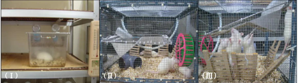
(Ⅰ) (Ⅱ) (Ⅲ)
Figure 1. Intervention used for different groups. (Ι) Rats in the control group could easily obtain the food in the concavity of the grids steel cover. Rats ER (Ⅱ) and rats ERM (Ⅲ) were placed into the ER cage with a ladder and grid lateral wall.
Rats in the ER group (Ⅱ) could easily obtain the food in the concavity of the grids steel cover, but rats in the ERM group (Ⅲ) must climb up to the cover of the cage for foods and water.
Condition Grade Evaluation
Normal 0 No observable deficits
Moderate 1 Forelimb flexion
Severe 2 Decreased resistance to lateral push(and forelimb flexion) without circling
3 Same behavior as grade 2, with circling
Table 1. Neurologic examination after MCAo
ladder or lateral wall up to the cover of the cage.
After the rats were put into the ERM cage 24 h re- covery post-MCAo induction, food induction would be given for the rats to be familiar with the location of the food. If all ERM rats in the cage have already known the location of the food and water, the food induction would be discontinued. Rats in the ER group went through three phases: (1) MCAo in- duction, (2) post-MCAo induction recovery and (3) feed in the ER cage 24 h after MCAo induction.
Each rat received 15 food granules each day at 10 a.m. Video records were made with a Sony Video1) from 10 to 11 a.m., and from 3 to 4 p.m. each day.
The temperature was 25±1℃, humidity was 55±10%, and light and darkness was given every 12 hours in the laboratory.2)3)
Induction of MCAo
Middle cerebral artery occlusion (MCAo) was exe- cuted according to Nagasawa and Kogure method (1989). After general inhalation anesthesia, the body was kept at a uniform temperature using a heating pad and a rectal thermometer. The right common carotid artery was separated from the vagus nerve and then the external and internal carotid arteries were separated from the right common carotid artery.
The common and external carotid arteries were li- gated loosely with a pre-hung thread and a micro- vascular clip was used for the branch of internal carotid artery. After the origin of the branch of in- ternal carotid artery was incised, the probe made of 4-0 nylon surgical suture2) of 1.5 ㎝ long coated with silicone was put into the internal carotid artery and fixed by tying the thread hung in the internal carotid artery and the microvascular clip was removed.
1) DCR-SR 87, Sony, Japan.
2) Xantopren, Bayer Dental, Germany.
Reperfusion was achieved by removing the suture except silicon two hours after MCAo.
Motor Function Test
Climbing test (CT)
CT applying Tang et al method (2007), the fre- quency of climbing up the ladder was measured by video records for 3, 7 and 14 days, two hours per day (10~11 a.m., 3~4 p.m.). The rat climbing to top of the ladder or to the cover of the cage was given 1 point and the rat climbing to the middle of the ladder or lateral wall was given .5 point. The fre- quency of climbing each hour was totaled and then divided by 2.
Modified limb placing tests (MLPTs)
MLPTs consisted of three tasks according to the methods described by Puurunen et al (2001a). First, the forward visual limb placing test observed the stretch of the forelimbs towards the table when the tester held the middle body of the rat and suspended it 10 cm over the table. The normal stretch of both forelimbs was scored as 0 point and the abnormal flexion of one forelimb was scored as 1 point.
Second, the proprioceptive limb placing test observed the retrieval and placement of each forelimb of the rat. The tester positioned the forelimbs of the rat on the edge of a table and turned its head at 45° angle to remove visual perception and tactile stimulation by whiskers and then gently pulled the forelimbs down and released to stimulate the joints and muscle of forelimbs. Finally, the lateral pushing limb placing test observed the lateral placement of the forelimb and the hindlimb of the rat. The tester placed the rat along the table edge and pushed its lateral body gently toward the table edge. The rat positioning its left and right forelimbs and forelimbs and hindlimbs normally was given 0 point, the rat reacting with a delay or incompletely was given 1, and the rate hav- ing no reaction was given 2. Each point was added.
Cognitive Function Test
Morris water maze acquisition test (MWMAT) The Morris water maze tests were conducted us- ing modified and complemented Fukunaga et al.
method (1999). The water tank used as the Morris water maze was a circular pool 160 ㎝ in diameter and 50 ㎝ high. The depth of water in the water tank was 30 cm and the temperature of water was 22±2℃. The circular platform was made of a circular transparent acrylic 12 ㎝ in diameter wrapped in the gauze for convenient lifting and was located 2 ㎝ below the water surface for rats not to see with the naked eye. The test table, chairs, and tester in the outside of the water maze needed to be always at the same place to be used as a cue. Because the water was made opaque by black ink, the circular platform was not seen and could not be used as a visual cue. The video tracking system was used to record the rats moving in the water maze. The cir- cular escape platform was placed in the center of the southeast quadrant and one of the last quadrant was used as the starting point. The rats entered water at the spot 5 cm away from the edge of the Morris water maze, facing the wall of the Morris water maze. The rats were given four trials a day for five days and the time of latency to acquire the sub- merged escape platform was measured using S-MART3) program. When the rats could not arrive at the circular escape platform after 60 seconds, then they were guided to the escape platform, stayed for 30 seconds, and tried again. The direction of putting rats in the Morris water maze varied every time us- ing Table of Random Numbers. 4)
Morris water maze retention test (MWMRT) In the 14th day at the end of test, the circular es- cape platform was removed and the rats were re- leased between west and north where the circular platform was located previously in the same way as the acquisition test. The trials lasted 60 seconds, and
3) S-MART, Pab Lab, Spain.
Figure 2. Comparison of climbing test frequency among the two groups of MCAo in rats. **p
<.01, ***p<.001, compared between the ER and ERM groups. The ERM group was significantly larger than the ER group.
Figure 3. Comparison of modified limb placing tests scores among the three groups of MCAo in rats.
**p<.01, ***p<.001, compared among the control, ER and ERM groups. The ERM group was significantly decreased compared to other groups.
Figure 4. Comparison of acquisition performances among the three groups of MCAo in rats. ***p<.001, compared among the control, ER and ERM groups. The time to find the circular platform in the ERM group significantly decreased compared to other groups.
their dwelling time around the quadrant where the circular escape platform was located previously.
Statistical Analysis
SPSS 14.0 ver. for windows® was used for all statistical tests and the results were expressed as the mean and standard deviation. All data were checked for normality by use of the Kolmogorov-Smirnov test. The differences of CT, MLPTs, MWMAT be- tween the three groups were analyzed using repeated measures analysis of variance (ANOVA). The differ- ences on CT was analyzed using two sample t-test and the differences on MLPTs, MWMAT and MWMRT between the three groups were analyzed using one-way ANOVA. The Tukey test was con- ducted for post hoc analysis. The alpha level of stat- istical significance was set at .05.
Results Motor Function Test
Climbing test (CT)
There was significant interaction between group and time in the result of the repeated measures ANOVA of CT change (F (1, 8)=36.790, p<.001).
While the ER group was increased gradually at the 3rd, 7th, and 14th day as 2.1, 3.2, and 5.4 re- spectively, the ERM group was increased rapidly as 7.1, 8.4, and 11. The result of two sample t-test for group comparison showed that the ERM group had a significant difference at the 3rd day (p<.001), 7th (p
<.01), and 14th (p<.001) compared with the ER group (Figure 2).
Modified limb placing tests (MLPTs)
There was significant interaction between group and time in the result of the repeated measures ANOVA of MLPTs change (F (2, 12)=37.899, p
<.001). The result of one-way ANOVA for group comparison showed that there was a significant dif-
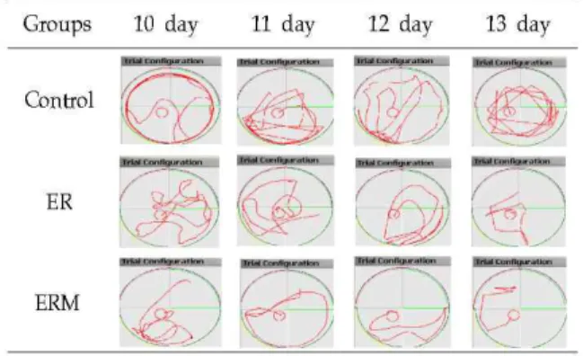
Figure 5. Comparison of acquisition performance among the three groups of MCAo in rats. Note thigmotaxis behaviour in MCAo in rats.
Figure 6. Comparison of dwelling times in target quadrant of retention test among the three groups of MCAo in rats. *significant difference in control, ER and ERM groups, †significant difference between the ER and ERM groups.
The time to dwell on the quadrant circular platform in the ERM group was significantly increased compared to other groups.
ference at the 3rd day (F (2, 12)=10.571, p<.01), 7th (F (2, 12)=24.500, p<.001), and 14th (F (2, 12)=48.857, p<.001). The result of post hoc analysis showed that there was a significant difference at the 3rd day between the control and ERM groups and 7th and 14th day between the control and ER groups, the ER and ERM groups, and the control and ERM groups (Figure 3).
Cognitive Test
Morris water maze acquisition test (MWMAT) There was significant interaction between group and time in the result of the repeated measures ANOVA of MWMAT change (F (2, 12)=44.365, p
<.001). The control group had a slow decrease pat- tern from 10th day (60.00 sec) to 13th day (45.20 sec); the ER group had a higher decrease pattern than the control group from 53.60 sec. to 16.40 sec.;
and the ERM group had a higher decrease pattern than the control and ER groups from 52.00 sec. to 10.20 sec. The result of one-way ANOVA for group comparison showed that there was a significant dif- ference at the 11th day (F (2, 12)=61.355, p<.001), 12th (F (2, 12)=70.711, p<.001), and 13th (F (2, 12)=70.711, p<.001). The result of post hoc analysis showed that there was a significant difference at the 11th, 12th, and 13th day between the control and ER groups, the ER and ERM groups, and the control and ERM groups (Figure 4) (Figure 5).
Morris water maze retention test (MWMRT) There was a significant difference in the changes in the time to dwell on the quadrant circle for MWMRT in the result of one-way ANOVA (F (2, 12)=30.176, p<.001). The result of post hock analysis showed that there was a significant difference in the time to dwell on the quadrant circular platform in all groups (the control group: 9.20 sec., the ER group:
4.60 sec., the ERM group: 18.20 sec.) and there was a significant difference between the ER and ERM groups (Figure 6).
Discussion
This study was intended to examine the effects on motor and cognitive functions of in the rats with fo- cal ischemic brain injury and provide clinical basic materials by conducting ERM using food for rats with focal ischemic brain injury to perform willed
target tasks.
The CT and MLPTs were conducted to assess the motor functions of forelimb and hindlimb. CT meas- ured the frequency of climbing up the ladder and lateral wall and MLPTs were used to assess the proprioceptive sensibility and tactile sensation of eyesight and limbs.
The first CT showed that the ERM group had more frequency of climbing the ladder at the 3rd, 7th, and 14th than the ER group because the ERM group climbed up the ladder and lateral wall will- ingly to eat food and water. On the other hand, the ER group showed much less frequency to climbing up the ladder and lateral wall than the ERM group because they could eat food and water without climbing up. This study showed that food and water was the motive power which induced behaviors for needs satisfaction, played a role of providing motiva- tion, and maintained and reinforced behaviors (Shontz, 1978). Based on this result, it is suggested that motivation-induced environment and active will is very important to perform target tests in rats with brain injury.
In the second MLPTs, the ERM group showed quicker recovery of sensory-motor ability at the 3rd day than the control group and at the 7th and 14th day than the control and ER groups. Biernask and Corbett (2001) reported that ER alone had no a great effect on the improvement of fine digital and fore- limb functions caused by brain injury, and only when ER and skilled-reach training was conducted together, motor functions caused by brain injury could be improved by changing neural plasticity with the increase in dendrite branches in brain. The quicker recovery of the ERM group compared to the ER group may result from the ERM group's willed repetition of climbing the ladder and lateral wall, which could improve proprioceptive sensibility and tactile sensation of forelimbs and hindlimbs.
Furthermore, they needed and trained forelimb's reaching and grasping to eat food and water. Tang et al (2007) also observed that the group conducting
both ER and willed movement therapy showed a significant difference at the 3rd, 7th, and 15th day in the results of CT compared with the the ER group and there was a significant difference in the forelimb placing test compared with the ER group. It agrees with the result of this study.
The MWMAT is intended to assess learning and memory. It is frequently used to test cognitive func- tions in rats and in the behavioral neuroscience field (Jolkkonen et al, 2003). The Morris water maze con- ducts acquisition training which enhances external visual cues and spatial location and a retention test which recollects stored spatial memory.
In MWMAT of this study, it was found that the control group had the deficit of spacial leaning ability of the Morris water maze tests because of hippo- campal damage caused by ischemic brain injury (Squire and Zola, 1996). The ER and ERM groups were found to be reduced in the time to find the circular platform over time. The ERM group had better performance to find the circular platform from the 11th day compared with the control group and ER groups. In MWART, the ERM group showed the longest dwelling time compared with the control and ER groups. Many animal experiments have reported that ER increases the recovery of memory loss and improves spatial learning ability in rates with brain injury (Dahlqvist et al, 2004; Puurunen et al, 2001b;
Ronnback et al, 2005). It may be partly because rats receiving ER have more chance of activity through a various of stimulation than those in the general cage (Belayev et al, 2003; Biernaskie and Corbett, 2001;
Dahlqvist et al, 2004) and it may increase brain plasticity and improve learning and memory ability.
Then it may increase the number of neurons and synapses of hippocampus and brain plasticity and as a result improve learning and memory ability (Briones et al, 2004; Johansson, 2004). The ERM group had better improvement in spatial learning and memory ability than the ER group because the ERM group had to repeatedly go up and down the ladder and lateral wall more frequently than the ER group
to eat food and water. Since they had to train skil- led-forelimb reaching and grasping to eat food and water which was placed in the high place, the ERM group had better improvement in spatial learning ability. It agrees with the research result by Wurm et al (2007) that skilled forelimb training increased neurogenesis of hippocampus and improved spatial learning ability. Based on this result, although envi- ronmental factors are important in physical therapy for a stroke, it is considered that motivation of treatment and active attitude toward treatment may play an important role in the treatment process and outcomes.
To sum up, the ERM in rats with focal ischemic brain injury has a more significant impact on restor- ing motor and cognitive functions. Further clinical research should focus on developing intervention methods which can continuously induce motivation in patients along with assessment of motivation before physical therapy.
Conclusion
The results of this study were as follows: 1. For motor function test there were significant interactions among the groups in the given time (p<.001). For the CT, the ERM group was significantly larger than the ER group. For the MLPTs, the ERM group was significantly decreased compared to other groups. 2.
For cognitive function test there were significant in- teractions among the groups in the given time (p
<.001). For the MWMAT, the time to find the cir- cular platform in the ERM group significantly de- creased compared to other groups. For the MWMRT, the time to dwell on the quadrant circular platform in the ERM group was significantly increased com- pared to other groups. These results suggested that the ERM could improve the motor and cognitive functions in the rats with focal ischemic brain injury.
References
Bederson JB, Pitts LH, Tsuji M, et al. Rat middle cerebral artery occlusion: Evaluation of the model and development of a neurologic examination. Stroke. 1986;17(3):472-476.
Belayev A, Saul I, Liu Y, et al. Enriched environ- ment delays the onset of hippocampal damage after global cerebral ischemia in rats. Brain Res.
2003;964(1):121-127.
Biernaskie J, Corbett D. Enriched rehabilitative train- ing promotes improved forelimb motor function and enhanced dendritic growth after focal ische- mic injury. J Neurosci. 2001;21(14):5272-5280.
Briones TL, Klintsova AY, Greenough WT. Stability of synaptic plasticity in the adult rat visual cor- tex induced by complex environment exposure.
Brain Res. 2004;1018(1):130-135.
Dahlqvist P, Ronnback A, Bergstrom SA, et al.
Environmental enrichment reverses learning im- pairment in the Morris water maze after focal cerebral ischemia in rats. Eur J Neurosci.
2004;19(8):2288-2298.
Fukunaga A, Uchida K, Hara K, et al. Differentiation and angiogenesis of central nervous system stem cells implanted with mesenchyme into ischemic rat brain. Cell Transplant. 1999;8(4):435-441.
Hafen K, Jastrebow J, Nubling R, et al. Development of a patient questionnaire for assessment of motivation for rehabilitation (PAREMO). Rehabilitation (Stuttg).
2001;40(1):3-11.
Johansson BB. Brain plasticity in health and disease.
Keio J Med. 2004;53(4):231-246.
Jolkkonen J, Gallagher NP, Zilles K, et al. Behavioral deficits and recovery following transient focal cerebral ischemia in rats: Glutamatergic and GABAergic receptor densities. Behav Brain Res.
2003;138(2):187-200.
Knapp ME. The contribution of Sister Elizabeth Kenny to the treatment of poliomyelitis. Arch Phys Med Rehabil. 1995;36(8):510-517.
Kotila M, Waltimo O, Niemi ML, et al. The profile
This article was received October 10, 2009, and was accepted November 5, 2009.
of recovery from stroke and factors influencing outcome. Stroke. 1984;15(6):1039-1044.
Nagasawa H, Kogure K. Correlation between cerebral blood flow and histologic changes in a new rat model of middle cerebral artery occlusion.
Stroke. 1989;20(8):1037-1043.
Pedersen PM, Jørgensen HS, Nakayama H, et al.
Orientation in the acute and chronic stroke pa- tient: Impact on ADL and social activities. The Copenhagen Stroke Study. Arch Phys Med Rehabil. 1996;77(4):336-339.
Perry CE. Principles and techniques of the Brunnstrom approach to the treatment of hemiplegia. Am J Phys Med. 1967;46(1):789-815.
Puurunen K, Jolkkonen J, Sirviö J, et al. An alpha(2)-a- drenergic antagonist, atipamezole, facilitates behav- ioral recovery after focal cerebral ischemia in rats.
Neuropharmacology. 2001a;40(4):597-606.
Puurunen K, Jolkkonen J, Sirviö J, et al. Selegiline combined with enriched-environment housing attenuates spatial learning deficits following fo- cal cerebral ischemia in rats. Exp Neurol.
2001b;167(2):348-355.
Ronnback A, Dahlqvist P, Svensson PA, et al. Gene ex- pression profiling of the rat hippocampus one month after focal cerebral ischemia followed by enriched environment. Neurosci Lett. 2005;385(2):173-178.
Semans S. The Bobath concept in treatment of neu- rological disorders: A neuro-developmental treatment. Am J Phys Med. 1967;46(1):732-788.
Shontz FC. Psychological adjustment to physical dis- ability: Trends in theories. Arch Phys Med
Rehabil. 1978;59(6):251-254.
Shumway-Cook A, Woollacott MH. Motor control:
Translating research into clinical practice. 3rd ed.
London, Lippincott Williams & Wilkins, 2006:16-17.
Sims NR, Anderson MF. Mitochondrial contributions to tissue damage in stroke. Neurochem Int.
2002;40(6):511-526.
Squire LR, Zola SM. Ischemic brain damage and memory impairment: A commentary. Hippocampus.
1996;6(5):546-552.
Stockmeyer SA. An interpretation of the approach of Rood to the treatment of neuromuscular dysfunction. Am J Phys Med. 1967;46(1):900-961.
Tang Q, Yang Q , Hu Z, et al. The effects of willed movement therapy on AMPA receptor properties for adult rat following focal cerebral ischemia.
Behav Brain Res. 2007;181(2):254-261.
Voss DE. Proprioceptive neuromuscular facilitation.
Am J Phys Med. 1967;46(1):838-899.
Wurm F, Keiner S, Kunze A, et al. Effects of skilled forelimb training on hippocampal neurogenesis and spatial learning after focal cortical infarcts in the adult rat brain. Stroke. 2007;38(10):2833-2840.
You YY, Ann CS. A study of the relationships between perceived rehabilitation-motivation and quality of life in patients after a cerebrovascular accident. J Korean Soc Occup Ther. 2009;17(2):1-16.