Background and Purpose Lacosamide (LCM) is an antiepileptic drug that enhances the slow inactivation of sodium channels and modulates collapsin response mediator protein-2.
LCM was recently demonstrated to exert a neuroprotective effect in a murine model of trau- matic brain injury and status epilepticus. Assuming the same underlying excitotoxicity-related brain injury mechanism, we hypothesized that LCM would have a neuroprotective effect in hypoxic-ischemic brain injury.
Methods We divided rats into three groups at each testing session: pre- or postfed with LCM, fed with normal saline, and sham. A hypoxic-ischemic brain injury was induced by subjecting 7-day-old rats to right carotid artery coagulation followed by 2.5 h of exposure to 8% oxygen.
The animals were killed on postnatal day 12 to evaluate the severity of brain damage. Open field testing was also performed between week 2 and week 6, and the Morris water maze test was performed in week 7 after hypoxia-ischemia.
Results The incidence of liquefactive cerebral infarction was lower in rats prefed with LCM at 100 mg/kg/dose, with the mortality rate being higher at higher doses (200 and 300 mg/kg/
dose). The infarct areas were smaller in LCM-prefed rats in several brain regions including the hemisphere, hippocampus, cortex, and striatum. Spatial learning and memory function were better in LCM-prefed rats (p<0.05). No effect was observed in postfed rats.
Conclusions This study suggests that LCM pretreatment exerts a neuroprotective effect on hypoxia-ischemia in neonatal rats. The obtained results suggest that LCM pretreatment could be used as an effective neuroprotective method for neonates under hypoxic-ischemic condi- tions including heart surgery.
Key Words brain, hypoxia-ischemia, lacosamide, neuroprotection, newborn, rat.
Neuroprotective Effect of Lacosamide on Hypoxic-Ischemic Brain Injury in Neonatal Rats
INTRODUCTION
Hypoxic-ischemic brain injury is a common cause of long-term disabilities in the pediatric population. Up to 25% of the survivors of neonatal hypoxia exhibit devastating neuropsy- chological handicaps, including developmental delay, cerebral palsy, epilepsy, and learning disability. The lifelong consequences and medical costs mean that neonatal hypoxia-isch- emia represents one of the highest burdens on society.
Excitotoxicity has been proposed as a major cause of neuronal death induced by ischemic brain injury, and hence it has been suggested that antiepileptic drugs could exert beneficial effects on ischemic brain insults.1-4 Lacosamide (LCM) is a new antiepileptic drug that has a unique dual mode of action: it enhances the slow inactivation of sodium channels and regulates the action of neurotrophins via interaction with collapsin response mediator protein-2 (CRMP-2). The neuroprotective effect of LCM has been demonstrated using in vivo animal models of status epilepticus5 and traumatic brain injury.6 We hypothesized Gun-Ha Kim
Jung Hye Byeon Baik-Lin Eun
Department of Pediatrics,
Korea University College of Medicine, Seoul, Korea
pISSN 1738-6586 / eISSN 2005-5013 / J Clin Neurol 2017;13(2):138-143 / https://doi.org/10.3988/jcn.2017.13.2.138
Received June 7, 2016 Revised October 15, 2016 Accepted October 18, 2016 Correspondence Baik-Lin Eun, MD, PhD Department of Pediatrics,
Korea University College of Medicine, 148 Gurodong-ro, Guro-gu, Seoul 08308, Korea Tel +82-2-2626-1229 Fax +82-2-2626-1224 E-mail [email protected]
cc This is an Open Access article distributed under the terms of the Creative Commons Attribution Non-Com- mercial License (http://creativecommons.org/licenses/by-nc/4.0) which permits unrestricted non-commercial use, distribution, and reproduction in any medium, provided the original work is properly cited.
JCN
Open Access ORIGINAL ARTICLEKim GH et al.
JCN
that LCM would have a neuroprotective effect on hypoxic- ischemic brain injury in neonatal rats by assuming the same underlying excitotoxicity-related brain injury mechanism.
METHODS
Animal model
On postnatal day 7, Sprague-Dawley rats were anesthetized with isoflurane, and the right common carotid artery was iso- lated and coagulated (UM880, Umeco, Seoul, Korea). After recovery in an incubator at 36.5°C for 1 h, the rats were ex- posed to 8% oxygen (balanced gas N2, 1 ppm H2O, Jung Ang Sanso, Seoul, Korea) for 2.5 h in a glass chamber to induce a hypoxic-ischemic brain injury. After a 30-min recovery peri- od, the rats were transferred to the dam. In this model, blood flow in the right hemisphere decreases during hypoxic ex- posure and recovers within 24 h after exposure, resulting in both focal brain ischemia and reperfusion.
Animal group and LCM administration
We used an oral preparation of LCM (Vimpat®, UCB Pharma, Smyrna, GA, USA). The rats were randomly divided into three groups: pre- or postfed with LCM, fed with normal saline, and sham. LCM was administered via an orogastric tube im- mediately before and after hypoxia-ischemia in the pretreat- ment group, and immediately after and 2 h later in the post- treatment group.
Neurohistopathologica findings Qualitative measure
On postnatal day 12, the brain was removed and evaluated for gross morphological damage. Ipsilateral liquefactive cere- bral infarction was evaluated by direct visual inspection (Fig.
1). The criteria for assessing cerebral infarction included pal- lor (loss of staining), atrophy, and tissue loss. The total num- bers of animals per group were as follows: 13 sham, 16 nor- mal-saline-fed, and 48 LCM-prefed rats at pretreatment; and 13 sham, 16 normal-saline-fed, and 25 LCM-postfed rats at
posttreatment.
Quantitative measure
After finishing the gross morphological evaluations, the brain samples were frozen at -70°C, sliced at 20 μm intervals, fixed in paraformaldehyde vapor, and stained with cresyl vi- olet. To quantify the severity of brain injury, we measured bi- lateral hemispheric cross-sectional areas in coronal sections at the level of the anterior commissure and mid-dorsal hip- pocampus using ImageJ software (US National Institutes of Health, Bethesda, MD, USA). The mean percentage of right- sided damage relative to the intact left side was compared between LCM-fed and control rats.
Behavioral tests Open field test
We performed the open field activity test from week 2 to week 7 after hypoxia-ischemia. The animals were placed in a black Plexiglas box (60×60×25 cm) for 10 min, during which their locomotive behaviors were monitored and ana- lyzed using an auto-tracking system (SmarTrack, Smartech, Madison, WI, USA). Sham (n=14), normal-saline-prefed (n=
16), and LCM-prefed (n=22, 100 mg/kg/dose) 8-week-old rats were included in the open field test.
Morris water maze test
The Morris water maze test was performed at week 8 after hypoxia-ischemia. The apparatus used for this experiment was a circular stainless steel tank (155 cm in diameter and 60 cm deep) containing water (24.0–26.0°C) to a depth of 40 cm. Skimmed milk was added to make the water opaque.
The extramaze cues were kept constant. Three groups of 8-week-old rats were included in the Morris water maze test:
sham (n=14), normal-saline-prefed (n=16), and LCM-prefed (n=22, 100 mg/kg/dose) rats. The rats learned to escape from the water by searching for a submerged (hidden) Plexiglas platform (10 cm in diameter and 38.5 cm high) in a fixed position. To familiarize them with the experimental situa- tion, before the actual training the rats swam for 90 s with the platform removed. During each trial, the animal was placed in the water facing the wall at one of five designated starting points. Animals that failed to find the platform with- in 60 s were placed onto the platform and kept there for 30 s.
The time to find the platform (called the escape latency) was measured. This test was performed five times per day for 5 days.
Statistical analysis
All calculations were performed using SPSS (version 20.0, Fig. 1. Example images of the rat brain. Left image shows brain tissue
loss, while the brain is intact in the right image.
Neuroprotective Effect of Lacosamide
JCN
SPSS Inc., Chicago, IL, USA). The proportion of subjects with brain infarction according to the LCM dose in the pretreat- ment and posttreatment groups was analyzed using Fisher’s exact test. The measured infarction volume and behavioral test results were analyzed using the Kruskal-Wallis test. Prob- ability values of p<0.05 were considered significant.
Ethics
This study was approved by The Animal Use Committee of the Korea University College of Medicine (approval no. KUI- ACUC-2012-87), and all procedures involving animals were performed in accordance with the National Institutes of Health guide for the care and use of laboratory animals.
RESULTS
Neuroprotective effect of LCM LCM pretreatment
The numbers of rats in the various study groups were as fol- lows: 13 sham, 16 normal-saline-treated, and 7, 22, 8, and 11 LCM-pretreated rats in the 50, 100, 200, and 300 mg/kg/
dose groups, respectively (Table 1). The percentage of sub- jects with brain infarction was 25.0% (4/16) for 100 mg/kg/
dose LCM, which was significantly lower than in the nor- mal-saline-treated group (100%, 10/10) (p<0.01). The mor- tality rate did not differ between the 100 mg/kg/dose LCM- pretreatment group and the sham group (p=1.00). However, the mortality rate increased with the LCM dose (p=0.04 and p<0.01 for 200 and 300 mg/kg/dose, respectively).
LCM posttreatment
The mortality rate increased with the LCM dose, while the infarction rate was not affected (Table 2). All survivors had infarction for the different doses of LCM, and so no effect was observed in the posttreatment group.
Comparison of tissue damage using quantitative measures The infarction area was quantified from cross-sectional im- ages at the level of the anterior commissure and mid-dorsal hippocampus, and the mean percentage of right-side dam- age was calculated. Pretreatment with LCM at 100 mg/kg/
dose (n=16) significantly decreased the infarcted areas in several brain regions including the hemisphere (10.1±3.4, mean±SEM; p=0.001), hippocampus (19.6±3.3, p=0.005), cortex (15.5±3.1, p<0.001), and striatum (14.5±2.4, p<0.001) compared to the normal-saline group (n=10): 24.1±5.4, 40.5±5.1, 38.4±3.9, and 40.2±4.6, respectively (Fig. 2).
Behavioral test
Behavioral tests were performed to test the efficacy of LCM at 100 mg/kg/dose in preventing functional impairment in- duced by the insult.
Open field test
To evaluate locomotor activities, the total running distance was repeatedly measured from week 2 to week 7 following injury. There was no difference observed in the total running distance among sham, and normal-saline-fed, and LCM- prefed (100 mg/kg/dose) rats, suggesting that neither hy- poxia-ischemia nor LCM affected the exploratory activity (p>
Table 1. Numbers of subjects with brain infarction on gross inspection in the pretreatment group
Treatment (mg/kg/dose LCM) Total (n) Survived (n) Died (n, %) Infarction (n, %)
Sham 13 10 3 (23.1) 0 (0)
NS 16 10 6 (37.5) 10 (100)
50 7 7 0 (0) 7 (100)
100 22 16 6 (27.3) 4 (25.0)*
200 8 3 5 (62.5)* 0 (0)
300 11 0 11 (100)* -
*p<0.05.
LCM: lacosamide, NS: normal-saline-treated group.
Table 2. Numbers of subjects with brain infarction on gross inspection in the posttreatment group
Treatment (mg/kg/dose LCM) Total (n) Survived (n) Died (n, %) Infarction (n, %)
Sham 13 10 3 (23.1) 0 (0)
NS 16 10 6 (37.5) 10 (100)
100 6 6 0 (0) 6 (100)
200 6 2 4 (66.7) 2 (100)
300 7 2 5 (71.4) 2 (100)
400 6 1 5 (83.3) 1 (100)
LCM: lacosamide, NS: normal-saline-treated group.
Kim GH et al.
JCN
0.05) (Fig. 3).
Morris water maze test
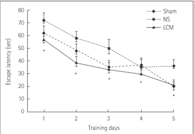
The Morris water maze test was performed at week 8 to com- pare the learning and spatial memory between the LCM- prefed and control rats. The mean escape latency was short- er for the LCM-prefed rats than for the normal-saline-prefed rats (Fig. 4), implying better spatial memory functioning in the LCM-pretreatment group (p<0.001, p=0.002, 0.002, 0.025, and 0.001 for five consecutive trials).
DISCUSSION
In this study, pretreatment with LCM at 100 mg/kg/dose sig- nificantly decreased the infarct areas in several brain regions and improved spatial learning and memory function. How- ever, no effect of LCM was observed in the postfed group.
Cellular and synaptic events are similar to those caused by ischemia or epilepsy. In both situations, acute membrane de- polarization is caused by sodium and calcium influx via volt- age-gated channels as well as ligand-gated channels.1,7-10 Accordingly, the following antiepileptic drugs that reduce
sodium influx exhibit demonstrated neuroprotective effects:
phenytoin,11 fosphenytoin,12 topiramate,13,14 and zonisamide.15 In addition, lamotrigine16 (which inhibits both N-type calci- um channels and sodium channels) and levetiracetam17 Fig. 2. Quantification of infarct areas after lacosamide (LCM) pretreatment at 100 mg/kg/dose. The mean percentage of right-side damage relative to the intact left side. Pretreatment with LCM at 100 mg/kg/dose (n=16) significantly decreased the infarcted areas in several brain regions (in- cluding the hemisphere, hippocampus, cortex, and striatum) relative to the normal-saline-treated group (NS; n=10). *p<0.05.
50
40
30
20
10
0
Hemisphere (%)
Sham NS
*
*
*
*
*
*
*
* LCM
50
40
30
20
10
0
Striatum (%)
Sham NS LCM
50
40
30
20
10
0
Cortex (%)
Sham NS LCM
50
40
30
20
10
0
Hippocampus (%)
Sham NS LCM
18,000 16,000 14,000 12,000 10,000 8,000 6,000 4,000 2,000 0
Distance (cm)
1 2 3 4 5 6 Test weeks
Sham NS LCM
Fig. 3. Open field activities test. Three groups were included in the open field activities test from week 2 to week 7 following hypoxia- ischemia: sham (n=14), normal-saline-treated (NS; n=16), and LCM- pretreated (100 mg/kg/dose, n=22) groups. No difference was found in exploratory behavior between the LCM-pretreatment group (100 mg/kg/dose) and the control group, suggesting that neither hypoxia- ischemia nor LCM affected exploratory activity. Data are mean and SEM values. LCM: lacosamide.
Neuroprotective Effect of Lacosamide
JCN
(which modulates N-type calcium channels) were also dem- onstrated to be neuroprotective against brain ischemia.
While the submolecular mechanism of slow inactivation is still poorly understood, LCM–as a selective modifier of slow inactivation–has been demonstrated to have addition- al benefits compared to classic sodium-channel-modulating anticonvulsants. Such anticonvulsants are relatively inactive in the 6-Hz psychomotor model of treatment-resistant sei- zures, whereas LCM has shown full effectiveness in the same test and also in the streptozotocin model for diabetic neu- ropathic pain. These findings also imply that the selective enhancement of slow inactivation in the voltage-gated so- dium channel and/or the interaction of LCM with CRMP-2 can result in a distinct pharmacological profile compared to classic sodium-channel-modulating anticonvulsants.18-22
CRMP-2 plays crucial roles in neuronal polarity, axonal guidance, and axonal degeneration.23 It also has roles in sev- eral neurodegenerative diseases, such as Alzheimer’s disease, epilepsy, and ischemia.24 Moreover, its expression has been clarified in several in vivo animal models.25 LCM binds to CRMP-2 to modulates its function and possibly exert a syn- ergistic effect with the slow inactivation of sodium channels as a neuroprotective strategy.
Only the pretreatment regimens in the present study showed a neuroprotective effect on hypoxia-ischemia. LCM pretreatment at 100 mg/kg/dose decreased both the inci- dence and volume of brain infarction. In addition, better spatial learning and memory function was observed in pre- treatment group, while no beneficial effect was observed in the posttreatment group. The early prevention of inflamma- tion and neuronal loss may reduce the overall CNS inflam-
matory response to injury and reduce oxidative stress, which in turn may reduce the development of chronic, long-term inflammation and associated neuronal degeneration. The early suppression of inflammation may also prevent synaptic remodeling, thereby improving functional outcomes.
A dose-dependent neuroprotective effect of LCM as shown in our study has also been reported for mouse models of traumatic brain injury and electrical status epilepticus.5,6 How- ever, those studies showed that only high-dose LCM exhibit neuroprotective effects.
While LCM has a wide safety margin and fewer fatal side effects,26 higher doses (i.e., above 200 mg/kg/dose) appear to be fatal in our animal model. The cause of death is suspect- ed to be a combined effect of respiratory depression by the sedative effect of LCM, compromised cardiac function, and prolonged hypoxic insult. The cause of death and the real safety margin could be clarified in future studies involving older rats that are large enough to allow the application of a ventilator device or blood pressure monitor.
In conclusion, the findings of this study suggest that LCM exerts a neuroprotective effect against hypoxia-ischemia in neonatal rats. This suggests that LCM pretreatment could be used as an effective neuroprotective method for neonates under hypoxic-ischemic conditions including heart surgery.
Further clinical studies are needed to confirm these preclini- cal observations and to define an appropriate dosing regimen.
Conflicts of Interest
The authors have no financial conflicts of interest.
Acknowledgements
This research was supported by a grant of the Korea Health Technology R & D Project through the Korea Health Industry Development Institute (KHIDI), funded by the Ministry of Health & Welfare, Republic of Korea (grant number: HI15C0810).
REFERENCES
1. Calabresi P, Cupini LM, Centonze D, Pisani F, Bernardi G. Antiepi- leptic drugs as a possible neuroprotective strategy in brain ischemia.
Ann Neurol 2003;53:693-702.
2. Liu Y, Barks JD, Xu G, Silverstein FS. Topiramate extends the thera- peutic window for hypothermia-mediated neuroprotection after stroke in neonatal rats. Stroke 2004;35:1460-1465.
3. Noh MR, Kim SK, Sun W, Park SK, Choi HC, Lim JH, et al. Neuro- protective effect of topiramate on hypoxic ischemic brain injury in neonatal rats. Exp Neurol 2006;201:470-478.
4. Schubert S, Brandl U, Brodhun M, Ulrich C, Spaltmann J, Fiedler N, et al. Neuroprotective effects of topiramate after hypoxia-ischemia in newborn piglets. Brain Res 2005;1058:129-136.
5. Licko T, Seeger N, Zellinger C, Russmann V, Matagne A, Potschka H.
Lacosamide treatment following status epilepticus attenuates neuro- nal cell loss and alterations in hippocampal neurogenesis in a rat elec- trical status epilepticus model. Epilepsia 2013;54:1176-1185.
6. Wang B, Dawson H, Wang H, Kernagis D, Kolls BJ, Yao L, et al.
Lacosamide improves outcome in a murine model of traumatic brain injury. Neurocrit Care 2013;19:125-134.
80 70 60 50 40 30 20 10 0
Escape latency (sec)
1 2 3 4 5 Training days
Sham NS LCM
Fig. 4. Morris water maze test. Three groups were included in the Morris water maze test: sham (n=14), normal-saline-treated (NS;
n=16), and LCM-pretreated (100 mg/kg/dose, n=22) 8-week-old rats.
The mean escape latency was shorter in the LCM-pretreatment group than in the NS, which implies better spatial memory function in the LCM-pretreatment group. Data are mean and SEM values.
*p<0.05. LCM: lacosamide.
*
* * *
*
Kim GH et al.
JCN
7. Arundine M, Tymianski M. Molecular mechanisms of calcium-de- pendent neurodegeneration in excitotoxicity. Cell Calcium 2003;34:
325-337.
8. Calabresi P, Centonze D, Bernardi G. Cellular factors controlling neuronal vulnerability in the brain: a lesson from the striatum. Neu- rology 2000;55:1249-1255.
9. Meldrum BS. Update on the mechanism of action of antiepileptic drugs. Epilepsia 1996;37 Suppl 6:S4-S11.
10. Xiong ZG, Zhu XM, Chu XP, Minami M, Hey J, Wei WL, et al. Neu- roprotection in ischemia: blocking calcium-permeable acid-sensing ion channels. Cell 2004;118:687-698.
11. Vartanian MG, Cordon JJ, Kupina NC, Schielke GP, Posner A, Raser KJ, et al. Phenytoin pretreatment prevents hypoxic-ischemic brain damage in neonatal rats. Brain Res Dev Brain Res 1996;95:169-175.
12. Chan SA, Reid KH, Schurr A, Miller JJ, Iyer V, Tseng MT. Fosphenyto- in reduces hippocampal neuronal damage in rat following transient global ischemia. Acta Neurochir (Wien) 1998;140:175-180.
13. Edmonds HL Jr, Jiang YD, Zhang PY, Shank R. Topiramate as a neuro- protectant in a rat model of global ischemia-induced neurodegenera- tion. Life Sci 2001;69:2265-2277.
14. Yang Y, Shuaib A, Li Q, Siddiqui MM. Neuroprotection by delayed administration of topiramate in a rat model of middle cerebral artery embolization. Brain Res 1998;804:169-176.
15. Owen AJ, Ijaz S, Miyashita H, Wishart T, Howlett W, Shuaib A.
Zonisamide as a neuroprotective agent in an adult gerbil model of global forebrain ischemia: a histological, in vivo microdialysis and be- havioral study. Brain Res 1997;770:115-122.
16. Wiard RP, Dickerson MC, Beek O, Norton R, Cooper BR. Neuropro- tective properties of the novel antiepileptic lamotrigine in a gerbil model of global cerebral ischemia. Stroke 1995;26:466-472.
17. Hanon E, Klitgaard H. Neuroprotective properties of the novel antiep-
ileptic drug levetiracetam in the rat middle cerebral artery occlusion model of focal cerebral ischemia. Seizure 2001;10:287-293.
18. Beydoun A, D’Souza J, Hebert D, Doty P. Lacosamide: pharmacology, mechanisms of action and pooled efficacy and safety data in partial- onset seizures. Expert Rev Neurother 2009;9:33-42.
19. Beyreuther BK, Freitag J, Heers C, Krebsfänger N, Scharfenecker U, Stöhr T. Lacosamide: a review of preclinical properties. CNS Drug Rev 2007;13:21-42.
20. Errington AC, Stöhr T, Heers C, Lees G. The investigational anticon- vulsant lacosamide selectively enhances slow inactivation of voltage- gated sodium channels. Mol Pharmacol 2008;73:157-169.
21. Halford JJ, Lapointe M. Clinical perspectives on lacosamide. Epilepsy Curr 2009;9:1-9.
22. Patsalos PN, Berry DJ. Pharmacotherapy of the third-generation AEDs: lacosamide, retigabine and eslicarbazepine acetate. Expert Opin Pharmacother 2012;13:699-715.
23. Wilson SM, Xiong W, Wang Y, Ping X, Head JD, Brittain JM, et al.
Prevention of posttraumatic axon sprouting by blocking collapsin re- sponse mediator protein 2-mediated neurite outgrowth and tubulin polymerization. Neuroscience 2012;210:451-466.
24. Hensley K, Venkova K, Christov A, Gunning W, Park J. Collapsin re- sponse mediator protein-2: an emerging pathologic feature and ther- apeutic target for neurodisease indications. Mol Neurobiol 2011;43:
180-191.
25. Touma E, Kato S, Fukui K, Koike T. Calpain-mediated cleavage of col- lapsin response mediator protein(CRMP)-2 during neurite degener- ation in mice. Eur J Neurosci 2007;26:3368-3381.
26. Thomas D, Scharfenecker U, Schiltmeyer B, Doty P, Cawello W, Horst- mann R. Low potential for drug-drug-interaction of lacosamide. Epi- lepsia 2006;47 Suppl 4:200.