INTRODUCTION
Promyelocytic leukemia protein (PML) was first identi- fied as the product of the promyelocytic leukemia gene (PML) that fuses with the retinoic acid receptor (RAR ) gene in the t (15;17) translocation of acute promyelocytic leukemia (APL) (1). PML-RAR is known to be a domi- nant negative oncoprotein that exerts its putative leuke- mogenic effect by inhibiting assembly of the distinctive nuclear structure, PML nuclear body (NB) (2). Meanwhile, immunolocalization studies using sera from patients with autoimmune diseases showed that the PML was diffusely present within the nucleus and/or localized to distinctive
“multiple nuclear dots”, which were originally described as an autoantigenic target in a patient with primary biliary cirrhosis (3). PML NBs vary in number between 10 and 30 per nucleus, and typically have a diameter of between 0.2 and 1 m. The number, size, and morphology of PML NBs alter throughout the cell cycle (4, 5) and are also dynamical- ly changed by diverse extracellular environment including DNA and RNA viral infection (6-8).
Although it is well documented the proper organization
of PML NBs is essential for normal cell proliferation and hematopoietic differentiation (9), the functions of PML or PML NBs still remain elusive in the pathogenesis of inflam- matory and neoplastic diseases except APL. The PML expre- ssion was considerably upregulated in inflammatory tissues such as hepatitis as well as during either normal or patho- logical proliferative states including benign or malignant tumors (4, 10-13). In most of malignant solid tumors examined, the PML was markedly overexpressed and orga- nization of PML NBs was not disturbed, unlike in cases of APL (13-15). Variable PML expression in human solid tumors suggests that the PML may play another role in addition to as a tumor suppressor, inducer of apoptosis and a depot of viral proteins (16, 17), and the mechanism of PML expres- sion can be different from that of leukemogenesis.
Among solid neoplasms in which the PML is overex- pressed, we selected hepatocellular carcinomas (HCCs) by the following indications: first, the expression of PML is quite different between non-neoplastic hepatocytes and HCC cells (13, 14); secondly, most of hepatocytes are in G0 phase of cell cycle, while a significant fraction of HCC cells are in actively proliferative stage; thirdly, hepatocellular car-
433
Ghil-Suk Yoon, Eunsil Yu
Department of Pathology, University of Ulsan College of Medicine Asan Medical Center, Seoul, Korea
Received : 29 January 2001 Accepted : 28 March 2001
Address for correspondence Eunsil Yu, M.D.
Department of Pathology, University of Ulsan College of Medicine Asan Medical Center, 388-1 Poongnap- dong, Songpa-gu, Seoul 138-736, Korea Tel : +82-2-2224-4552, Fax : +82-2-472-7898 E-mail : [email protected]
Overexpression of Promyelocytic Leukemia Protein and Alteration of PML Nuclear Bodies in Early Stage of Hepatocarcinogenesis
Promyelocytic leukemia protein (PML) is a major component of PML nuclear bodies (PML NBs). Fusion of promyelocytic leukemia gene (PML) with retinoic acid receptor gene with the t (15;17) translocation causes disassembly of PML NBs, leading to development of acute promyelocytic leukemia. In contrast, PML overexpression as well as different morphological changes of PML NBs were described in a few solid tumors. In this study, the expression of PML through the multistep hepatocarcinogenesis was analyzed in 95 cases of human hepatocel- lular carcinomas (HCCs) for comparison along with dysplastic nodules (DNs) and background liver cirrhosis (LC) or chronic hepatitis by immunohistochem- istry and immunoblot. In addition, cases of HCCs were further evaluated accord- ing to their histologic grade and etiology. The amount of PML as well as the num- ber and size of PML NBs increased gradually through the progression from LC, DNs to HCCs. The overexpression of PML in HCCs was much more closely associated with HBV infection than HCV infection or alcoholic liver disease. The PML expression, however, was not correlated with histologic grade of HCCs.
These results suggest that PML is involved in the early stage of multistep hepa- tocarcinogenesis, and HBV infection may be associated with the overexpression of PML and the morphological alteration of PML NBs.
Key Words : Leukemia, Promyelocytic; Nuclear Body; Carcinoma, Hepatocellular; Dysplastic Nodule;
Liver Cirrhosis; Hepatitis B Virus
cinogenesis is typical of a multistage process from chronic liver disease including liver cirrhosis (LC) through dysplas- tic nodule (DN) to HCCs (18), thus the PML expression can be properly correlated with the corresponding patho- logic conditions.
MATERIALS AND METHODS Tissue samples
Ninety-five cases of surgically resected HCCs were select- ed from the surgical pathology files of the Department of Pathology, Asan Medical Center, Seoul, Korea or Depart- ment of Pathology, Mt. Sinai Hospital, New York Universi- ty, NY, U.S.A. Seventy-five cases were positive for HBV DNA or HBs antigen. Ten cases of HCCs were positive for HCV RNA and had no evidence of HBV infection and another 10 cases had past medical history of chronic alco- holism and were negative for HCV RNA, HBs or HBc antigens. Six cases of HCCs developed within a DN and three cases had a separate DN each. Serial sections of forma- lin-fixed and paraffin-embedded tissues from HCCs and non-neoplastic livers were cut for hematoxylin and eosin stain and immunohistochemistry.
Histologic features of HCCs were classified according to the grading system of Edmondson and Steiner (19) and those of non-neoplastic livers were reviewed.
Among 75 cases, fresh frozen tissues from HCCs and cor- responding nontumor livers were available in 9 cases, one of which had both DN and HCC.
Cell culture
HepG2, HepG2.2.15 (20) and Hep3B cells were main- tained in DMEM (Gibco, Gaithersburg, MD), supplement- ed with 10% fetal calf serum at 37℃in a humidified atmosphere containing 5% CO2.
Immunohistochemical and immunofluorescent staining
Immunohistochemistry was done following streptavidin biotin complex method after antigen retrieval by boiling twice in citrate buffer (pH 6.0) for 5 min as previously described (21). The primary antibody for PML used in this study was PG-M3 (Santa Cruz, California, U.S.A., 1:100 dilution). For immunofluorescent staining, cultured cells on coverslips were fixed in cold methanol for 10 min. Fluores- cein-conjugated anti-mouse immunoglobulin was com- plexed with the primary antibodies for 1 hr. The tissue sec- tions or cells were mounted with Fluoromount G (Fischer Scientific Co., Pittsburgh, PA) and examined by using Olympus light microscope, Olympus Venox fluorescent microscope or Zeiss confocal laser scanning microscope.
Expression of the PML was described as follows: 1) aver- age number of PML NB-positive cells per 1,000 hepato- cytes or HCC cells: 2) average number of PML NBs in one nucleus of hepatocytes or HCC cells: 3) shape of PML NBs as fine dots, large dots greater than 1.0 m in diameter, or ball-shaped NBs.
Immunoprecipitation and immunoblotting
Preparation of nuclear proteins was done as previously described (21). Protein amount was calibrated by using BCA protein assay. Twenty g from the supernatant of each sample was incubated with the anti-PML antibody, PG-M3 at 1:200 dilution coupled to protein A-sepharose for 1 hr at 4℃. After extensive washing with the buffer, bound pro- teins were separated by SDS-PAGE and transferred to nitro- cellulose papers which were allowed to react with the anti- PML antibodies for 1 hr. Immune complexes were detected by the chemiluminescence detection system (Amersham Pharmacia Biotech LtD, UK).
Statistical analysis
The significance of differences in the expression of PML of HCCs, DNs, LC and chronic hepatitis (CH) was estimat- ed by paired t-test, Two-sample test, one way analysis of variance (ANOVA) procedure. The level of statistical signif- icance was set at 95% for all evaluations.
RESULTS PML expression in non-neoplastic liver
Seventy-one cases of HCCs were associated with LC and 24 cases occurred in the background of chronic hepatitis (CH). In non-neoplastic liver, rare hepatocytes showed one or two PML NBs that were slightly variable in size and shape (Fig. 1A). The difference of average number of PML NB- positive hepatocytes between CH and LC was not statistical- ly significant (Table 1). Most of the reactive hepatocytes in CH and regenerating hepatocytes in LC that revealed PML NBs were unevenly distributed and closely associated with portal inflammatory cells. Average number of PML NBs in one hepatocyte nucleus was also higher in LC than that in CH, which was not statistically significant (Table 1).
PML expression in dysplastic nodules
Nine cases of DN were associated with HCCs in the back- ground of LC. Average number of PML NB-positive hepa- tocytes was significantly increased in DNs compared with that in adjacent LC or CH (p<0.05) (Table 1). Six of nine cases of DN were classified low grade and three were high
grade. There was also a difference in number of PML NB- positive hepatocytes between low (31.27±43.26/1,000 cells) and high grade DNs (72.72±29.69/1,000 cells) (p<0.05 by paired t-test). The number of PML NBs in one hepatocyte nucleus was higher in DN than in LC, which was not statistically significant. Most of PML NBs were enlarged and some of them were ball-shaped with an empty center (Fig. 1B).
PML expression in hepatocellular carcinomas
The average number of PML NB-positive HCC cells per 1,000 HCC cells was markedly increased. The differences in average number of PML NB-positive cells between HCC, DN and LC were statistically significant (Table 1). The number of PML NBs per nucleus was also higher in HCC than in DN, LC and CH, which was not statistically signifi- cant (Table 1). Shape of PML NBs was quite different as well: greater than 50% of PML NBs in HCCs were large
solid dots or ball-shaped with an empty core (Fig. 1C).
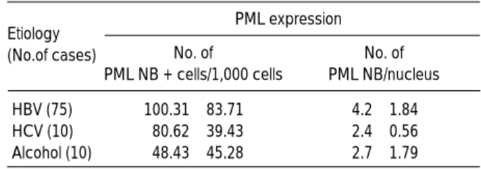
The average number of PML NB-positive HCC cells was greater in HCCs associated with HBV than in HCCs associ- ated with HCV or alcohol (Table 2). The number of PML NBs per one tumor cell nucleus was also higher in HBV- associated HCCs than in HCV-associated HCCs or alcohol- associated HCCs (Table 2). The shape of PML NBs, howev-
*HCC, hepatocellular carcinoma; DN, dysplastic nodule; LC, liver cirrho- sis; CH, chronic hepatitis; PML, promyelocytic leukemia protein; PML NB, PML nuclear body; �HBV vs HCV and HBV vs alcoholic, p<0.05 according to one way Analysis of Variance (ANOVA) procedure Etiology
(No.of cases)
PML expression No. of PML NB/nucleus No. of
PML NB + cells/1,000 cells
HBV (75) 100.31±83.71� 4.2±1.84
HCV (10) 80.62±39.43 2.4±0.56
Alcohol (10) 48.43±45.28 2.7±1.79
Table 2.Expression of PML in 95 cases of hepatocellular carci- noma according to etiologies
*HCC, hepatocellular carcinoma; DN, dysplastic nodule; LC, liver cir- rhosis; CH, chronic hepatitis; PML, promyelocytic leukemia protein;
PML NB, PML nuclear body; �HCC vs DN, HCC vs LC and HCC vs CH, p< 0.05 according to paired T-test; �DN vs LC and DN vs CH, p<0.05 according to Analysis of Variance(ANOVA) procedure: Duncan's multi- ple range test
Diagnosis (No.of cases)
PML expression No. of PML NB/nucleus No. of
PML NB + cells/1,000 cells
HCC (95) 92.81±78.31� 3.8±1.86
DN (9) 45.09±46.43� 2.8±1.26
LC (71) 8.95±7.66 1.8±1.54
CH (24) 4.70±6.15 0.5±0.50
Table 1.Expression of the PML in hepatocellular carcinomas, dysplastic nodules, liver cirrhosis and chronic hepatitis
*HCC, hepatocellular carcinoma; DN, dysplastic nodule; LC, liver cir- rhosis; CH, chronic hepatitis; PML, promyelocytic leukemia protein;
PML NB, PML nuclear body Histologic grade
(No.of cases)
PML expression No. of PML NB/nucleus No. of
PML NB + cells/1,000 cells
1 (12) 93.75±106.5 4.5±2.40
2 (40) 99.37±80.93 3.5±1.72
3 (33) 88.43±68.25 3.8±1.80
4 (10) 80.62±65.31 4.6±1.64
Total cases (95) 92.81±78.31 3.8±1.86 Table 3.Expression of the PML in 95 cases of hepatocellular carcinomas according to histologic grade
A B C
Fig. 1.PML expression in liver cirrhosis (A), dysplastic nodule (B), and hepatocellular carcinoma (C): A, Only one or two dots of PML NBs (arrows) are noted in regenerating hepatocytes. B, PML NBs are enlarged and some of them are ball-shaped (arrow). C, Most of PML NBs in HCC cells are markedly enlarged with empty cores (original magnification, ×1,000).
er, was similar in all HCCs. There was no significant corre- lation between the PML expression and histologic grade (Table 3). Pleomorphic nucleus in high grade HCCs, how- ever, had greater number of much larger or ball-shaped PML NBs than in lower grade HCCs.
Differential overexpression of the PML in HCC compared with those in LC and DN was confirmed by immunoblot- ting in a case that had both DN and HCC in the back- ground of LC (Fig. 2).
PML expression in hepatocellular carinoma cell lines
To examine the effect of HBV infection, we compared the expressions of PML in HepG2, HepG2.2.15, and Hep3B cells. HepG2.2.15 is a stable cell line that has been trans- fected by whole HBV genome in HepG2 cells and produce HBV DNA (20). HepG2 was established from a human hepatoblastoma and is not associated with HBV infection.
Hep3B is a human hepatocellular carcinoma cell line pro- ducing HBV DNA and antigens. Five to ten PML NBs were detected in all HepG2 cells in interphase. Their shape was monotonous and about 0.5 m in size (Fig. 3A). In HepG2.2.15 and Hep3B cells, the number of PML NBs was similar, but more than 50% of PML NBs were enlarged up to 1-2 m with an empty core (Fig. 3B).
DISCUSSION
The development and progression of HCC is typical of a multistep process from chronic liver disease caused by viral infection or toxic substance through DN to early or advan- ced HCC with genetic and/or epigenetic alterations. Thus, we analyzed the PML expression in HCCs as well as DNs and adjacent non-neoplastic liver tissues in the present study. Although the PML is known to be present diffusely in the nucleoplasm or in PML NBs, the PML was localized mainly to PML NBs as revealed by immunohistochemical staining with the monoclonal antibody, PG-M3. Thus, we described the PML expression according to the number of PML NB-positive hepatocytes or HCC cells per 1,000 hep- atocytes or HCC cells and the number of PML NBs in one nucleus of hepatocyte or HCC cell.
The differential overexpression of the PML in DN and HCC in this study indicates that the PML expression is dif- ferentially regulated during the multistep hepatocarcino- genesis, and the alteration of the PML expression may occur first in the early stage of hepatocarcinogenesis, from LC to DN formation. In contrast to our results, Chan et al. (15) described overexpression of the PML in the PML NBs in 50% of human HCCs and most of non-tumorous cirrhotic liver, thus indicated that the PML overexpression was asso- ciated with both LC and HCC formation. Gambacorta et al.
(13) reported the PML expression in 10/10 liver tumors, but neither histologic grading of the HCCs nor compara- tive analysis between preneoplastic lesions and HCCs has been made. The immunoprecipitation with immunoblot analysis in a few representative cases of HCCs supported also a distinct overexpression of the PML in DN and HCC compared with non-neoplastic liver. To clarify a possibility of transcriptional upregulation, northern blot analysis using RT-PCR products of the PML in LC, DN and HCC has been undergoing. At this moment, however, it can not be determined whether the overexpression of the PML in DNs
250
98 64
50
36
30 PML
Mol. wt
(kDa) Normal
Human Liver
HCC DN LC
*
Fig. 2. Immunoprecipitation and immunoblot analysis for the PML in a case of hepatocellular carcinoma (HCC) with dysplas- tic nodule (DN) in a background of liver cirrhosis (LC). Note dif- ferential overexpression of the PML (arrow) in LC, DN and HCC.
*, immunoglobulin heavy chain.
Fig. 3.PML NBs in HepG2 (A) and HepG2.2.15 cells (B) by confocal scanning microscope. Note enlarged ball-shaped PML NBs in HepG2.2.15 compared with a few fine dots in HepG2 cells (original magnification, ×1,000).
A B
and HCCs is a cause for hepatocellular carcinogenesis or an epiphenomenon during hepatocarcinogensis.
Statistically significant difference of the PML expression between HBV-associated HCCs and HCV- or alcohol-associ- ated HCCs and morphological characteristics of PML NBs in HepG2.2.15, a stably transfected cell line by HBV genome, suggest a strong relationship between HBV infection and PML NBs. PML NBs are known to be the site of DNA virus transcription and replication (8), thus HBV infection may affect the size or shape of PML NBs. Aoki et al. (22) reported that a subgenomic HBV DNA sequence (15 AB) is a hot spot for genomic recombination and a portion of 15AB-like sequence is homologous to break-point clusters of the human PML gene. Considering a close relationship between HBV infection and HCCs, it may be suggested that the PML might be a recombinogenic candidate triggering genomic instability in HBV-associated hepatocarcinogenesis. The structure of ball- shaped PML NBs is known to be associated with a recruit- ment of other PML NB-associated proteins such as SUMO-1 (23, 24) and TRF1 or TRF2 (25), and assembly with PML within PML NBs. Thus a further analysis of coimmunopre- cipitates with PML and PML isoforms in LC, DN and HCC using two-dimensional gel electrophoresis may elucidate an alternative mechanism of the PML overexpression and the morphological alteration of PML NBs.
ACKNOWLEDGMENTS
This work was supported by Korea Research Foundation Grant (KRF-1998-021-F000139). We are grateful to Ms.
Ki Won Lee and So Young Oh for their technical assistance, and to Prof. Swan N. Thung for providing cases of hepato- cellular carcinomas.
REFERENCES
1. de The H, Chomienne C, Lanotte M, Degos L, Dejean A. The t(15;
17) translocation of acute promyelocytic leukaemia fuses the retinoic acid receptor alpha gene to a novel transcribed locus. Nature 1990;
347: 558-61.
2. Dyck JA, Maul GG, Miller WH Jr, Chen JD, Kakizuka A, Evans RM. A novel macromolecular structure is a target of the promyelo- cyte-retinoic acid receptor oncoprotein. Cell 1994; 76: 333-43.
3. Szostecki C, Guldner HH, Will H. Autoantibodies against “nuclear dots”in primary biliary cirrhosis. Semin Liver Dis 1997; 17: 71-8.
4. Terris B, Baldin V, Dubois S, Degott C, Flejou JF, Henin D, Dejean A. PML nuclear bodies are general targets for inflammation and cell proliferation. Cancer Res 1995; 55: 1590-7.
5. Koken MH, Linares-Cruz G, Quignon F, Viron A, Chelbi-Alix MK, Sobczak-Thepot J, Juhlin L, Degos L, Calvo F, de The H. The PML growth-suppressor has an altered expression in human oncogene- sis. Oncogene 1995; 10: 1315-24.
6. Maul GG. Nuclear domain 10, the site of DNA virus transcription and replication. Bioessays 1998; 20: 660-7.
7. Bell P, Brazas R, Ganem D, Maul GG. Hepatitis delta virus replica- tion generates complexes of large hepatitis delta antigen and anti- genomic RNA that affiliate with and alter nuclear domain 10. J Virol 2000; 74: 5329-36.
8. Maul GG, Yu E, Ishov A, Epstein AL. Nuclear domain 10 (ND10) associated proteins are also present in nuclear bodies and redis- tribute to hundreds of nuclear sites after stress. J. Cell Biochem 1995; 59: 498-513.
9. Wang ZG, Delva L, Gaboli M, Rivi R, Giorgio M, Cordon-Cardo C, Grosveld F, Pandolfi PP. Role of PML in cell growth and the retinoic acid pathway. Science 1998; 279: 1547-51.
10. Sterndorf T, Grotzinger T, Jensen K, Will H. Nuclear dots: actors on many stages. Immunobiology 1997; 198: 307-31.
11. Lam YW, Ammerlaan W, O WS, Kroese F, Opstelten D. Cell type- and differentiation stagedependent expression of PML domains in rat, detected by monoclonal antibody HIS55. Exp Cell Res 1995;
221: 344-56.
12. Aractingi S, de The H, Gluckman E, Le Goue C, Carosela ED.
PML is expressed in chronic graftversus-host disease lesions. Bone Marrow Transplant 1997; 19: 1125-8.
13. Gambacorta M, Flenghi L, Fagioli M, Pileri S, Leoncini L, Bigerna B, Pacini R, Tanci LN, Pasqualucci L, Ascani S, Mencarelli A, Liso A, Pelicci PG, Falini B. Heterogeneous nuclear expression of the promyelocytic leukemia (PML) protein in normal and neoplastic human tissues. Am J Pathol 1996; 149: 2023-35.
14. Cho Y, Lee I, Maul GG, Yu E. A novel nuclear substructure, ND10: distribution in normal and neoplastic human tissues. Int J Mol Med 1998; 1: 717-24.
15. Chan JY, Chin W, Liew CT, Chang KS, Johnson PJ. Altered expression of the growth and transformation suppressor PML gene in human hepatocellular carcinomas and in hepatitis tissues. Eur J Cancer 1998; 34: 1015-22.
16. Zhong S, Salomoni P, Pandolfi PP. The transcriptional role of PML and the nuclear body. Nat Cell Biol 2000; 2: E85-90.
17. Maul GG, Negorev D, Bell P, Ishov AM. Review: properties and assembly mechanisms of ND10, PML bodies, or PODs. J Struct Biol 2000; 129: 278-87.
18. International Working Party. Terminology of nodular hepatocellu- lar lesions. Hepatology 1995; 22: 983-93.
19. Edmondson HA, Steiner PE. Primary carcinoma of the liver; a study of 100 cases among 48,900 necropsies. Cancer 1954; 7: 462-503.
20. Block TM, Lu X, Platt FM, Foster GR, Gerlich WH, Blumberg BS, Dwek RA. Secretion of human hepatitis B virus is inhibited by the imino sugar N-butyldeoxynojirimycin. Proc Natl Acad Sci USA 1994; 91: 2235-9.
21. Yu E, Lee KW, Lee HJ. Expression of promyelocytic leukaemia protein in thyroid neoplasms. Histopathology 2000; 36: 1-8.
22. Aoki H, Kajino K, Arakawa Y, Hino O. Molecular cloning of a rat chromosome putative recombinogenic sequence homologous to the hepatitis B virus encapsidation signal. Proc Natl Acad Sci USA 1996; 93: 7300-4.
23. Muller S, Matunis MJ, Dejean A. Conjugation with the ubiquitin-
related modifier SUMO-1 regulates the partitioning of PML within the nucleus. EMBO J 1998; 17: 61-70.
24. Sternsdorf T, Jensen K, Will H. Evidence for covalent modification of the nuclear dot-associated proteins PML and Sp100 by PIC1/
SUMO-1. J Cell Biol 1997; 139: 1621-34.
25. Yeager TR, Neumann AA, Englezou A, Huschtscha LI, Noble JR, Reddel RR. Telomerasenegative immortalized human cells contain a novel type of promyelocytic leukemia (PML) body. Cancer Res 1999; 59: 4175-9.