INTRODUCTION
Stem cells represent populations of cells that can give rise to all kinds of tissue types necessary to constitute an organ.
Traditional understandings on stem cells were mainly derived from hematopoietic stem cells in the model where aplastic bone marrow cells damaged by destructive radiation was re- populated using bone marrow transplantation (1). Over the last decades, studies on this cell population, namely hemato- poietic stem cells have revealed much of unique properties not found in other cell types, such as self-renewal division, a mitotic division leading to a production of same stem cells, or asymmetric division, a unique division leading to un-equal production of daughter cells from same mother cells (2).
Although the regulatory mechanisms controlling the self- renewing process or asymmetric division might have the key to more efficient use of stem cell in expansion culture or gene- tic modification, they still remain largely unknown await- ing further research. Another characteristic of stem cells in- ferred from hematopoietic stem cell is their extensive hetero- geneity even after the highest purification process that cur- rently available. The most important character of these stem cells, however, is their life-long reconstitutive activity as de- monstrated by the long-term repopulating ability in animal transplantation model and specific cultures designed for in
vitro assay (3). While the regulatory mechanisms for hema- topoietic stem cells have been under active investigation, unexpected breakthroughs were made in other aspects of stem cell biology. One is the finding that adult hematopoietic stem cells give rise to many other tissue type in addition to blood cells, such as neuronal or muscle cells. Similar surprising find- ings continue to unveil the previously hidden pluripotency of adult stem. A series of these new findings in stem cell dif- ferentiation initially provoked a big chaos in the classical con- cept of cell development and differentiation. New concepts of retro-differentiation, plasticity in differentiation, and exis- tence of very primitive pluripotent stem cells are emerging.
Furthermore, a novel issue on stem cell identity has been ad- dressed as to whether stem cells exist as a distinct clone in each organ and maintained throughout the development (clon- al nature) or they are rather product of organ function to main- tain integrity of each organ (functional nature) (4).
Another breakthrough in the stem cell area is the success in establishing human embryonic stem cells (5), which has triggered a vigorous debate between ethics and scientific merit of their use. Human embryonic stem cells can give rise to a greater numbers of tissue type from single cell nature (6-12) and continue to self-renew to the extent that adult stem cells can never achieve. Despite these attractive features in embry- onic stem cells, still many hurdles ahead before clinical use
Il-Hoan Oh, Dong-Wook Kim*
Cell & Gene Therapy Institute, Catholic Research Institute of Medical Science, Catholic Hematopoietic Stem Cell Transplantation Center*, The Catholic University of Korea, Seoul, Korea
Address for correspondence Il-Hoan Oh, M.D.
Cell & Gene Therapy Institute, Catholic Research Institute of Medical Science, 505 Banpo-dong, Seocho-gu, Seoul 137-040, Korea
Tel : +82.2-590-2591, Fax : +82.2-591-3994 E-mail : [email protected]
*This study was supported by a grant of the Korea Health 21 R&D Project, Ministry of Health & Welfare, Republic of Korea (01-PJ1-PG3-20600-0007).
151
Three-Dimensional Approach to Stem Cell Therapy
Recent progress in stem cell research is opening a new hope for cell therapy in regenerative medicine. Two breakthroughs were made in the stem cell era, one, new discoveries in multipotentiality of adult stem cells beyond the traditionally appreciated extent, and the other, establishment of pluripotent stem cell from human embryo. In addition to the newly identified multipotentiality of adult stem cells, their ability to be trans-differentiated toward other tissue types (stem cell plasticity) as well as to migrate toward the site of tissue damage make adult stem cells particularly attractive choice for stem cell based therapy. Stem cell therapy for organ regeneration, therefore, could be approached from three distinct dimen- sions: first, direct differentiation of multi-potent stem cells toward desired tissue types; secondly, regeneration of specific tissues through in vivo stem cell plas- ticity, and lastly, by tissue-specific stem cells from many types of organs. While each approach in stem cell therapy poses distinctive limitations for their success- ful clinical applications, understanding regulatory mechanisms of stem cell self- renewal and their in vivo engraftment will mostly extend their medical efficacy of stem cell based therapy.
Key Words : Stem Cells; Tissue Therapy; Stem Cell Therapy; Multipotentiality; Plasticity; Tissue Spe- cific Stem Cell
� REVIEW �
Received : 21 March 2002 Accepted : 26 March 2002
Fig. 1. Multipotentiality of bone marrow stem cell at single cell level.
Sca-1+
CD34+
PKH label
48 hr
PKH-labeled cell in each organ
Lung Esophagus
Stomach Colon
Skin Bile duct cyst
etc.
BM
such as immune rejection by difference in histocompatibili- ty between donor and recipients, possible tumor formation after in vivo transplantation, and problem of potential inap- propriate/improper differentiation. While embryonic stem cells are at an emerging stage in the avenue of cell therapy, adult stem cells have been intensively used for hematological and cancer-related managements in clinical practice. Further- more, recent studies are still expanding their use in many clin- ical situations that previously thought unrelated, such as me- tabolic diseases, bone diseases, or autoimmune diseases. There- fore, this review, focusing on stem cell-based cell therapy, will address discussions mostly to adult stem cells, rather than covering both types of stem cells, which should be beyond the current extent of scope.
MULTIPOTENTIALITY OF ADULT STEM CELLS
It has been a general concept that adult stem cells, in con- trast to embryo-derived stem cells that have a totipotent dif- ferentiation potential, are limited in their cell types that can be derived from a given source of adult stem cells. In addi- tion, it has been well accepted that this limitation is princi- pally determined by their developmental origin, in such a way that the ectoderm-derived cells give rise to cells of ecto- dermal origin and those from the mesoderm give rise to cells of mesodermal origin. Furthermore, the developmental pro- cess has been thought to be irreversible process associated with lineage determination. However, series of new discov- eries prompted the change of these classical concept await- ing emerge of new concept for cell development. In 1998, Geiger et al. (13) performed an experiment as to whether the adult cell would become like embryonic cells in a microen- vironment that normal developmental process is occurring.
They harvested bone marrow hematopoietic stem cells derived from transgenic mice for human beta globin gene and inject- ed into blastocysts of developing mice. The resulting mice demonstrated a developmental chimerism, i.e., existence of donor-derived cells at various stages of development, includ- ing york sac, fetal liver, and adult bone marrow. The notion
from this remarkable observation was that, given a certain microenvironment, the adult cells could also participate in the developmental process going backward in their develop- mental clock. Similarly, erythrocytes derived from adult donor did express the embryo-type hemoglobin ( -globin and - globin), suggesting that the gene expression program in adult genome could be reprogrammed in fetal microenvironment toward that in fetal genomic program. This intriguing obser- vation of developmental plasticity of adult cells was rapidly extended to other models of developmental plasticity to inves- tigate the extent of plasticity that adult stem cells can have.
From early 2000, such trials brought up several remarkable observations that adult stem cells indeed have the differen- tiation potential beyond the developmental origin. The first evidence was obtained injecting neuronal precursor cells into blastocyst of developing mice (14). In this experiment, adult transgenic mice expressing -galactosidase (lacZ) gene pro- vided neural progenitor cells in the form of collection of im- mature cells, called neurosphere. After injection into blasto- cysts, the donor-derived neurosphere was tracked for their contribution to various types of cells. Surprisingly, the neu- rosphere, which was of ectodermal origin, was found to con- tribute to most of the tissues including intestine, heart, liver, mesonephron, as well as brain and notocord. This was the first demonstration that adult stem cells have a higher differenti- ation potential than previously thought beyond the develop- mental barrier, although, some criticisms were raised for pos- sible contamination of other primitive stem cell population.
However, on May 1991, Krause et al. (15) provided even stronger observations using single cell suspensions. In their experiment (schematically illustrated in Fig. 1), hematopoietic stem cells in bone marrow was purified using surface mark- ers (CD34+ Sca-1+). The purified cells then were labeled with a lipid membrane-binding dye, PKH26, and transplanted into another mouse. Forty eight hours after transplantation, the bone marrow of primary transplanted mice were harvest- ed and the labeled cells were isolated at a single-cell level under microscopic guidance. These single cells were inocu- lated into blastocysts for further development of the embryo, then tracked down for the distribution of the labeled cells
throughout the whole embryos. Again, the labeled single cells of hematopoietic origin were found to give rise to almost all kinds of tissues including skin, epithelial cells of the gastroin- testinal tract, bile duct cyst, liver, and lung, further establish- ing the notion that adult stem cells could be as pluripotent as embryonic stem cells if placed in a specific permissive micro- environment.
PLASTICITY OF STEM CELLS
In concordance with the new recognition of multipotent differentiation potential of adult stem cells, many observa- tions were made for their differentiation toward other type tissue cells out of normal differentiation program. While the ability of a cell to give rise to a variety of different cell types is referred as “multipotentiality”, ability of a particular cell to become different cell types is commonly referred to as
“plasticity of differentiation”. One typical experiment show- ing the plasticity of adult stem cells was made by Lagasse et al. (1). In their experiment, an animal model of type 1 tyro- sinemia with fumaryl acetoacetate hydrolase deficiency (FAH-/-) (16) was employed as a test model, which is characterized by hepatotoxicity due to accumulation of toxic metabolite caused by lack of FAH and hence, their dependence on 2- (2-nitro-4-trifluoro-methylbenzyol)-1,2,-cyclohexanedione (NTBC) for survival. When unpurified bone marrow cells from Rosa 26 mice (wild type for the FAH and transgenic for the -galactosidase gene) were transplanted into lethally irradiated FAH-/-mice and NTBC was withdrawn, four out of nine mice remained healthy, while all of the control group mice died of hepatotoxicity. Exploration of the surviving mice after 7 months revealed hundreds of regenerating hepatic nodule in the transplanted mice, primarily consisted of hep- atocytes with wild type FAH (FAH+/+) and -galactosidase gene, which should have been derived from transplanted bone marrow cells from the donor. Further studies showed that hematopoietic stem cells (KTLS, c-kit+ thylow Lin- Sca-1+) (17, 18) and CD45+ was the only population responsible for hepatic regeneration, with no similar phenomenon for more differentiated cells such as Lin+ or c-kit- cells. Thus it has become clear that primitive hematopoietic stem cells with all the surface markers to become blood cells, could give rise to hepatocytes in a certain in vivo condition that hepatocytes are in emergency state.
Similar observations were made (19, 20) even in a human model where a liver transplantation or bone marrow trans- plantation was performed in cross sexual matching. In case of bone marrow transplantation, where the donor was male and the recipient was female, Y-chromosome-positive hepa- tocytes were observed in the female recipient’s liver, suggest- ing that a part of male donor’s bone marrow cells contributed to the hepatogenesis in the female recipient. In contrast, in case of female-to-male liver transplantation Y chromosome-
positive hepatocytes were observed in the transplanted liver, indicating that non-hepatic cells of the male recipient had contributed to the hepatogenesis.
Another interesting observation regarding stem cell plas- ticity was the regeneration of myocardium using bone mar- row cells. In 2001, Orlic et al. (21) tried the first experiment exploring the possible conversion of hematopoietic stem cells to myocardium. In the model, coronary arteries of mice were ligated to induce myocardial infarction. Shortly after infarc- tion, lineage-negative bone marrow cells from transgenic mice expressing enhanced green fluorescent protein (EGFP) were sorted into c-kit+ and c-kit- populations and injected into the peri-necrotic region. When the heart injected with cells were inspected 9 days after injection, 68% of infarcted region of myocardium in mice injected with c-kit+ lineage-negative cells, but not the myocardium in mice injected with c-kit- lineage-negative cells showed regeneration across the three layers of myocardium. Surprisingly again, most of the regen- erated cells were EGFP+ suggesting that the bone marrow derived hematopoietic stem cells were recruited to the necrotic region and participated in the de novo regeneration of myo- cardium. Furthermore, with anatomical regeneration of myo- cardium, the functional aspect of the heart was also concomi- tantly improved both in systolic pressure (about 40% increase) and diastolic pressure (about 36% lower). Interestingly, in addition to regeneration of myocardium, there was simulta- neous regeneration of endocardium and vessels, thus raising a hope that hematopoietic stem cell plasticity could be suit- able for both repair of necrotic region and redistribution of blood flows around the coronary vessel occlusion.
An even more interesting observation made by the same group (22) was that the general increase in the circulating number of c-kit+lin- cells using granulocyte-colony stimu- lating factor (G-CSF) and stem cell factor (SCF) led to an in- crease in the availability of circulating HSC to repair the in- farcted myocardium. According to the report, 70% of cyto- kine-mobilized mice survived 27 days post infarct, while only 17% of sham-operated control mice survived the same period.
In addition, cytokine-induced cardiac repair decreased the infarct size by 40%, cavity dilation by 26%, and diastolic stress by 70%.
In concordance with the re-vascularization described by Orlic et al., Kocher et al. (23) demonstrated that intravenous injection of CD34+CD117bright cells resulted in infiltration of vascular endothelial cells around the infarct zone within 48 hr of coronary artery ligation, but such a phenomenon was not observed in unaffected myocardium or myocardium of sham- operated rats. In addition, the injection of CD34+CD117 bright cells resulted in a 3-5 fold increase in neovasculariza- tion around the infarct area associated with concomitant in- crease in myocardial function compared to those resulting from injection of CD34+CD117dim.
Similarly, bone marrow cells could be differentiated into skeletal muscles (24, 25). In a mice model of Duchenne’s
muscular dystrophy (DMD), transplantation of hematopoi- etic stem cells as well as muscle stem cells (SP cells, see below for description) would reconstitute the dystrophin-positive muscle cells by 10-30% when examined 12 weeks after trans- plantation. This observation is particularly interesting in that stem cell transplantation could be potentially used for sys- temic delivery of therapeutic cells to broad areas of injury in the body.
Many similar observations were made for the plasticity of adult stem cells. In addition to the listed examples, many other tissues such as neuronal tissue (26, 27), renal tissue (28- 30), cartilage and bone (31-33) have been shown to be derived from in vivo transplanted bone marrow cells.
Furthermore, in most of cases, the stem cell plasticity is bi-directional, i.e., bone marrow cells can differentiate into other tissues, and vice versa. For example, muscle stem cells, certain portion of hepatic tissues and neuronal tissues could differentiate into blood etc. (14, 19-21, 25, 26, 28, 33-39) (summarized in Fig. 2).
TISSUE-SPECIFIC STEM CELLS
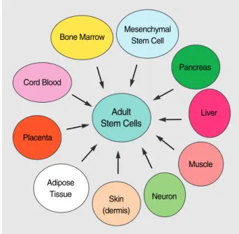
In addition to the multipotentiality of stem cells that can give rise to various tissue types and their plasticity that can lead to different tissue types, adult stem cells provide addi- tional potential way of tissue regeneration, i.e., through tissue- specific stem cells. It has been shown that many of adult or- gans have their own stem cells that retain some multipoten- tiality, albeit to a variable extent depending on the organ type. These cells include those from the pancreas, neuron, bone and cartilage, liver, skin, and even adipose tissues (sum-
marized in Fig. 3).
It is, however, important to note that the limited ranges of differentiation potential does not necessarily mean their limitation for used in cell therapy. Rather, it could be a bet- ter source for stem cell therapy if it is more committed to a specific lineage of tissue when purity of cell type are to be taken.
Pancreatic stem cell
It has been known from traditional observation that the pancreatic ductal epithelium is the source of various islet-asso- ciated endocrine cell populations including alpha, beta, and delta cells in the islets of Langhans. Therefore, the pancreat- ic ductal epithelium has been believed to contain stem cells responsible for pancreatic endocrine cells but to easily differ- entiate upon in vitro culture, thereby losing the insulin-secret- ing ability (40, 41). In 2000, Ramiya et al. and Bonner-Weir et al. simultaneously developed series of culture method by which pancreatic ductal stem cells can proliferate maintain- ing their ability to differentiate into islet-progenitor cells (IPC) and accordingly ability to differentiate into insulin- secreting beta cells (42, 43). In these reports, the islet-pro- ducing cells were developed from crude ductal pancreatic epithelium and thus obtained IPCs were maintained in up to 150 serial passages (42) retaining their ability to secrete insulin and glucagons upon terminal differentiation in vitro.
Subsequent injection of these islet cells into the renal capsule demonstrated that thus prepared islet cells led to neovascu- larization in the local environment, and secrete insulin in vivo.
According to the report, the blood glucose levels of diabetic mice (non-obese diabetic: NOD) were maintained up to 5
Fig. 2. Stem cell plasticity.
Kidney
Muscle
Brain Liver
Heart Myocardium
& vessel
Bone Marrow Stem Cell
BM Stroma
Osteoblast Chondrocyte Poulsom et al.
(2001)
Bjornson et al. (1999) Brazelton et al. (2000) Clarke et al. (2000)
Prockop (1997) Orlic et al.(2001)
Peterson et al.
(1999)
Jankson et al. (1999) Gussoni et al. (1999)
Ferrari et al. (1998)
Fig. 3. Diverse source of adult stem cells.
Mesenchymal Stem Cell Bone Marrow
Pancreas
Liver
Muscle Placenta
Cord Blood
Neuron Skin
(dermis) Adipose
Tissue
Adult Stem Cells
months even in the absence of exogenous insulin administra- tion, while the control group showed hyperglycemia (700 mg/mL) in two weeks. Interestingly, the islet cells that were used for transplantation were obtained from same strain (pre- diabetic NOD mice), leaving a possibility that such islet cells could again become the target of autoimmune attack as in type 1 diabetic mice. However, surprisingly, the cells protect- ed by a polymer capsule (to protect from immune attack) and those without any polymer showed similar maintenance of transplanted cells thus suggesting possible extension of this therapeutic model to autologous transplantation settings for human diabetes.
Neuronal stem cells
Most neuronal cells are formed during the embryonic and postnatal period, but some neurons continue to proliferate in adult mammalian brain. Since 1992, Reynolds and Weis have observed that adult neuronal cells have the self-renewal capacity and that they can proliferate and differentiate into all three components of the nervous system (neurons, astro- cytes, and oligodendrocytes) (44, 45). Recent progress in neu- ronal stem cell research found that these cells are mostly de- rived from ependymal cells lining the ventricle of the nervous system. These cells then proliferate in the subventricular re- gion and migrate to olfactory bulb, where they differentiate and integrate into each neuronal structure (46). More recent report also suggests that, in adult mouse brain, the neural stem cells reside both in ependymal and subventricular zone (47). Interestingly, these neural stem cells could be identi- fied by surface expression markers in addition to their char- acteristic marker “nestin”. These includes their expression of notch-1, low levels of PNA (peanut agglutinin binding), and HAS (heat stable antigen) which make it possible to puri- fy neuronal stem cells using surface marker.
Neural stem cells have well characterized advantage for cell therapeutic application, that is, they have an intrinsic ability to migrate toward the injured site as well as their capacity to renew various neuronal cells. During the brain or spinal cord injury these neural stem cells undergo extensive prolif- eration and migrate towards the site, either in a dorsal or lat- eral direction, over the 4 week period and form a scar that persists up to 1 yr. However, ependymal cell do not appear to be the only cells that have a healing effect, since in the above model of neural injury, most astrocytes have been de- rived from the ependymal area (Dil-postive), while oligoden- drocytes and neuronal cells were Dil-negative, when ependy- mal cells were pre-labeled with Dil before injury (46)
In another model using neural stem cell for cell therapy, neural stem cells served as a therapeutic vehicle to deliver a therapeutic gene to the target site. In 2000, both Aboody et al.
(48) and Benedetti et al. (49) demonstrated that an exogenous- ly implanted glioblastoma, which is characterized by rapid and diffuse infiltration over the brain area and poor progno-
sis, was thoroughly entrapped with simultaneously admin- istered neural stem cells. In this experiment the neural stem cells administered were cells immortalized from fetal brain by expressing c-myc. Despite the fact that they had been im- mortalized, the cells integrated into a neuronal structure, and stopped their proliferation, hence without causing a tumor in vivo. Interestingly, the expression of IL-4 gene (49) exerted a therapeutic effect on the glioblastoma comparable to unmod- ified neural stem cells, suggesting that the surrounding stem cells recruit a certain local effecter molecule which act target- ing the tumor cells. More interestingly, when the immortal- ized neural stem cells were equipped with the gene encod- ing cytosine deaminase (48), the neural stem cells surround- ing the tumor released this enzyme, and when 5-fluorocyto- sine was added systemically, the enzyme converted 5-fluoro- cytosine into 5-fluorouracil, and exerted a selective cytotoxic effect, resulting in up to 80% reduction in the tumor volume.
Hematopoietic stem cells
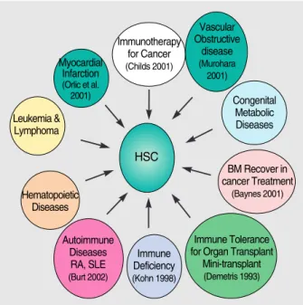
As described above, hematopoietic stem cells can give rise to all kinds of blood cells including myeloid and lymphoid cells, as well as their plasticity-related organogenesis. In addi- tion to the organogenesis by plasticity, their tissue-specific nature itself has enabled extensive application of this stem cells in medicine (21, 50-55) (summarized in Fig. 4).
However, the more efficient use of hematopoietic stem cells (HSC) would require strategy to preserve stem cell properties, i.e., self-renewing capacity because they are highly prone to differentiation during in vitro manipulative process. Under- standing of the self-renewing mechanism of HSC will enable further applications including gene therapy using HSC, cord
Fig. 4. Application of hematopoietic stem cells (HSCs).
Immunotherapy for Cancer (Childs 2001)
Vascular Obstructive
disease (Murohara 2001)
Congenital Metabolic Diseases
Immune Deficiency (Kohn 1998) Hematopoietic
Diseases Leukemia &
Lymphoma Myocardial
Infarction (Orlic et al.
2001)
Autoimmune Diseases RA, SLE (Burt 2002)
BM Recover in cancer Treatment
(Baynes 2001)
Immune Tolerance for Organ Transplant Mini-transplant (Demetris 1993)
HSC
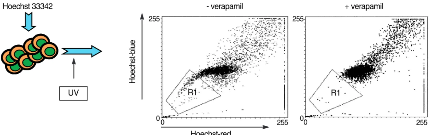
Fig. 5. Identification of SP (side population) cells as stem cell source. Cells were stained with Hoechst 33342 dye and subject to UV excitation in flowcytometer. The UV-excited Hoechst dye emits two different waves, one with >650 nm (red), and 488 nm (blue). These two lights are split into vertically arranged independent detectors. The tail part (Hoechst dim) is formed due to dye efflux mechanism of stem cells, which is dependent on the verapamil-sensitive efflux pump. The SP cells are, therefore, verified by loss of the same pop- ulation by verapamil treatment.
UV
Hoechst 33342 - verapamil
R1 R1
255
00 255
255
00 255 Hoechst-red
+ verapamil
Hoechst-blue
blood expansion, ex vivo expansion and efficient tumor purg- ing (2).
Purification of HSC:CD34 has been a gold standard marker for primitive stage HSCs and many clinical applications in- cluding tumor purging have been focused on the selective purification of CD34+ cells. Further studies revealed that still a major heterogeneity existed in the CD34+ population by CD38, AC133, and Thy-1 expression (56, 57). For exam- ple, CD34+CD38- cells are mostly enriched with most prim- itive stage HSCs which can be read out either by long-term in vitro culture (long-term culture initiating cells: LTC-IC) (3) or long-term in vivo NOD/SCID (non-obese diabetic/
severe combined immune deficiency) repopulating cells (CRU:
competitive repopulating unit) (58). In contrast, CD34+
CD38+ cells are more enriched with progenitor cells restrict- ed in their potential spectrum of lineages and in their self- renewing potential, which are often read out either by CFU- S12, CFU-14, or colony-forming assay in semi-solid medi- um (59). However, recent evidence revealed that additional populations that had been previously neglected (i.e., primi- tive CD34- cell populations) could be engrafted in NOD/
SCID mice with low clonogenicity in long-term culture, sug- gesting that this population could be an even more primitive cell population (60).
In addition to purification of HSCs by cell surface markers, functional characteristics of HSCs using their intrinsic dye- efflux effect were also described (61). These dye-effluxing cell population, called side population (SP) cell, are charac- terized by dim Hoechst 33342 staining when activated by UV light due to verapamil-sensitive dye efflux function (Fig. 5).
The SP cells were weak in CD34 expression, and lacked most of lineage-specific markers. Interestingly, like HSCs, multi- potent stem cells from other tissues such as muscle and liver shares common phenotype, suggesting that the SP cell phe-
notype might be a universal stem cell marker (62).
ONTOLOGICAL DIFFERENCE IN HSCS
HSCs have been found to exist in different forms of hema- topoietic organs throughout the ontological difference, i.e., adult bone marrow, neonatal cord blood, and fetal liver. Each stage of HSCs is characterized by differential functional char- acteristics in terms of in vivo self-renewal capacity, in vitro proliferation potential, and optimal growth factor require- ment (63-65). For example, fetal liver HSCs were character- ized by the highest in vitro proliferation potential and in vivo self-renewing capacity, while adult bone marrow cells have the lowest position in both terms, and umbilical cord blood is in the intermediate position (66). The basis for these func- tional differences among ontologically different populations remains unknown. Previously we have performed a series of gene expression studies to investigate the distinct gene expres- sion patterns among different stages of ontology (67). We found that series of gene expression pattern is conserved dur- ing in vivo differentiation from CD34+CD38- cells to CD34+
CD38+ cells and during in vitro differentiation mediated by growth factor stimulation. Interestingly, similar difference was also conserved during ontology-related differences in gene expression in such a way that the gene expression pattern in ontologically earlier stage HSC is more close to the patterns in growth factor-stimulated cells. These findings led us to speculate that there is a certain stage of HSC activation com- mon to in vitro stimulation and in vivo activation called
“priming”and according to this hypothesis, fetal liver and umbilical cord blood HSC mimic the state already growth factor-stimulated and primed in the activation process, when compared to adult bone marrow stem cells (schematically illustrated in Fig. 6).
THERAPEUTIC APPLICATION OF UMBILICAL CORD BLOOD
Previous bone marrow transplantations were mostly per- formed using adult bone marrow stem cells, often hard to find the donor in allogeneic transplantations, or often contam- inated with tumor cells in autologous transplants. In addi- tion to their advantage in finding a donor and their safety from infection or tumor cell contamination, the umbilical cord blood has many functional advantages over adult bone marrow stem cells (63, 68, 69). Accordingly, cord blood stem cell transplantation has become a world-wide trend as a new alternative way of bone marrow reconstitution in many dis- ease conditions including genetic diseases and malignant tumors, evidenced by the simultaneous clinical trials both in American blood bank and ‘Eurocord’(70-72).
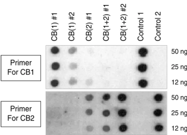
However, despite many advantages of cord blood in trans- plantation, the total cell number available has been the major limitation for a broader ranges of recipients in addition to the current applications mainly limited to children. Recently we have been trying to overcome this limit in the total cell number by co-transplantation of double unit cord blood to determine if any additive effect could be seen. When using several sets of cord blood pair varying in the HLA matching (from full 6-loci match to full mismatch), there was a consis- tent deviation toward a one single unit (>9:1 ratio) out of two independent donors (illustrated in Fig. 7). Similar devi- ations were also observed in a human model, where mixed double cord transplantation was done in one leukemic patient,
with engraftment pattern predominantly contributed by a single donor (manuscript in preparation). Therefore addition- al strategies to overcome these limitations need to be devel- oped for wider application of umbilical cord blood.
CONCLUSION
As discussed so far, stem cell therapy is a powerful tool for organ regeneration and de novo production of cells to replace damaged tissues. The organ regeneration based upon stem cell therapy could be approached in a three-dimensional way.
The first dimension is using multi-potential and/or pluripo- tent stem cells such as embryonic stem cells or multipotent adult stem cells. However, the use of adult stem cells is lim- ited by the extremely low frequency and the amount avail- able in a given organ, although it is advantageous in that it seldom forms a tumor and that it shows organ-specific dif- ferentiation. In contrast, embryonic stem cells should over- come the hurdles ahead. They should be driven down to spe- cific differentiated cells before transplantation in order to pre- vent tumor formation in vivo, and that therapeutic cloning is almost inevitable in order to overcome the immune medi- ated rejections, which is a very inefficient process with low success rate of normal development. Overcoming these hur- dles in embryonic stem cells, like the strategy to expand adult stem cells, will broaden the potential choice of the source in cell therapy.
The second dimension of cell therapy would be through
Intermediate priming 34+38-
Quiescent Unresponsive
Decisive commitment
Adult BM
Cytokine
FL CB 34+38+
Primer For CB2
CB(1) #1 CB(1) #2 CB(2) #1 CB(1+2) #1 CB(1+2) #2 Control 1 Control 2
50 ng 25 ng 12 ng Primer
For CB1
50 ng 25 ng 12 ng
Fig. 7. Differential contribution of mixed cord blood (CB) transplan- tation. Shown above is the representative data obtained from full six-loci matched (HLA-A,B,DR) double cord blood mixing trans- plantation into the NOD/SCID mice. The polymorphism in HLA-DP locus was used to discriminate the cells derived from each donor (CB (1) and CB (2)). The intensity of each dot is related to the amount of engraftment from specific donor cells as hybridized by donor specific probe corresponding to the distinctive sequence in HLA-DP region. # represents number of transplanted NOD/
SCID mice in each group and control 1 and 2 are the lanes con- taining purified DNA as a positive control.
Fig. 6. Hypothetical illustration of stem cell priming and the onto- logical difference characterized by different priming. A series of gene expression changes were identified during ontological devel- opment from fetal liver cells to adult bone marrow cells (Oh et al., 2000). The pattern of expression changes is conserved during changes from quiescent (CD34+CD38-) population to mitotically activated (CD34+CD38+ or growth factor stimulated) populations.
Therefore, we set a hypothesis that there is a certain stage of gene expression status in stem cell i.e., “intermediate priming”that a suppressed quiescent stem cell needs to pass through in order to become mitotically active and sensitive to extra cellular signal and, and after that, more decisive commitment to lineage-restrict- ed cells is occurring. According to this hypothesis, fetal liver cells and cord blood cells are already in a state that is primed in the intermediate stage compared to adult bone marrow cells, and hence show more immediate and higher proliferation potential.
the stem cell plasticity, which can regenerate many tissues using different types of stem cells. The problem of this ap- proach is that we can not answer such questions as ‘what is the controlling mechanisms?’or‘how does this process oc- cur?’To be useful for cell therapy, these phenomenological descriptions of plasticity should be further dissected into the regulatory mechanisms so that the efficiency of organ regen- eration by the process could reach a therapeutic level.
The third dimension of stem cell therapy would be through tissue-specific stem cells, such as pancreatic stem cells, hema- topoietic stem cells for lympho-myeloid reconstitution, or liver stem cells. The advantage of the tissue-specific stem cells is that it can produce a highly homogenous population of the differentiated cells unlike pluripotent embryonic stem cells, where the possibility of improper or inappropriate dif- ferentiation remains to be cleared. Again, however, the major obstacle of this approach is that the cell number is limited for a medically effective cell therapeutic dose.
Therefore, molecular mechanisms for the expansion of adult stem cells and differentiation of pluripotent stem cells should be elucidated before major benefit from stem cell therapy is envisioned.
REFERENCES
1. Lagasse E, Connors H, Al-Dhalimy M, Reitsma M, Dohse M, Osborne L, Wang X, Finegold M, Weissman IL, Grompe M. Puri- fied hematopoietic stem cells can differentiate into hepatocytes in vivo. Nat Med 2000; 6: 1229-34.
2. Ogawa M. Differentiation and proliferation of hematopoietic stem cells. Blood 1993; 81: 2844-53.
3. Sutherland HJ, Eaves CJ, Eaves AC, Dragowska W, Lansd orp PM.
Characterization and partial purification of human marrow cells capable of initiating long-term hematopoiesis in vitro. Blood 1989;
74: 1563-70.
4. Blau HM, Brazelton TR, Weimann JM. The evolving concept of a stem cell: entity or function? Cell 2001; 105: 829-41.
5. Thomson JA, Itskovitz-Eldor J, Shapiro SS, Waknitz MA, Swiergiel JJ, Marshall VS, Jones JM. Embryonic stem cell lines derived from human blastocysts. Science 1998; 282: 1145-7.
6. Itskovitz-Eldor J, Schuldiner M, Karsenti D, Eden A, Yanuka O, Amit M, Soreq H, Benvenisty N. Differentiation of human embryonic stem cells into embryoid bodies compromising the three embryonic germ layers. Mol Med 2000; 6: 88-95.
7. Kaufman DS, Hanson ET, Lewis RL, Auerbach R, Thomson JA.
Hematopoietic colony-forming cells derived from human embryonic stem cells. Proc Natl Acad Sci USA 2001; 98: 10716-21.
8. Kawasaki H, Suemori H, Mizuseki K, Watanabe K, Urano F, Ichinose H, Haruta M, Takahashi M, Yoshikawa K, Nishikawa S, Nakatsuji N, Sasai Y. Generation of dopaminergic neurons and pigmented epithelia from primate ES cells by stromal cell-derived inducing activi- ty. Proc Natl Acad Sci USA 2002; 99: 1580-5.
9. Kehat I, Kenyagin-Karsenti D, Snir M, Segev H, Amit M, Gepstein
A, Livne E, Binah O, Itskovitz-Eldor J, Gepstein L. Human embry- onic stem cells can differentiate into myocytes with structural and functional properties of cardiomyocytes. J Clin Invest 2001; 108:
407-14.
10. Liu S, Qu Y, Stewart TJ, Howard MJ, Chakrabortty S, Holekamp TF, McDonald JW. Embryonic stem cells differentiate into oligo- dendrocytes and myelinate in culture and after spinal cord trans- plantation. Proc Natl Acad Sci USA 2000; 97: 6126-31.
11. Lumelsky N, Blondel O, Laeng P, Velasco I, Ravin R, McKay R.
Differentiation of embryonic stem cells to insulin-secreting structures similar to pancreatic islets. Science 2001; 292: 1389-94.
12. McDonald JW, Liu XZ, Qu Y, Liu S, Mickey SK, Turetsky D, Got- tlieb DI, Choi DW: Transplanted embryonic stem cells survive, dif- ferentiate and promote recovery in injured rat spinal cord. Nat Med 1999; 5: 1410-2.
13. Geiger H, Sick S, Bonifer C, Muller AM. Globin gene expression is reprogrammed in chimeras generated by injecting adult hematopoi- etic stem cells into mouse blastocysts. Cell 1998; 93: 1055-65.
14. Clarke DL, Johansson CB, Wilbertz J, Veress B, Nilsson E, Karlstrom H, Lendahl U, Frisen J. Generalized potential of adult neural stem cells. Science 2000; 288: 1660-3.
15. Krause DS, Theise ND, Collector MI, Henegariu O, Hwang S, Gard- ner R, Neutzel S, Sharkis SJ. Multi-organ, multi-lineage engraftment by a single bone marrow-derived stem cell. Cell 2001; 105: 369-77.
16. Grompe M, al-Dhalimy M, Finegold M, Ou CN, Burlingame T, Kennaway NG, Soriano P. Loss of fumarylacetoacetate hydrolase is responsible for the neonatal hepatic dysfunction phenotype of lethal albino mice. Genes Dev 1993; 7: 2298-307.
17. Morrison SJ, Lagasse E, Weissman IL. Demonstration that Thy (lo) subsets of mouse bone marrow that express high levels of lineage markers are not significant hematopoietic progenitors. Blood 1994;
83: 3480-90.
18. Morrison SJ, Weissman IL. The long-term repopulating subset of hematopoietic stem cells is deterministic and isolatable by phenotype.
Immunity 1994; 1: 661-73.
19. Alison MR, Poulsom R, Jeffery R, Dhillon AP, Quaglia A, Jacob J, Novelli M, Prentice G, Williamson J, Wright NA. Hepatocytes from non-hepatic adult stem cells. Nature 2000; 406: 257.
20. Petersen BE, Bowen WC, Patrene KD, Mars WM, Sullivan AK, Murase N, Boggs SS, Greenberger JS, Goff JP. Bone marrow as a potential source of hepatic oval cells. Science 1999; 284: 1168-70.
21. Orlic D, Kajstura J, Chimenti S, Jakoniuk I, Anderson SM, Li B, Pickel J, McKay R, Nadal-Ginard B, Bodine DM, Leri A, Anversa P. Bone marrow cells regenerate infarcted myocardium. Nature 2001;
410: 701-5.
22. Orlic D, Kajstura J, Chimenti S, Limana F, Jakoniuk I, Quaini F, Nadal-Ginard B, Bodine DM, Leri A, Anversa P. Mobilized bone marrow cells repair the infarcted heart, improving function and sur- vival. Proc Natl Acad Sci USA 2001; 98: 10344-9.
23. Kocher AA, Schuster MD, Szabolcs MJ, Takuma S, Burkhoff D, Wang J, Homma S, Edwards NM, Itescu S. Neovascularization of ischemic myocardium by human bone-marrow-derived angioblasts prevents cardiomyocyte apoptosis, reduces remodeling and improves cardiac function. Nat Med 2001; 7: 430-6.
24. Pennisi E. Bone marrow cells may provide muscle power. Science 1998; 279: 1456.
25. Gussoni E, Soneoka Y, Strickland CD, Buzney EA, Khan MK, Flint AF, Kunkel LM, Mulligan RC. Dystrophin expression in the mdx mouse restored by stem cell transplantation. Nature 1999; 401: 390-4.
26. Brazelton TR, Rossi FM, Keshet GI, Blau HM. From marrow to brain: expression of neuronal phenotypes in adult mice. Science 2000; 290: 1775-9.
27. Mezey E, Chandross KJ, Harta G, Maki RA, McKercher SR. Turn- ing blood into brain: cells bearing neuronal antigens generated in vivo from bone marrow. Science 2000; 290: 1779-82.
28. Poulsom R, Forbes SJ, Hodivala-Dilke K, Ryan E, Wyles S, Navarat- narasah S, Jeffery R, Hunt T, Alison M, Cook T, Pusey C, Wright NA. Bone marrow contributes to renal parenchymal turnover and regeneration. J Pathol 2001; 195: 229-35.
29. Ito T, Suzuki A, Imai E, Okabe M, Hori M. Bone marrow is a reser- voir of repopulating mesangial cells during glomerular remodeling.
J Am Soc Nephrol 2001; 12: 2625-35.
30. Imasawa T, Utsunomiya Y. Stem cells in renal biology: bone mar- row transplantation for the treatment of IgA nephropathy. Exp Nephrol 2002; 10: 51-8.
31. Horwitz EM, Prockop DJ, Fitzpatrick LA, Koo WW, Gordon PL, Neel M, Sussman M, Orchard P, Marx JC, Pyeritz RE, Brenner MK.
Transplantability and therapeutic effects of bone marrow-derived mesenchymal cells in children with osteogenesis imperfecta. Nat Med 1999; 5: 309-13.
32. Pereira RF, Halford KW, O’Hara MD, Leeper DB, Sokolov BP, Pol- lard MD, Bagasra O, Prockop DJ. Cultured adherent cells from mar- row can serve as long-lasting precursor cells for bone, cartilage, and lung in irradiated mice. Proc Natl Acad Sci USA 1995; 92: 4857- 61.
33. Prockop DJ. Marrow stromal cells as stem cells for nonhematopoi- etic tissues. Science 1997; 276: 71-4.
34. Jackson KA, Mi T, Goodell MA. Hematopoietic potential of stem cells isolated from murine skeletal muscle. Proc Natl Acad Sci USA 1999; 96: 14482-6.
35. Kawada H, Ogawa M. Bone marrow origin of hematopoietic progen- itors and stem cells in murine muscle. Blood 2001; 98: 2008-13.
36. McKinney-Freeman SL, Jackson KA, Camargo FD, Ferrari G, Mav- ilio F, Goodell MA. Muscle-derived hematopoietic stem cells are hematopoietic in origin. Proc Natl Acad Sci USA 2002; 99: 1341-6.
37. Uchida N, Fujisaki T, Eaves AC, Eaves CJ. Transplantable hema- topoietic stem cells in human fetal liver have a CD34(+) side popu- lation (SP)phenotype. J Clin Invest 2001; 108: 1071-7.
38. Ferrari G, Cusella-De Angelis G, Coletta M, Paolucci E, Stornaiuolo A, Cossu G, Mavilio F. Muscle regeneration by bone marrow-derived myogenic progenitors. Science 1998; 279: 1528-30.
39. Bjornson CR, Rietze RL, Reynolds BA, Magli MC, Vescovi AL.
Turning brain into blood: a hematopoietic fate adopted by adult neural stem cells in vivo. Science 1999; 283: 534-7.
40. Bonner-Weir S, Baxter LA, Schuppin GT, Smith FE. A second path- way for regeneration of adult exocrine and endocrine pancreas. A possible recapitulation of embryonic development. Diabetes 1993;
42: 1715-20.
41. Gu D, Sarvetnick N. Epithelial cell proliferation and islet neogenesis in IFN- transgenic mice. Development 1993; 118: 33-46.
42. Ramiya VK, Maraist M, Arfors KE, Schatz DA, Peck AB, Cornelius JG. Reversal of insulin-dependent diabetes using islets generated in vitro from pancreatic stem cells. Nat Med 2000; 6: 278-82.
43. Bonner-Weir S, Taneja M, Weir GC, Tatarkiewicz K, Song KH, Sharma A, O’Neil JJ. In vitro cultivation of human islets from expand- ed ductal tissue. Proc Natl Acad Sci USA 2000; 97: 7999-8004.
44. Reynolds BA, Weiss S. Generation of neurons and astrocytes from isolated cells of the adult mammalian central nervous system. Sci- ence 1992; 255: 1707-10.
45. Weiss S, Dunne C, Hewson J, Wohl C, Wheatley M, Peterson AC, Reynolds BA. Multipotent CNS stem cells are present in the adult mammalian spinal cord and ventricular neuroaxis. J Neurosci 1996;
16: 7599-609.
46. Johansson CB, Momma S, Clarke DL, Risling M, Lendahl U, Frisen J. Identification of a neural stem cell in the adult mammalian central nervous system. Cell 1999; 96: 25-34.
47. Rietze RL, Valcanis H, Brooker GF, Thomas T, Voss AK, Bartlett PF. Purification of a pluripotent neural stem cell from the adult mouse brain. Nature 2001; 412: 736-9.
48. Aboody KS, Brown A, Rainov NG, Bower KA, Liu S, Yang W, Small JE, Herrlinger U, Ourednik V, Black PM, Breakefield XO, Snyder EY. From the cover: neural stem cells display extensive tro- pism for pathology in adult brain: evidence from intracranial gliomas.
Proc Natl Acad Sci USA 2000; 97: 12846-51.
49. Benedetti S, Pirola B, Pollo B, Magrassi L, Bruzzone MG, Rigamonti D, Galli R, Selleri S, Di Meco F, De Fraja C, Vescovi A, Cattaneo E, Finocchiaro G. Gene therapy of experimental brain tumors using neural progenitor cells. Nat Med 2000; 6: 447-50.
50. Childs R, Barrett J. Nonmyeloablative stem cell transplantation for solid tumors: Expanding the application of allogeneic immunother- apy. Semin Hematol 2002; 39: 63-71.
51. Murohara T. Therapeutic vasculogenesis using human cord blood- derived endothelial progenitors. Trends Cardiovasc Med 2001; 11:
303-7.
52. Baynes RD, Dansey RD, Klein JL, Hamm C, Campbell M, Abella E, Peters WP. High-dose chemotherapy and hematopoietic stem cell transplantation for breast cancer: past or future? Semin Oncol 2001;
28: 377-88.
53. Demetris AJ, Murase N, Fujisaki S, Fung JJ, Rao AS, Starzl TE.
Hematolymphoid cell trafficking, microchimerism, and GVH reac- tions after liver, bone marrow, and heart transplantation. Transplant Proc 1993; 25: 3337-44.
54. Kohn DB, Hershfield MS, Carbonaro D, Shigeoka A, Brooks J, Smogorzewska EM, Barsky LW, Chan R, Burotto F, Annett G, Nolta JA, Crooks G, Kapoor N, Elder M, Wara D, Bowen T, Mad- sen E, Snyder FF, Bastian J, Muul L, Blaese RM, Weinberg K, Parkman R. T lymphocytes with a normal ADA gene accumulate after transplantation of transduced autologous umbilical cord blood CD34+ cells in ADA-deficient SCID neonates. Nat Med 1998; 4:
775-80.
55. Burt RK, Slavin S, Burns WH, Marmont AM. Induction of tolerance in autoimmune diseases by hematopoietic stem cell transplantation:
getting closer to a cure? Blood 2002; 99: 768-84.
56. Gallacher L, Murdoch B, Wu DM, Karanu FN, Keeney M, Bhatia M.
Isolation and characterization of human CD34(-)Lin(-) and CD34(+) Lin(-) hematopoietic stem cells using cell surface markers AC133 and CD7. Blood 2000; 95: 2813-20.
57. Mayani H, Dragowska W, Lansdorp PM. Characterization of func- tionally distinct subpopulations of CD34+ cord blood cells in serum- free long-term cultures supplemented with hematopoietic cytokines.
Blood 1993; 82: 2664-72.
58. Bhatia M, Wang JC, Kapp U, Bonnet D, Dick JE. Purification of primitive human hematopoietic cells capable of repopulating immune- deficient mice. Proc Natl Acad Sci USA 1997; 94: 5320-5.
59. Wolf NS, Priestley GV. Kinetics of early and late spleen colony devel- opment. Exp Hematol 1986; 14: 676-82.
60. Bhatia M, Bonnet D, Murdoch B, Gan OI, Dick JE. A newly discov- ered class of human hematopoietic cells with SCID-repopulating activity. Nat Med 1998; 4: 1038-45.
61. Goodell MA, Rosenzweig M, Kim H, Marks DF, DeMaria M, Par- adis G, Grupp SA, Sieff CA, Mulligan RC, Johnson RP. Dye efflux studies suggest that hematopoietic stem cells expressing low or unde- tectable levels of CD34 antigen exist in multiple species. Nat Med 1997; 3: 1337-45.
62. Wulf GG, Jackson KA, Goodell MA. Somatic stem cell plasticity:
current evidence and emerging concepts. Exp Hematol 2001; 29:
1361-70.
63. Holyoake TL, Nicolini FE, Eaves CJ. Functional differences between transplantable human hematopoietic stem cells from fetal liver, cord blood, and adult marrow. Exp Hematol 1999; 27: 1418-27.
64. Nicolini FE, Holyoake TL, Cashman JD, Chu PP, Lambie K, Eaves CJ. Unique differentiation programs of human fetal liver stem cells shown both in vitro and in vivo in NOD/SCID mice. Blood 1999; 94:
2686-95.
65. Wang JC, Doedens M, Dick JE. Primitive human hematopoietic cells are enriched in cord blood compared with adult bone marrow or mobilized peripheral blood as measured by the quantitative in vivo SCID-repopulating cell assay. Blood 1997; 89: 3919-24.
66. Rebel VI, Miller CL, Eaves CJ, Lansdorp PM. The repopulation po- tential of fetal liver hematopoietic stem cells in mice exceeds that of their liver adult bone marrow counterparts. Blood 1996; 87: 3500-7.
67. Oh IH, Lau A, Eaves CJ. During ontogeny primitive (CD34(+) CD38(-)) hematopoietic cells show altered expression of a subset of genes associated with early cytokine and differentiation responses of their adult counterparts. Blood 2000; 96: 4160-8.
68. Kim DK, Fujiki Y, Fukushima T, Ema H, Shibuya A, Nakauchi H.
Comparison of hematopoietic activities of human bone marrow and umbilical cord blood CD34 positive and negative cells. Stem Cells 1999; 17: 286-94.
69. Leung W, Ramirez M, Civin CI. Quantity and quality of engrafting cells in cord blood and autologous mobilized peripheral blood. Biol Blood Marrow Transplant 1999; 5: 69-76.
70. Gluckman E. Current status of umbilical cord blood hematopoietic stem cell transplantation. Exp Hematol 2000; 28: 1197-205.
71. Rubinstein P, Carrier C, Scaradavou A, Kurtzberg J, Adamson J, Migliaccio AR, Berkowitz RL, Cabbad M, Dobrila NL, Taylor PE, Rosenfield RE, Stevens CE. Outcomes among 562 recipients of placental-blood transplants from unrelated donors. N Engl J Med 1998; 339: 1565-77.
72. Gluckman E, Rocha V, Boyer-Chammard A, Locatelli F, Arcese W, Pasquini R, Ortega J, Souillet G, Ferreira E, Laporte JP, Fernandez M, Chastang C. Outcome of cord-blood transplantation from relat- ed and unrelated donors. Eurocord Transplant Group and the Euro- pean Blood and Marrow Transplantation Group. N Engl J Med 1997;
337: 373-81.