INTRODUCTION
CD99 is an ubiquitous 32 kDa transmembrane protein encoded by the mic2 gene. It is expressed on most of human tissues, especially on cortical thymocytes, pancreatic islet cells, and Leydig and Sertoli cells. So far, several anti-CD99 anti- bodies such as 12E7, O662, L129, D44, HBA71, O13, and DN16 have been reported, and 12E7, HBA71, O13, and DN16 were revealed to be useful for diagnosis of tumor on routine paraffin sections (1-5). Identification of the CD99 ex- pression in formalin-fixed, paraffin-embedded tissues is par- ticularly useful in differentiating Ewing's sarcoma/primitive neuroectodermal tumors (PNETs) from other types of small round cell tumors (1-3, 6). In addition, its expression has been reported in lymphoblastic lymphoma/leukemia (7) and some rhabdomyosarcoma (8). Immunohistochemical detection of the CD99 expression has been reported in a number of other tumors, including mesenchymal chondrosarcomas (4), chloro- mas (9), acute myelogenous leukemia (9), some of ovarian tumors (10), and ependymomas (5) by the antigen retrieval technology. However, the functional significance of the CD99 expression in these tumors has not yet been known.
Recently, another isoform of CD99 (CD99 type II) was re- ported (11). CD99 type II is a truncated isoform that is pro- duced by alternative splicing of the CD99 gene transcript and its molecular weight is 28 kDa. In contrast to CD99 type I, transcripts of CD99 type II were detected at lower levels in a cell type-specific manner (11). Since there are no anti-CD99 antibodies that are able to discriminate the two isoforms of CD99, the tissue distribution pattern of CD99 type II has not been reported.
CD99 type II was known to be able to functionally coun- teract with the type I. It has been reported that the engage- ment of CD99 induces homotypic aggregation (12), up-reg- ulation of T cell receptor and major histocompatibility com- plex molecules (13), and apoptosis of immature thymocytes (14). In contrast, overexpression of the CD99 type II inhibited the homotypic aggregation of IM-9 cells by CD99 engage- ment (11). In addition, a recent study revealed that the trans- fection of CD99 type II variant in carcinoma cell line enhanced the in vitro invasiveness and matrix metalloproteinase (MMP) activity, together with inhibition of homotypic cell adhesion (15).
In the present study, we evaluated the expression of CD99 Kyeong Cheon Jung, Weon Seo Park¶, Young Mee Bae¶, Jang-Hee Hahn*, Kyuhyoung Hahn�, Hansoo Lee�, Hae Wan Lee�, Hyung-Jin Koo, Hai-Jeong Shin, Hyung Sik Shin, Young Euy Park, Seong Hoe Park‖
Department of Pathology, General Surgery�, Genetic Engineering�, Hallym University, Chunchon;
Department of Pathology¶, Anatomy*, College of Medicine; Vascular System Research Center�, Division of Life Sciences�, College of Natural Science, Kangwon National University, Chunchon;
Department of Pathology‖, Seoul National University College of Medicine, Seoul, Korea
Address for correspondence Kyeong Cheon Jung, M.D.
Department of Pathology, Hallym University College of Medicine, 1 Okchon-dong, Chunchon 200-702, Korea
Tel : +82.33-240-1631, Fax : +82.33-241-8250 E-mail : jungkc@hallym.ac.kr
*This work was supported by Korea Research Foundation Grant (2000-015-DP0311).
483
Immunoreactivity of CD99 in Stomach Cancer
CD99 is characteristically expressed in Ewing's sarcoma/primitive neuroectoder- mal tumor. Recently its immunoreactivity has also been reported in other tumors.
However, the significance of CD99 isoforms expressed in these tumors has not been elucidated. In this study, we evaluated the expression of CD99 isoforms and its relationship with histopathologic parameters in gastric adenocarcinomas.
Paraffin sections of 46 gastric adenocarcinomas were stained with an anti-CD99 monoclonal antibody, YG32. Twelve (26.1%) cases of 46 gastric adenocarcinomas showed immunoreactivity to YG32. The CD99 expression was also seen both in non-neoplastic foveolar epithelial cells and infiltrating lymphocytes. In addition, Western blot and RT-PCR analyses revealed that the type I is the predominant isoform of CD99 in non-neoplastic and neoplastic gastric tissues. The CD99 ex- pression was usually seen in the intestinal type adenocarcinoma, while rarely in the diffuse type. The CD99 immunoreactivity decreased in MMP-2-overexpress- ing adenocarcinomas (p=0.028). Our results suggest that the type I is the major isoform of CD99 expressed in non-neoplastic gastric mucosa and gastric adeno- carcinomas and its downregulation in gastric adenocarcinoma may be associat- ed with cellular dedifferentiation and/or MMP-2 overexpression.
Key Words : CD99; Stomach Neoplasms; Adenocarcinoma; Matrix Metalloproteinases
Received : 26 February 2002 Accepted : 2 April 2002
antigen in gastric adenocarcinoma, which is one of the most common cancers in Korea and also compared the CD99 im- munoreactivity with histopathologic parameters and the ex- pression of MMPs.
MATERIALS AND METHODS Tissue
The tissues from 46 cases of gastric adenocarcinoma were obtained from patients who had undergone gastrectomy at Chunchun Sacred Heart Hospital, Chunchun, Korea, between 1999 and 2001. The gross appearance of early gastric cancer (EGC) was classified according to the criteria by the Japanese Gastroenterological Endoscopy Society (16). Macroscopic clas- sification of advanced gastric cancer (AGC) was carried out according to the Borrmann's classification (17). Each tumor was classified on the basis of the modified WHO's classifica- tion system (18) as follows: tubular (well differentiated, mod- erately differentiated, and poorly differentiated), mucinous, or signet-ring-cell carcinoma. Histological type was also classi- fied according to the Lauren's classification (19).
Immunohistochemistry
Formalin-fixed, paraffin-embedded tissues were cut into 5- m serial sections, attached to silane-coated slides, deparaf- finized in xylene, and redehydrated in phosphate-buffered saline (PBS), pH 7.4. The deparaffinized sections were boiled for 10 min in 0.01 M citrate buffer, pH 6.0, for antigen-re- trieval and endogenous peroxidase activity was blocked with 0.3% hydrogen peroxide for 30 min. Immunohistochemical staining was performed using the LSAB kit (DAKO, Carpin- teria, CA, U.S.A.). The deparaffinized sections were preincu- bated with normal goat serum to prevent nonspecific binding, and then incubated overnight at 4℃with an optimal dilu- tion of the primary antibodies. The sections were rinsed with PBS and incubated with biotinylated anti-mouse immunoglob- ulin for 1 hr at room temperature and then with streptavidin- horseradish peroxidase conjugate. The enzyme reaction was developed with 0.03% 3′3-diaminobenzidine tetrahydrochlo- ride containing 0.006% hydrogen peroxide. As a negative control, the primary antibodies were replaced by an irrelevant isotype-matched antibody. YG32 (anti-CD99) antibody was purchased from DiNonA (Seoul, Korea), and A-Gel VC2 (anti-MMP-2), SL-1 IID4 (anti-MMP-3), and IIA5 (anti- MMP-9) antibodies were purchased from Pharmingen (San Diego, CA, U.S.A.).
Western blotting
Frozen tissues were lysed with 1% Triton X-100 in 50 mM Tris-HCl, pH 7.4, 50 mM EDTA, and 1 mM PMSF. The ly-
sates were mixed gently by slowly inverting the tube, incu- bated at 4℃for 30 min and centrifuged at 13,000 g for 15 min for removal of nuclei. The supernatant was used for SDS- PAGE under the reduced conditions. The acrylamide concen- tration of the separation gels was 12.5% and appropriate mo- lecular weight markers were used. The electrophoretic transfer of proteins to nitrocellulose was done at 30 V for 16 hr. After protein transfer, the nonspecific binding sites of the nitrocel- lulose were blocked by a solution of 5% skimmed milk. The antigens were detected by incubating the paper with primary antibodies. Horseradish peroxidase-conjugated antibody (DA- KO) was used as a secondary antibody, followed by enhanced chemiluminescence assay (ECL, Pharmacia, Uppsala, Sweden) for visualization. YG32 antibody was used to detect both iso- forms of CD99 and anti-human - actin antibody (ACTN05;
Lab Vision, Fremont, CA, U.S.A.) was used to normalize the amount of protein loaded.
Competitive RT-PCR
Total RNA was isolated from the frozen tissues using an RNA extraction kit (Bioneer, Daejon, Korea) and cDNA was first prepared by reverse transcription of 20 g RNA in a 50 L reaction volume containing PCR buffer, 1 mM DTT, 200 M dNTPs, 200 pmol oligo-dT18. The competitor for competitive RT-PCR was prepared using a competitive DNA construction kit (Takara, Shiga, Japan). Equal volumes of cDNA and DNA competitor were amplified in a PCR mas- ter mix composed of Tris-HCl, pH 9.0, 2.5 U Taq DNA polymerase, 250 U dNTP, 40 mM KCl, and 1.5 mM MgCl2 (Bioneer, Daejon, Korea). Primers were as follows; human CD99 type I & type II sense, 5′-GGTTTCGATTTATCTG ATGCC-3′, type I antisense, 5′-CTATTTCTCTAAAAGAG TACG-3′, type II antisense, 5′- CCCTAGGTCTTCAGCC AT-3′; human -actin sense, 5′-CAAGAGATGGCCACGG CTGCT-3′, antisense, 5′-TCCTTCTGCATCCTGTCGGC A-3′. The CD99 type I and type II primers were predicted to amplify 485 and 420 bp DNA fragments, respectively, while -actin primers generated a 275 bp-product. Samples were heated to 94℃for 30 sec, followed by either 55℃for 1 min (for the CD99 primers) or 68℃for 1 min (for the actin primers) and then 72℃for 1 min for 35 cycles. The PCR products were resolved by 2% agarose gel electrophore- sis and stained with ethidium bromide.
RESULTS
Immunoreactivity of CD99 in Non-neoplastic and Neo- plastic Gastric Tissues
Of the 46 patients with gastric carcinomas, 27 were male and 19 were female. The median age was 61 yr (rage, 37 to 79). Histologically, there were 23 AGCs and 23 EGCs. The
histopathologic features of the cases are summarized in Table 1.
To detect the CD99 expression in non-neoplastic and neo- plastic tissues, formalin-fixed, paraffin-embedded tissue sec- tions were stained with an anti-CD99 antibody, YG32. Non- neoplastic foveolar epithelial cells showed a weak immunore- activity to CD99 (Fig. 1A). The CD99 immunoreactivity was characteristically membranous. Strong expression of CD99 was also identified in infiltrating lymphocytes as previously reported (20). In contrast, the CD99 protein was stained only in 7 of 23 AGCs (30.4%) and 5 of 23 EGCs (21.7%). The immunoreactivity in carcinoma cells was usually stronger than that in non-neoplastic epithelium. Characteristically, CD99 immunoreactivity was detected only in the intestinal- and mixed-type adenocarcinomas except for one case (Fig. 1B, Table 2). In the three CD99-poisitive adenocarcinomas of mixed types, the CD99 immunoreactivity was mostly detect- ed in the glandular components, while CD99 was rarely ex- pressed in the diffuse component (Fig. 1C and 1D). In addi- tion, highly anaplastic tumor cells were not stained by anti- CD99 antibody, either (Fig. 1E). Among adenocarcinomas of the diffuse type, CD99-immunoreactivity was found in only
Histologic type (WHO classification) Tubular
Well differentiated Moderately differentiated Poorly differentiated Mucinous
Signet ring cell
Histologic type (Lauren's classification) Intestinal
Mixed Diffuse Depth of invasion
Mucosa Submucosa Proper muscle Subserosa
Serosa and omentum Lymph node metastasis
Present Absent
9 13 1
7 6 10
4 1 18
17 6
6 9 5
3
15
8
13 10
0 23 AGC (n=23) EGC (n=23) Table 1.Clinicopathological features of 46 gastric adenocarci- nomas
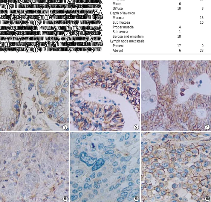
Fig. 1.Immunohistochemistry for CD99 in gastric adenocarcinoma. CD99 is expressed in the non-neoplastic gastric mucosa (A).
Among the carcinomas, the expression of CD99 is identified predominantly in the intestinal (B) and mixed types (C). In the cases of CD99-positive carcinomas of the mixed type, the CD99 expression is positive in the glandular component (C) but negative in the dif- fuse component (D). In addition, highly atypical cells are not stained by an anti-CD99 antibody (E). Among adenocarcinomas of the diffuse type, only one case shows immunoreactivity for CD99 (F). (×200).
D E F
A B C
one case (Fig. 1F).
Isotypes of CD99 Expressed in Gastric Cancer
So far there has been no anti-CD99 antibody that can dis- criminate CD99 type II from CD99 type I. The two isoforms of CD99 could thus be identified only by Western blotting or RT-PCR. To determine which isoform of CD99 antigens is expressed in gastric tissues, cancerous and noncancerous tissues in the 25 gastric adenocarcinomas were analyzed by Western blot analysis. Use of human thymus as a positive control confirmed that the YG32 antibody was reactive with both long (32 kDa) and short forms (28 kDa) of CD99 (Fig.
2A). Cell extract from tumor and normal tissues produced a band at 32 kDa, which indicated the CD99 type I, whereas the short form of CD99 was not detected in any case. How-
ever, the CD99 type I band was detected both in CD99-posi- tive and CD99-negative tumor tissues and the band intensity was not correlated with immunoreactivity in paraffin-embed- ded tissue. This may be due to the CD99 antigen expressed in reactive lymphocytes (Fig. 2A).
We also performed competitive RT-PCR by using primers specific for the CD99 type I and type II transcripts to detect the expression of CD99 isoforms even at a very low level. In agreement with results from Western blot analysis, the tran- scripts of CD99 type I were detected in all cases, and the expression level of CD99 type I mRNA was not correlated with immunoreactivity in paraffin-embedded tissue (data not shown). In addition, the transcripts of CD99 type II were detected in both CD99-positive and CD99-negative tumor tissues (Fig. 2B), indicating the low level expression of CD99 type II in stomach tissues. However, there was no difference in the transcript level of CD99 type II between CD99-posi- tive and CD99-negative tumor tissues. These results suggest that the immunoreactivity of CD99 in gastric carcinoma cells may not be due to the overexpression of CD99 type II.
Lauren's classification Intestinal
Mixed Diffuse
46 22 6 18
34 (73.9%) 14 (63.6%) 3 (50.0%) 17 (94.4%)
12 (26.1%) 8 (36.4%) 3 (50.0%) 1 (5.6%) n
Table 2. Immunoreactivity of CD99 in gastric adenocarcinomas CD99
- +
CD99 + 8 6 2* 4 4 3 5
CD99 - 17 4 13* 10 7 10 7
Total 25 10 15 14 11 13 12
n
Table 3. MMP Immunoreactivity of tumor cells in gastric adeno- carcinomas
MMP-2
- +
MMP-3
- +
MMP-9
- +
*p=0.028; Fisher's exact test.
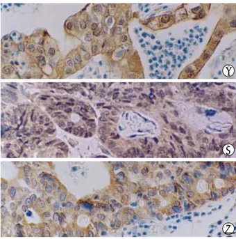
Fig. 3.Immunohistochemistry for MMP-2 (A), MMP-3 (B), and MMP- 9 (C) in advanced gastric adenocarcinoma. MMPs are expressed in the cytoplasm of tumor cells (×200).
Fig. 2.Identification of the CD99 isoforms expressed in non-neo- plastic and carcinoma tissues. (A) Western blot analysis. Total lysates were prepared from non-neoplastic (N) and tumor (T) tis- sues and the CD99 expression was detected using an anti-CD99 antibody, YG32. Type I CD99 (32 kDa) is detected in both CD99- negative (left four lanes) and CD99-positive (right four lanes) cases, while the type II isoform (28 kDa) is not detected in any case. The amount of protein in samples was normalized by Western blot- ting of the human -actin protein (42 kDa). (B) Competitive RT- PCR analysis of CD99 type II mRNA expression in CD99-nega- tive (left four lanes) and CD99-positive (right four lanes) tumor tis- sues using the specific primer pairs predicted to amplify the type II isoform of CD99 mRNA. The amount of mRNA in samples was normalized by competitive RT-PCR of human -actin.
A
B
C A
B
42 kDa CD99-
N T N T
CD99+
Thymocyte N T N T
CD99- CD99+
Competitor -Actin 32 kDa 28 kDa
Competitor CD99 II
Expression of MMP-2, MMP-3, and MMP-9 in Gastric Cancer
Although various functions of CD99 in lymphoid cells have been reported, the function of CD99 in tumorigenesis has not been elucidated. Only a recent research using carcinoma cell lines suggested that the overexpression of CD99 type II enhances the in vitro invasiveness and MMP activity (15). In addition, it has been reported that the expression of MMP-2 and MMP-9 was increased in gastric cancers (21-24). There- fore, in order to analyze the possible relationship between CD99 immunoreactivity and the expression of MMPs in gas- tric adenocarcinomas, immunohistochemical analysis was performed. MMP-2 was not expressed in any of the normal gastric epithelium studied. In contrast, 15 of 25 cases (60.0%) of gastric carcinoma showed immunoreactivity for MMP-2 in the carcinoma cells (Fig. 3A, Table 3). In 8 CD99-positive cases, MMP-2 immunoreactivity was detected only in 2 cases (25.0%), while 13 (76.5%) of 17 CD99-negative cases showed MMP-2 immunoreactivity. These results showed an inverse relationship between MMP-2 overexpression and CD99 im- munoreactivity (p=0.028). MMP-3 and MMP-9 immuno- reactivities were also detected in tumor cells of gastric adeno- carcinoma (in 11, 12 cases, respectively; Fig. 3, Table 3).
However, there was no relation between the MMP-3 or MMP- 9 expression and CD99 immunoreactivity.
DISCUSSION
In soft tissue tumors, the CD99 expression has long been used as a diagnostic marker for Ewing's sarcoma/PNET (1- 3, 6), and recently, many other tumors have been addeded to the list of tumors with CD99 immunoreactivity (4, 5, 7- 10). Nevertheless, the isoforms of CD99 expressed in these tumors have not yet been determined. Thus, we examined the expression of each type of CD99 in non-neoplastic and neoplastic gastric tissues and analyzed the relationship be- tween CD99 expression and histopathologic parameters and MMP immunoreactivity.
Advances in antigen retrieval technology as well as the de- velopment of new antibodies against the CD99 antigen have contributed to the detection of CD99 in more tumor cases.
Thus, we performed immunohistochemical analysis in sto- mach cancer tissues after microwave treatment in citrate buf- fer, through which we could detect the expression of CD99 antigen in normal foveolar epithelial cells and infiltrating lymphocytes. The CD99 immunoreactivity was characteris- tically membranous in these cells. In contrast, only 23%
cases of gastric adenocarcinoma showed CD99 immunoreac- tivity. The immunoreactivity of CD99 was not correlated with age, depth of invasion, or lymph node metastasis. How- ever, CD99 immunoreactivity was mostly detected in ade- nocarcinomas of the intestinal type or glandular component
of the mixed type. In addition, highly anaplastic tumor cells did not express the CD99 antigen. Thus, these results imply that CD99 is expressed both in normal gastric epithelum and adenocarcinoma cells which show glandular differentiation (intestinal type), while CD99 expression is decreased in ade- nocarciconomas of diffue type, in which tumor cells are less differentiated. To identify the isoform of CD99 expressed in non-neoplastic and neoplastic gastric tissues, Western blot- ting was performed and only the type I isoform of 32 kDa in molecular weight was detected. Although the transcripts of CD99 type II were detected in non-neoplastic and neoplas- tic tissues by RT-PCR, there was no difference in the expres- sion level of mRNA of CD99 type II between CD99-posi- tive and CD99-negative cases. Thus, all these features suggest that the CD99 type I is the predominant isoform that is ex- pressed both in non-neoplastic and neoplastic gastric tissues and the loss of CD99 immunoreactivity in gastric adenocar- cinomas may be associated with dedifferentiaton of tumor cells.
Previous studies showed that the balance between the two CD99 isoforms affect the cell adhesion (11, 15). In addition, overexpression of CD99 type II enhanced the MMP activity in a carcinoma cell line (15). These results raised the possi- bility that the downregulation of CD99 type I expression or upregulation of CD99 type II expression may enhance the invasiveness of tumor cells by affecting cell adhesion or MMP activity. Thus, much attention has focused on the difference in the level of MMP expression between CD99-positive and CD99-negative cases.
MMPs are zinc-dependent enzymes that are involved in the degradation of the extracellular matrix. On the basis of their substrate specificities and structural homology, MMPs can be classified into subgroups of collagenases (MMP-1, MMP-8, and MMP-13), gelatinases (MMP-2 and MMP-9), stromelysins (MMP-3, MMP-10, MMP-12, and MMP-7), membrane-type MMPs (MT-MMPs) and other MMPs (25- 26). MMPs have been considered to play significant roles in tumor invasion and metastasis (25). Especially, MMP-2 (gela- tinase A, 72-kDa type IV collagenase) is well known as a de- composing enzyme of type IV collagen, laminin, and fibro- nectin, all of which are components of the basement mem- brane, and its relationship to the depth of invasion, metasta- sis in regional lymph nodes, and prognosis of gastric cancer has been reported (22, 27). In our study, MMP-2 was detected in 15 (60.0%) out of 25 gastric adenocarcinomas. Among the 8 CD99-positive cases, only two (25.0%) were MMP-2- positive, while MMP-2 immunoreactivity was detected in 13 (76.5%) out of 17 CD99-negative cases. These results sug- gest that MMP-2 overexpression in gastric adenocarcinoma may be associated with the downregulation of CD99 type I and thus support the in vitro data that the balance between the two CD99 isoforms contributes to the MMP activity.
In addition to MMP-2, MMP-3 and MMP-9 are involved in carcinogenesis and tumor invasion. MMP-9 (gelatinase B,
92-kDa type IV collagenase) have a gelatin-binding domain and its structure is similar to that of MMP-2 (25), wherease MMP-3 (stromelysin-1) is a member of stromelysins and can degrade collagens III, IV, V, IX, X, and XI, laminins, elastin, entactin, fibronectin, fibrin, fibrillins, fibulin, link protein, osteonectin, tenascin, vitronectin, and ECM proteoglycans (28). The expression of MMP-9 is associated with lymph node metastasis in gastric cancer (24), and the immunoreactivity for MMP-3 has also been reported in some cases of gastric cancers (21). In our study, 11 (44.0%) of 25 gastric carcinoma cases showed immunoreactivity for MMP-3 in tumor cells and MMP-9 was overexpressed in 12 cases (48.0%). How- ever, MMP-3 or MMP-9 immunoreactivity was not corre- lated with CD99 immunoreactivity.
CD99 is expressed on most of human tissues and the sig- nificant role of CD99 in tumorigenesis was only documented in Hodgkin's disease, in which the loss of CD99 has been shown to play a critical role in the formation of Hodgkin's and Reed-Sternberg cells (29-31). The results from our study also suggest that the downregulation of CD99 type I could be associated with dedifferentiation of tumor cells in gastric adenocarcinomas. Thus, it seems that the downregulation of CD99 expression rather than its overexpression may play a functionally significant role in tumorigenesis or tumor pro- gression.
In conclusion, our findings suggest that the downregula- tion of CD99 type I expression may be associated with de- differentiation of tumor cells and overexpression of MMP-2 in gastric adenocarcinomas.
ACKNOWLEDGMENTS
The authors wish to express deep thanks to Jae Nam Seo, Jin Sil Choi, Eun Suk Seong, and Seung Hee Lee (Department of Pathology, Hallym University College of Medicine) for their technical assistance.
REFERENCES
1. Kovar H, Dworzak M, Strehl S, Schnell E, Ambros IM, Ambros PF, Gadner H. Overexpression of the pseudoautosomal gene MIC2 in Ewing's sarcoma and peripheral primitive neuroectodermal tumor.
Oncogene 1990; 5: 1067-70.
2. Ambros IM, Ambros PF, Strehl S, Kovar H, Gadner H, Salzer-Kunts- chik M. MIC2 is a specific marker for Ewing's sarcoma and periph- eral primitive neuroectodermal tumors. Evidence for a common his- togenesis of Ewing's sarcoma and peripheral primitive neuroectoder- mal tumors from MIC2 expression and specific chromosome aberra- tion. Cancer 1991; 67: 1886-93.
3. Perlman EJ, Dickman PS, Askin FB, Grier HE, Miser JS, Link MP.
Ewing's sarcoma: routine diagnostic utilization of MIC2 analysis: a Pediatric Oncology Group/Children's Cancer Group Intergroup
Study. Hum Pathol 1994; 25: 304-7.
4. Granter SR, Renshaw AA, Fletcher CD, Bhan AK, Rosenberg AE.
CD99 reactivity in mesenchymal chondrosarcoma. Hum Pathol 1996;
27: 1273-6.
5. Choi YL, Chi JG, Suh YL. CD99 immunoreactivity in ependymoma.
Appl Immunohistochem Mol Morphol 2001; 9: 125-9.
6. Weidner N, Tjoe J. Immunohistochemical profile of monoclonal anti- body O13: antibody that recognizes glycoprotein p30/32MIC2 and is useful in diagnosing Ewing's sarcoma and peripheral neuroepithe- lioma. Am J Surg Pathol 1994; 18: 486-94.
7. Dworzak MN, Fritsch G, Fleischer C, Printz D, Froschl G, Buchinger P, Mann G, Gadner H. CD99 (MIC2) expression in paediatric B-lin- eage leukaemia/lymphoma reflects maturation-associated patterns of normal B-lymphopoiesis. Br J Haematol 1999; 105: 690-5.
8. Ramani P, Rampling D, Link M. Immunocytochemical study of 12E7 in small round-cell tumours of childhood: an assessment of its sensi- tivity and specificity. Histopathology 1993; 23: 557-61.
9. Zhang PJ, Barcos M, Stewart CC, Block AW, Sait S, Brooks JJ. Im- munoreactivity of MIC2 (CD99) in acute myelogenous leukemia and related diseases. Mod Pathol 2000; 13: 452-8.
10. Choi YL, Kim HS, Ahn G. Immunoexpression of inhibin alpha sub- unit, inhibin/activin betaA subunit and CD99 in ovarian tumors. Arch Pathol Lab Med 2000; 124: 563-9.
11. Hahn JH, Kim MK, Choi EY, Kim SH, Sohn HW, Ham DI, Chung DH, Kim TJ, Lee WJ, Park CK, Ree HJ, Park SH. CD99 (MIC2) regulates the LFA-1/ICAM-1-mediated adhesion of lymphocytes, and its gene encodes both positive and negative regulators of cellular adhesion. J Immunol 1997; 159: 2250-8.
12. Bernard G, Zoccola D, Deckert M, Breittmayer JP, Aussel C, Bernard A. The E2 molecule (CD99) specifically triggers homotypic aggre- gation of CD4+ CD8+ thymocytes. J Immunol 1995; 154: 26-32.
13. Choi EY, Park WS, Jung KC, Kim SH, Kim YY, Lee WJ, Park SH.
Engagement of CD99 induces up-regulation of TCR and MHC class I and II molecules on the surface of human thymocytes. J Immunol 1998; 161: 749-54.
14. Bernard G, Breittmayer JP, de Matteis M, Trampont P, Hofman P, Senik A, Bernard A. Apoptosis of immature thymocytes mediated by E2/CD99. J Immunol 1997; 158: 2543-50.
15. Kim E., Lee H-J, Hahn J-H, Park SH, Lee H. Expression of a spliced variant of CD99 membrane protein increases motility, matrix degra- dation, and invasiveness of human breast carcinoma cells. Proc Amer Assoc Cancer Res 2000; 41: 231-2.
16. Murakami T. Pathomorphological diagnosis: definition and gross classification of early gastric cancer. In; Murakami T, editor, Early Gastric Cancer. Tokyo: University of Tokyo Press, 1971: 53-5.
17. Borrmann R, Geschwulste des Magens und Duodenums. In: Henke F, Lubarsch O, editors. Handbuck der Spezieller Pathologischen Anato- mie und Histologie. Berlin: Springer Verlag, 1926: 825.
18. Watanabe H, Jass JR, Sobin LH. Histological typing of esophageal and gastric tumors. In: Watanabe H, Jass JR, Sobin LH, editors.
World Heath Organization Histological Classification of Tumors, 2nd edition. Berlin: Springer Verlag, 1989; 18: 20-6.
19. Lauren P. The two histological main types of gastric carcinoma; dif- fuse and so-called intestinal type carcinoma. An attempt at a histo-
logical classification. APMIS 1965; 64: 31-49.
20. Park CK, Shin YK, Kim TJ, Park SH, Ahn GH. High CD99 expres- sion in memory T and B cells in reactive lymph nodes. J Korean Med Sci 1999; 14: 600-6.
21. Murray GI, Duncan ME, Arbuckle E, Melvin WT, Fothergill JE.
Matrix metalloproteinases and their inhibitors in gastric cancer.
Gut 1998; 43: 791-7.
22. Allgayer H, Babic R, Beyer BC, Grutzner KU, Tarabichi A, Schild- berg FW, Heiss MM. Prognostic relevance of MMP-2 (72-kD col- lagenase IV) in gastric cancer. Oncology 1998; 55: 152-60.
23. Inoue T, Yashiro M, Nishimura S, Maeda K, Sawada T, Ogawa Y, Sowa M, Chung KH. Matrix metalloproteinase-1 expression is a prognostic factor for patients with advanced gastric cancer. Int J Mol Med 1999; 4: 73-7.
24. Kabashima A, Maehara Y, Kakeji Y, Baba H, Koga T, Sugimachi K. Clinicopathological features and overexpression of matrix met- alloproteinases in intramucosal gastric carcinoma with lymph node metastasis. Clin Cancer Res 2000; 6: 3581-4.
25. Stetler-Stevenson WG, Yu AE. Proteases in invasion: matrix met- alloproteinases. Semin Cancer Biol 2001; 11: 143-52.
26. Shiozawa J, Ito M, Nakayama T, Nakashima M, Kohno S, Sekine I.
Expression of matrix metalloproteinase-1 in human colorectal car- cinoma. Mod Pathol 2000; 13: 925-33.
27. Sundblad A, Ricci L. MMP-2 expression (type IV collagenase) in gastric cancer. Acta Gastroenterol Latinoam 1998; 28: 287-90.
28. Sternlicht MD, Lochter A, Sympson CJ, Huey B, Rougier JP, Gray JW, Pinkel D, Bissell MJ, Werb Z. The stromal proteinase MMP3/
stromelysin-1 promotes mammary carcinogenesis. Cell 1999; 98:
137-46.
29. Kim SH, Shin YK, Lee IS, Bae YM, Sohn HW, Suh YH, Ree HJ, Rowe M, Park SH. Viral latent membrane protein 1 (LMP-1)-induced CD99 down-regulation in B cells leads to the generation of cells with Hodgkin's and Reed-Sternberg phenotype. Blood 2000; 95: 294-300.
30. Lee I, Kim MK, Choi EY, Mehl A, Jung KC, Gil MC, Rowe M, Park SH. CD99 expression is positively regulated by Sp1 and is negative- ly regulated by Epstein-Barr virus latent membrane protein 1 through nuclear factor-kappaB. Blood 2001; 97: 3596-604.
31. Lee IS, Shin YK, Chung DH, Park SH. LMP1-induced downregula- tion of CD99 molecules in Hodgkin and Reed-Sternberg cells. Leuk Lymphoma 2001; 42: 587-94.