INTRODUCTION
Endothelin has been identified as a neurohormone that is activated during heart failure, and also by renin-angiotensin- aldosterone, epinephrine-norepinephrine, and natriuretic pep- tide. In addition, a number of studies have shown that endo- thelin-1 and its receptors over ischemic myocardial tissue are increased during ischemia-reperfusion injury. The endothe- lin-1 is the principal isoform in disease states and also is the most potent vasoconstrictor. It induces myocardial injury during ischemia and reperfusion as a contributor (1).
There are numerous reports about the relationship between endothelin and congestive heart failures (CHF). Some authors showed that endothelin-1 is an independent, noninvasive pre- dictor of functional and hemodynamic response to therapy in patients with CHF. The majority of heart failure is caused by cardiomyopathy, and the increased endothelin levels have also been observed in the cardiomyopathy heart such as car- diomyopathic Syrian hamsters (2) or in the experimental Cha- gas’ cardiomyopathic hearts infected with Trypanosoma cruzi (3).
Tezosentan, the first intravenous endothelin A and B recep- tor antagonist, has been evaluated as the treatment for acute and chronic heart failure. There are two large randomized con- trolled studies about tezosentan on acute heart failure syn- drome-RITZ (randomized intravenous tezosentan study) and VERITAS (value of endothelin receptor inhibition with tezo- sentan in acute heart failure studies) (4, 5). Although these studies demonstrated mixed results of tezosentan as the drug of treatment for decompensated heart failure, the roles of the myocardial protective effect of tezosentan in various heart fail- ure models have not yet been defined. The objectives of this experiment are to evaluate the myocardial protective effects of tezosentan in various experimental heart failure models.
MATERIALS AND METHODS Animals
Total 90 Sprague-Dawley male rats (6-8 weeks old, 200-
782
Se Min Ryu1, Hark Jei Kim2, Kyu Ran Cho3, and Won-Min Jo4
Department of Thoracic and Cardiovascular Surgery1, College of Medicine, Kangwon National University, Chuncheon; Department of Thoracic and Cardiovascular Surgery2, Korea University Guro Hospital, Korea University College of Medicine, Seoul;
Department of Radiology3, Korea University Anam Hospital, Korea University College of Medicine, Seoul;
Department of Thoracic and Cardiovascular Surgery4, Korea University Ansan Hospital, Korea University College of Medicine, Ansan, Korea
Address for correspondence Won-Min Jo, M.D.
Department of Thoracic and Cardiovascular Surgery, Korea University Ansan Hospital, Korea University College of Medicine, 516 Gojan-dong 1-ga, Danwon-gu, Ansan 425-707, Korea Tel : +82.31-412-5060, Fax : +82.31-414-3249 E-mail : [email protected]
This study was supported by Korea University College of Medicine Grant (K0500141).
DOI: 10.3346/jkms.2009.24.5.782
Myocardial Protective Effect of Tezosentan, an Endothelin Receptor Antagonist, for Ischemia-Reperfusion Injury in Experimental Heart Failure Models
The myocardial protective effects of endothelin antagonist in ischemic cardiomyopa- thy (ICMP), doxorubicin-induced cardiomyopathy (DOX) and pressure-overload hypertrophy by transverse aortic constriction (TAC) models have been predicted to be different. The objective of this experiment, therefore, is to evaluate the myocar- dial protective effect of tezosentan, an endothelin receptor antagonist, in various experimental heart failure models. Sprague-Dawley rats (6-8 weeks old, 200-300 g) were randomized to three experimental groups (n=30 each): ICMP; DOX; and TAC group. Each of these groups was randomly assigned further to the following subgroups (n=10 each): sham-operated ischemia-reperfusion subgroup (SHAM);
tezosentan treated ischemia-reperfusion subgroup (Tezo); and tezosentan non-treat- ed ischemia-reperfusion subgroup (N-Tezo). Total circulatory arrest was induced for 1 hr, followed by 2 hr of reperfusion. The left ventricular developed pressure, peak positive and negative first derivatives, and coronary blood flow were significantly dif- ferent (P<0.05) among the SHAM, Tezo, and N-Tezo subgroups of the ICMP group at 30 min of reperfusion, but there were no statistically significant differences among the subgroups of the DOX and TAC groups. In conclusion, tezosentan, an endothe- lin receptor antagonist, showed myocardial protection effects only on the ischemic cardiomyopathy rat model, but not in the non-ischemic heart failure rat models.
Key Words : Cardiomyopathies; Receptors, Endothelin; Myocardial Ischemia; Myocardial Reperfusion Injury
Received : 25 September 2008 Accepted : 4 November 2008
300 g) were housed at 20±2℃and 55±20% humidity with 12-hr light/dark cycles, and free access to food and water in the Animal Care Facility at the Ansan Hospital, Korea Uni- versity, Korea. This study was approved by the Committee of Animal care of Korea University and conforms with the Guide for the Care and Use of Laboratory Animals published by the US National Institutes of Health (NIH Publication No. 85-23, revised 1996).
Ischemic cardiomyopathy model (ICMP)
Rats were anesthetized with ketamine (100 mg/kg) and xylazine (10 mg/kg), intraperitoneally. The endotracheal intu- bation was performed, and positive pressure ventilation was given using a mechanical ventilator (Harvard Apparatus, South Natick, MA, U.S.A.) with room air at a respiratory rate of 55-65 strokes/min and tidal volume of 10 mL/kg body weight to maintain normal PO2, PCO2and pH of blood.
Under the right lateral decubitus position, the left anterior descending coronary artery was ligated with a 6-0 silk suture through left anterior thoracotomy. The lungs were then inflat- ed and the chest incision was closed. After recovery of self respiration, extubation was performed.
Doxorubicin-induced cardiomyopathy model (DOX)
Doxorubicin hydrochloride (D1515, Sigma Chemical Co, St. Louis, MO, U.S.A.) was administrated in 6 equal injec- tions (each intraperitoneal injection containing 2.5 mg/kg in saline, for a total dose of 15 mg/kg) during 2 weeks. Rats were observed for their general appearance, behavior, and mor- tality for 4 weeks after the final injection.
Pressure-overload hypertrophy model by transverse aortic constriction (TAC)
Under sterile condition, the midline cervical incision was performed; 1-2 cm incision was made at the level of cricoid process and dissection down to the clavicle. After cutting of the right clavicle, the thymus was retracted gently, and the aortic arch was exposed. A constriction of the aortic arch was conducted between the both carotid arteries using a 16 gauze angiocatheter (internal diameter 0.6 mm).
Experimental design
We divided the experimental animals into three cardiomyo- pathy groups; ischemic dilated cardiomyopathy group (ICMP, n=30), doxorubicin-induced cardiomyopathy group (DOX, n=30) and pressure-overload hypertrophy group by transverse aortic constriction method (TAC, n=30). One month after the construction of experimental rat groups, each group was fur- ther randomly subdivided to a sham-operated ischemia-reper- fusion subgroup (SHAM) operation (SHAM, n=10), tezosen- tan treatment ischemia-reperfusion subgroup (Tezo, n=10), or tezosentan non-treatment ischemia-reperfusion subgroup (N-Tezo, n=10) (Fig. 1A). In the SHAM subgroup, only open and closure procedures or injection of saline instead of dox- orubicin were performed.
Ischemia-reperfusion protocols
Four weeks after the construction of cardiomyopathy mod- els, the rats were anesthetized with sodium pentobarbital (50 mg/kg, intraperitoneally). Hearts were rapidly excised, con- nected via the aorta to Langendorff apparatus (size 3, type 830, Hugo Sachs Elektronik, March-Hugstetten, Germany)
Sprague-Dawley
(200-300 gm, n=90) Total Circulatory Reperfusion
Arrest Langendorff
perfusion
30 min 90 min 150 min 210 min
Ischemic Dilated CMP (ICMP, n=30)
Sham-operation (Control group, n=10)
Saline-injection (Control group, n=10)
Sham-operation (Control group, n=10) ICMP with
Tezosentan (n=10)
DOX with Tezosentan
(n=10)
TAC with Tezosentan
(n=10) ICMP without
Tezosentan (n=10)
DOX without Tezosentan (n=10)
TAC without Tezosentan (n=10) Hypertrophic CMP
By TAC (TAC, n=30) Doxorubicin-induced
Non-ischemic Dilated CMP (DOX, n=30)
A
B
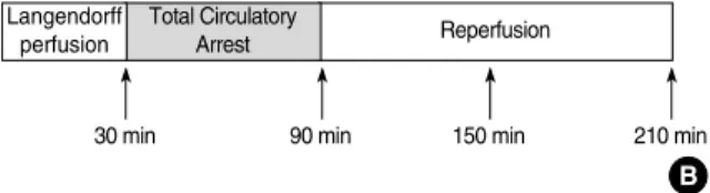
Fig. 1. (A) Experimental designs: there are three cardiomy- opathy rat groups needed and each group was divided into three sub-groups. As mentioned previously, three groups, including ischemic dilated cardiomyopathy group (ICMP, n=30), doxorubicin-induced non-ischemic dilated cardiomy- opathy group (DOX, n=30) and hypertrophic cardiomyopa- thy group by Transverse Aortic Constriction method (TAC, n=30) are prepared. After 1 month following constructions of cardiomyopathic rat groups, each group was then random- ly assigned to a SHAM operation (SHAM; control group, n=10) subgroup or tezosentan treatment ischemia-reperfusion subgroup (Tezo, n=10) or tezosentan non-treatment ischemia-reperfusion subgroup (N-Tezo, n=10). (B) Ischemia-reperfusion injury experimental protocol (isolated heart perfusion via Langendorff technique): after the stabilization period of 20-30 min, total circulatory arrest for ischemia about 1 hr was performed. After then, reperfusion was performed during 2 hr. In Tezo subgroup, the tezosentan, each 10-5M/L concentrations, is added to the cardioplegic solution and the perfusate.
and perfused retrogradely at a constant pressure of 80 mmHg with the perfusate (Krebs-Henseleit solution) of the follow- ing composition: NaCl, 118.1 mM; KCl, 4.6 mM; CaCl2, 2.5 mM; MgSO4, 1.2 mM; KH2PO4, 1.2 mM; NaHCO3, 24.8 mM; and glucose, 10 mM. The perfusate was continu- ously bubbled with a gas mixture of 95% O2/5% CO2(pH 7.4), and temperature was maintained at 37℃throughout the experiment. A latex balloon filled with water was insert- ed into the left ventricle through the left atrium and attached to a pressure transducer (DX-360; Nihon Kohden, Tokyo, Japan). After 30 min of stabilization period, total circulato- ry arrest was performed with cardioplegia and coldness about 1 hr. After then, reperfusion was continued for 2 hr. In Tezo subgroup, tezosentan 10-5M/L was added to the cardioplegic solution and the perfusate (Fig. 1B), but not to N-Tezo sub- group.
Drugs
Tezosentan (Ro 47-0203, Actelion Pharmaceuticals Ltd, Allschwil, Switzerland) is a non-selective specially formulated intravenous dual (endothelin-A and -B) endothelin receptor antagonist. Unlike its sister compound (bosentan), tezosen- tan is easily soluble in water and stable in solution at physi- ologic pH, therefore, usable clinically. Another drug, doxoru- bicin hydrochloride (D1515, Sigma Chemical Co) is well known for cardioselective toxicity and induces congestive heart failure.
Hemodynamic studies for cardiac performance
The left ventricular pressure was monitored with a pressure transducer to obtain the peak positive and negative first de- rivatives (dP/dTmaxand dP/dTmin), and the left ventricular devel- oped pressure (LVDP) was calculated as the difference bet- ween the left ventricular (LV) systolic and diastolic pressures.
These parameters were measured by an amplifier (AP601G;
Nihon Kohden) and also recorded using PowerLab/4sp soft- ware (AD Instruments, Mountain View, CA, U.S.A.). Coro- nary flow was also determined by collecting the coronary effluent from the hearts.
Statistical analysis
All data are presented as means±standard error of the mean.
The comparisons among three groups were carried out by Kruskal-Wallis Test. Differences were considered significant at P<0.05.
RESULTS
Left ventricular developed pressure (LVDP)
LVDP of Tezo subgroup was significantly higher than that of N-Tezo subgroup in the ICMP group after 30 min of reper- fusion (64.3±7.1 mmHg vs. 41.4±4.3 mmHg; P<0.05).
In the DOX or TAC model, LVDPs were not significantly different between Tezo and N-Tezo subgroup during the entire reperfusion period (Fig. 2A, Table 1).
dP/dTmax
In the ICMP group, dP/dTmax was significantly different among the three subgroups at 30 min of reperfusion (SHAM, 3,491±21 mmHg/sec; Tezo, 2,821±13 mmHg/sec; N- Tezo, 2,282±182 mmHg/sec; P<0.05). In the DOX or TAC groups, there were no statistical differences among three sub- groups (Fig. 2B, Table 1).
dP/dTmin
In the ICMP group, dP/dTminwas significantly different among the three subgroups at 30 min of reperfusion (SHAM, -2,382±114 mmHg/sec; Tezo, -1,638±130 mm Hg/sec;
N-Tezo, -1,387±81; P<0.05), similar to the above dP/dTmax. In the DOX or TAC groups, there were no statistically sig- nificant differences among the three subgroups (Fig. 2C, Table 1).
Coronary blood flow (CBF)
Only in the ICMP group, there were significant differences
*Hemodynamic variables are significantly different among SHAM, Tezo and N-Tezo subgroups at 30 min of reperfusion (P<0.05).
ICMP, ischemic cardiomyopathy rat group; DOX, doxorubicin treated non-ischemic dilated cardiomyopathy rat group; TAC, hypertrophic cardiomyopathy rat group by transverse aortic constriction; SHAM, Sham operated ischemia-reperfusion subgroup; Tezo, tezosentan treated ischemia-reperfusion subgroup; N-Tezo, tezosentan non-treatment ischemia-reperfusion subgroup; LVDP, left ventricular developed pressure; CBF, coronary blood flow.
ICMP*
SHAM Tezo N-Tezo
DOX
SHAM Tezo N-Tezo
TAC
SHAM Tezo N-Tezo
LVDP (mmHg) 75.2±5.3 64.3±7.1 41.4±4.3 46.8±3.2 45.1±2.9 45.6±3.8 75.2±4.2 56.2±3.2 58.7±2.9 dP/dTmax(mmHg/sec) 3,491±21 2,821±13 2,282±182 2,095±11 2,320±13 2,559±18 3,491±9 2,100±11 1915±7 dP/dTmin(mmHg/sec) -2,382±114 -1,638±130 -1,387±81 -1,639±184 -1,745±97 -2,085±81 -2,382±97 -1,532±93 -1,432±76 CBF (mL/min) 33.9±4.0 26.3±3.3 19.1±2.1 32.1±2.4 24.2±2.3 20.1±1.9 33.4±2.1 24.5±4.2 22.4±3.6 Table 1. Variable hemodynamic parameters at the reperfusion 30 min
Control Teso N-Teso
Fig. 2. The results of cardiac performance. (A) LV developed pressure (LVDP) during ischemia-reperfusion injury. (B) dP/dTmaxduring ische- mia-reperfusion injury. (C) dP/dTminduring ischemia-reperfusion injury. (D) Coronary blood flow (CBF) during ischemia-reperfusion injury.
*P<0.05 at 30 min of reperfusion among SHAM, Tezo and N-Tezo subgroups.
NS, there is no statistical significance among subgroups; ICMP, ischemic cardiomyopathy rat group; DOX, doxorubicin treated non-ischemic dilated cardiomyopathy rat group; TAC, hypertrophic cardiomyopathy rat group by transverse aortic constriction; SHAM, Sham operated ischemia-reperfusion subgroup; Tezo, tezosentan treated ischemia-reperfusion subgroup; N-Tezo, tezosentan non-treatment ischemia- reperfusion subgroup.
LVDP (mmHg)
100 80 60 40 20
0 Pre 10 30 50
Reperfusion Min
ICMP model
*P<0.05
LVDP (mmHg)
80
60
40
20
0
Pre 10 30 50
Reperfusion Min
DOX model
NS
LVDP (mmHg)
100 80 60 40 20
0 Pre 10 30 50
Reperfusion Min
TAC model
NS
A
CBF (mL/min)
50 40 30 20 10
0 0 10 20 30 40 50 60
Reperfusion Min
ICMP model
*P<0.05
CBF (mL/min)
40
30
20
10
0 0 10 20 30 40 50 60
Reperfusion Min
DOX model
NS
CBF (mL/min)
40
30
20
10
0 0 10 20 30 40 50 60
Reperfusion Min
TAC model
NS
D 5,000
4,000 3,000 2,000 1,000
0 Pre 10 30 50
Reperfusion Min
ICMP model
*P<0.05 5,000 4,000 3,000 2,000 1,000
0 Pre 10 30 50
Reperfusion Min
DOX model
NS 5,000
4,000 3,000 2,000 1,000
0 Pre 10 30 50
Reperfusion Min
TAC model
NS
B
0
-1,000
-2,000
-3,000
-4,000
Pre 10 30 50
Min
ICMP model
*P<0.05
C Reperfusion
0
-1,000
-2,000
-3,000
-4,000
Pre 10 30 50
Min
DOX model
NS Reperfusion
0
-1,000
-2,000
-3,000
-4,000
Pre 10 30 50
Min
TAC model
NS Reperfusion
dp/dTmax(mmHg/sec) dp/dTmax(mmHg/sec) dp/dTmax(mmHg/sec)
dp/dTmax(mmHg/sec) dp/dTmax(mmHg/sec) dp/dTmax(mmHg/sec)
among three subgroups after 30 min of reperfusion in the CBF (SHAM; 33.9±4.0 mL/min vs. Tezo; 26.3±3.3 mL/min vs. N-Tezo; 19.1±1.1 mL/min; P<0.05) (Fig. 2D, Table 1).
DISCUSSION
The effect of endothelin-1, the principal isoform of endothe- lin, is mediated by 2 types of receptor, endothelin-A and en- dothelin-B. The endothelin-A receptor appears to mediate vasoconstriction and stimulate the secretion of atrial natriuret- ic peptide, while the endothelin-B receptor mediates endo- thelin-induced vasodilatation and activation of the renin-an- giotensin-aldosterone system. Through these effects, endo- thelin has been demonstrated to be not only one of the most potent vasoconstrictors known, but also to mediate patho- logic hypertrophy and fibrosis of both ventricular and vas- cular tissues, thus promoting progression of atherosclerosis, ventricular and vascular remodeling (6-8).
Tezosentan, a dual inhibitor of endothelin A and B recep- tors, is weakly acidic, highly soluble in water and stable in physiologic pH solution. These properties make it an ideal candidate for intravenous use, and are the reason of why we used tezosentan in our study.
Heart failure has been associated with elevated plasma lev- els of endothelin-1, and the magnitude of the elevation with the severity of disease, the incidence of arrhythmia, and a worse prognosis. Increased endothelin levels have been described in patients with acute and chronic CHF that are predictive of increased mortality risk (9). Furthermore, increased endothe- lin levels have been suggested to play an important role in the increased systemic vascular resistance seen in CHF (10).
Therefore, endothelin seems to play a major role in the patho- genesis of experimental and clinical heart failure (11, 12).
Although the endothelin receptor antagonists may have the ability to improve the outcome of heart failure by reduc-
ing preload and afterload, increasing the contractility of fail- ing myocardium, delaying myocyte hypertrophy, and reduc- ing the incidence of arrhythmias, some studies demonstrat- ed no benefit of the endothelin receptor antagonists for myo- cardial ischemia-reperfusion heart models (13-15). Therefore, we aimed to investigate the myocardial protective effects of tezosentan for the ischemia-reperfusion injury in various heart failure models.
Before analyzing our experimental data, it was necessary to confirm the in-depth of animal heart failure models. There- fore, one month after cardiomyopathic animal models had been constructed, they were verified with pathologic speci- mens during euthanasia of the experimental animals. In the ICMP models, staining of triphenyltetrazolium chloride with evans blue was used for visualizing ischemia-infarct area of myocardium. In the DOX models, dilated myocardium was confirmed with pathologic specimens and hypertrophied myo- cardium was also confirmed in the TAC models (Fig. 3).
We also believe that hemodynamic parameters such as LVDP, dP/dTmax, dP/dTminand CBF are adequate in our pre- sent study to verify myocardial protective effect of tezosen- tan in various rat cardiomyopathy models. It should be men- tioned that these parameters have been used for proving drug efficacy in Langendorff technique (16).
As a result, our present study did not reveal any significant effect of tezosentan on DOX and TAC models. There are some hypotheses and limitations on this result in our study. First, although heart failure has been associated with elevated plas- ma levels of endothelin-1, the density of endothelin and en- dothelin binding sites had been produced in the heart has not been uniform among different animal heart failure models (17). In myocardial infarct model, it is well established that the infarcted myocardium induces the myocardial necrosis, and increases endothelin-1 levels many fold not only in the post-acute myocardial infarct scar tissues but also in the healed infarct scars after 4 and 13 weeks (18-20). Therefore, endothe-
Fig. 3. (A) In the ICMP models, staining of triphenyltetrazolium chloride with evans blue was used for visualizing ischemia-infarct area of myocardium (black dash). (B) In the DOX models, dilated myocardium was confirmed with pathologic specimens (white arrow-enlarged left ventricle). (C) In the TAC models, hypertrophied myocardium was confirmed with pathologic specimens (white arrow).
*The scale of the ruler in the photo is millimeter (mm).
ICMP, ischemic cardiomyopathy rat group; DOX, doxorubicin treated non-ischemic dilated cardiomyopathy rat group; TAC, hypertrophic cardiomyopathy rat group by transverse aortic constriction.
A B C
lin-1, a recognized fibrogenic factor, plays a crucial role in the stabilization of the necrotic area and in the healing of the scar (20). However, the most prominent histological feature of DOX is not the myocyte necrosis, but the loss of myofib- rils and cytoplasmic vacuolization caused by dilatation of the sarcoplasmic reticulum in the myocardial cells (21), and there is a report that high dose of endothelin antagonist is needed to block endothelin induced hypertension in the dilated car- diomyopathy rat models (22). In addition, sustained pressure overload stimulate pathological cardiac hypertrophy and dys- function (23) and hypertrophy progresses to decompensated phase with cardiac dilatation and contractile impairment dur- ing chronic pressure overload (24, 25). Some authors showed that the production of endothelin-1 increased in the hyper- trophied left ventricle (26), but it depends on the time of the disease progress. Ventricular endothelin-1 levels and the den- sity of endothelin-1 binding sites were increased significant- ly, without affecting their binding affinity, on day 8 of pres- sure overload (27), after then, the production of endothelin- 1 is decreased and endothelin receptors are down-regulated in hypertrophied ventricles 8 weeks after aortic coarctation (28). However, it is also reported that endothelin-1 levels in subjects with heart failure in whom they are found to be ele- vated in the late, decompensated phase of the disease (29).
This time-dependant up- and down-regulations of endothe- lin and endothelin receptors are also another proof for our hypothesis. That is, because of the differences of pathophys- iology among our study group, cardiac endothelin-1 and en- dothelin receptors might not uniformly be induced in vari- ous heart failure models.
Second, in addition to endothelin, renin-angiotensin-aldos- terone, epinephrine-norepinephrine, and natriuretic peptide have also been identified as neurohormones to be activated in cardiomyopathy (30). It is well known that doxorubicin administration has been shown to activate the renin-angio- tensin system (31, 32) and the atrial natriuretic peptide level has also been increased relative to the severity of the cardiac dysfunction in human (32). And, there is a study on angio- tensin and angiotensin receptor antagonist in the hypertro- phic cardiomyopathy (33), showing that stimulation of the angiotensin receptor has an important role in disease process during pressure overload. Therefore, these neurohormones might be more important disease mediators in the DOX or TAC models than in the ICMP model.
There was a limitation to our study. Endothelin is a stim- ulator of polymorphonuclear leukocyte (PMN) aggregation (34) and adhesion as well as a potent vasoconstrictor. Myocar- dial ischemia initiates an acute inflammatory response in which PMNs are of importance (35). Because we had used isolated heart perfusion using Langendorff technique, how- ever, most of the PMNs were diluted in the buffer perfused isolated heart. In other words, the effect of PMNs might be restricted and it can affect the results in our study.
In conclusion, our result reveals that tezosentan has myocar-
dial protective effects for the ischemia-reperfusion injury in the ischemic cardiomyopathy model, but not in the doxoru- bicin-induced cardiomyopathy and pressure-overload hyper- trophic heart failure models.
ACKNOWLEDGEMENTS
We thank ACTELION, Ltd, Allschwil, Switzerland for providing tezosentan.
REFERENCES
1. Parker JD, Thiessen JJ, Reilly R, Tong JH, Stewart DJ, Pandey AS.
Human endothelin-1 clearance kinetics revealed by a radiotracer technique. J Pharmacol Exp Ther 1999; 289: 261-5.
2. Inada T, Fujiwara H, Hasegawa K, Araki M, Yamauchi-Kohno R, Yabana H, Fujiwara T, Tanaka M, Sasayama S. Upregulated expres- sion of cardiac endothelin-1 participates in myocardial cell growth in Bio14.6 Syrian cardiomyopathic hamsters. J Am Coll Cardiol 1999;
33: 565-71.
3. Wittner M, Christ GJ, Huang H, Weiss LM, Hatcher VB, Morris SA, Orr GA, Berman JW, Zeballos GA, Douglas SA, Tanowitz HB. Try- panosoma cruzi induces endothelin release from endothelial cells. J Infect Dis 1995; 171: 493-7.
4. Cheng JW. Tezosentan in the management of decompensated heart failure. Cardiol Rev 2005; 13: 28-34.
5. Teerlink JR, McMurray JJ, Bourge RC, Cleland JG, Cotter G, Jon- deau G, Krum H, Metra M, O’Connor CM, Parker JD, Torre-Amione G, Van Veldhuisen DJ, Frey A, Rainisio M, Kobrin I. Tezosentan in patients with acute heart failure: design of the Value of Endothelin Receptor Inhibition with Tezosentan in Acute heart failure Study (VE- RITAS). Am Heart J 2005; 150: 46-53.
6. Battistini B, Chailler P, D’Orleans-Juste P, Briere N, Sirois P. Growth regulatory properties of endothelins. Peptides 1993; 14: 385-99.
7. Kaddoura S, Firth JD, Boheler KR, Sugden PH, Poole-Wilson PA.
Endothelin-1 is involved in norepinephrine-induced ventricular hyper- trophy in vivo. Acute effects of bosentan, an orally active, mixed en- dothelin ETA and ETB receptor antagonist. Circulation 1996; 93:
2068-79.
8. Teerlink JR. The development of new medical treatments for acute decompensated heart failure. Heart Fail Monit 2002; 2: 129-37.
9. Fishman WH, Kaur S, Singh I, Tamirisa P. Endothelin as a thera- peutic target in the treatment of cardiovascular disease. In: Frish- man WH, Sonnenblick EH, Sica DA, editors, Cardiovascular phar- macotherapeutics. 2nd ed. New York: McGraw-Hill, Medical Pub.
Division, 2003; 527-43.
10. Webb DJ. Evidence for endothelin-1-mediated vasoconstriction in severe chronic heart failure. Endothelin antagonism in heart failure.
Circulation 1995; 92: 3372.
11. Margulies KB, Hildebrand FL Jr, Lerman A, Perrella MA, Burnett JC Jr. Increased endothelin in experimental heart failure. Circula- tion 1990; 82: 2226-30.
12. Omland T, Lie RT, Aakvaag A, Aarsland T, Dickstein K. Plasma endothelin determination as a prognostic indicator of 1-year mortal- ity after acute myocardial infarction. Circulation 1994; 89: 1573-9.
13. Duchman SM, Thohan V, Kalra D, Torre-Amione G. Endothelin-1:
a new target of therapeutic intervention for the treatment of heart failure. Curr Opin Cardiol 2000; 15: 136-40.
14. McMurdo L, Thiemermann C, Vane JR. The effects of the endothe- lin ETA receptor antagonist, FR 139317, on infarct size in a rabbit model of acute myocardial ischaemia and reperfusion. Br J Phar- macol 1994; 112: 75-80.
15. Richard V, Kaeffer N, Hogie M, Tron C, Blanc T, Thuillez C. Role of endogenous endothelin in myocardial and coronary endothelial injury after ischaemia and reperfusion in rats: studies with bosentan, a mixed ETA-ETB antagonist. Br J Pharmacol 1994; 113: 869-76.
16. Xia Z, Kuo KH, McNeill JH, Ansley DM. Endothelin A and B recep- tor antagonist bosentan reduces postischemic myocardial injury in the rat: critical timing of administration. Can J Physiol Pharmacol 2005; 83: 259-66.
17. Sakai S, Yorikane R, Miyauchi T, Sakurai T, Kasuya Y, Yamaguchi I, Sugishita Y, Goto K. Altered production of endothelin-1 in the hy- pertrophied rat heart. J Cardiovasc Pharmacol 1995; 26 (Suppl 3):
S452-5.
18. Cernacek P, Stewart DJ, Monge JC, Rouleau JL. The endothelin sys- tem and its role in acute myocardial infarction. Can J Physiol Phar- macol 2003; 81: 598-606.
19. Nguyen QT, Cernacek P, Calderoni A, Stewart DJ, Picard P, Sirois P, White M, Rouleau JL. Endothelin A receptor blockade causes adverse left ventricular remodeling but improves pulmonary artery pressure after infarction in the rat. Circulation 1998; 98: 2323-30.
20. Nguyen QT, Cernacek P, Sirois MG, Calderone A, Lapointe N, Stew- art DJ, Rouleau JL. Long-term effects of nonselective endothelin A and B receptor antagonism in postinfarction rat: importance of tim- ing. Circulation 2001; 104: 2075-81.
21. Arola OJ, Saraste A, Pulkki K, Kallajoki M, Parvinen M, Voipio- Pulkki LM. Acute doxorubicin cardiotoxicity involves cardiomyocyte apoptosis. Cancer Res 2000; 60: 1789-92.
22. Watanabe K, Ohta Y, Kouda T, Sekiguchi T, Sato S, Nakazawa M, Hasegawa G, Naito M, Fuse K, Ito M, Hirono S, Tanabe N, Hanawa H, Kato K, Kodama M, Aizawa Y. Acute effects of endothelin-1 and TAK-044 (ET(A) and ET(B) receptor antagonist) in rats with dilat- ed cardiomyopathy. J Cardiovasc Pharmacol 2000; 36 (Suppl 2):
S49-54.
23. Heineke J, Molkentin JD. Regulation of cardiac hypertrophy by intra- cellular signalling pathways. Nat Rev Mol Cell Biol 2006; 7: 589-600.
24. Houser SR, Margulies KB. Is depressed myocyte contractility cen- trally involved in heart failure? Circ Res 2003; 92: 350-8.
25. Olson EN. A decade of discoveries in cardiac biology. Nat Med 2004;
10: 467-74.
26. Yorikane R, Sakai S, Miyauchi T, Sakurai T, Sugishita Y, Goto K.
Increased production of endothelin-1 in the hypertrophied rat heart due to pressure overload. FEBS Lett 1993; 332: 31-4.
27. Arai M, Yoguchi A, Iso T, Takahashi T, Imai S, Murata K, Suzuki T.
Endothelin-1 and its binding sites are upregulated in pressure over- load cardiac hypertrophy. Am J Physiol 1995; 268: H2084-91.
28. Sirvio ML, Uhlenius N, Stewen P, Metsarinne K, Fyhrquist F. The effect of aortic coarctation on expression of endothelin-1 and endothe- lin receptors in heart and lungs. Blood Press 1995; 4: 320-3.
29. Wei CM, Lerman A, Rodeheffer RJ, McGregor CG, Brandt RR, Wright S, Heublein DM, Kao PC, Edwards WD, Burnett JC Jr. En- dothelin in human congestive heart failure. Circulation 1994; 89:
1580-6.
30. Hayashi M, Tsutamoto T, Wada A, Maeda K, Mabuchi N, Tsutsui T, Horie H, Ohnishi M, Kinoshita M. Intravenous atrial natriuretic pep- tide prevents left ventricular remodeling in patients with first anterior acute myocardial infarction. J Am Coll Cardiol 2001; 37: 1820-6.
31. Arnolda L, McGrath B, Cocks M, Sumithran E, Johnston C. Adri- amycin cardiomyopathy in the rabbit: an animal model of low out- put cardiac failure with activation of vasoconstrictor mechanisms.
Cardiovasc Res 1985; 19: 378-82.
32. Bauch M, Ester A, Kimura B, Victorica BE, Kedar A, Phillips MI.
Atrial natriuretic peptide as a marker for doxorubicin-induced car- diotoxic effects. Cancer 1992; 69: 1492-7.
33. Lako-Futo Z, Szokodi I, Sarman B, Foldes G, Tokola H, Ilves M, Leskinen H, Vuolteenaho O, Skoumal R, deChatel R, Ruskoaho H, Toth M. Evidence for a functional role of angiotensin II type 2 recep- tor in the cardiac hypertrophic process in vivo in the rat heart. Cir- culation 2003; 108: 2414-22.
34. Gomez-Garre D, Guerra M, Gonzalez E, Lopez-Farre A, Riesco A, Caramelo C, Escanero J, Egido J. Aggregation of human polymor- phonuclear leukocytes by endothelin: role of platelet-activating fac- tor. Eur J Pharmacol 1992; 224: 167-72.
35. Hansen PR. Role of neutrophils in myocardial ischemia and reper- fusion. Circulation 1995; 91: 1872-85.