Schisandra Chinensis Inhibits Oxidative DNA Damage and Lipid Peroxidation Via Antioxidant Activity
Jin Boo Jeong, Hyung Jin Jeong*
School of Bioresources, Andong National University, Andong 760749, Korea
Abstract - Schisandra chinensis have been traditionally used in Asia for the treatment of dyspnea, cough, mouth dryness, spontaneous diaphoresis, nocturnal diaphoresis, nocturnal emission, dysentery, insomnia and amnesia. The purpose of this study is to evaluate the protective effects of Schisandra chinensis on oxidative DNA damage and lipid peroxidation induced by ROS in non cellular and cellular system. DPPH radical, hydroxyl radical and hydrogen peroxide scavenging assay were used to measure the antioxidant activities. Phi X-174RF I plasmid DNA cleavage assay and intracellular DNA migration assay were used to evaluate the protective effect on oxidative DNA damage. MTT assay and lipid peroxidation assay were used for evaluating the protective effect on oxidative cell damage. It was found to scavenge DPPH radical, hydrogen peroxide and hydroxyl radical and it inhibited oxidative DNA damage, lipid peroxidation and cell death induced by hydroxyl radical.
These data indicate that Schisandra chinensis possesses a spectrum of antioxidant and DNA-protective properties
Key words - Lipid peroxidation, Oxidative DNA damage, Oxidative cell death, Reactive oxygen species (ROS), Schisandra chinensis
*
Corresponding author. E-mail : [email protected]Introduction
Every cell in prokaryote or eukaryote is continuously exposed to exogenous and endogenous agents that damage its DNA (Barzilai and Yamamoto, 2004). Among DNA damage causing cancer development, approximately 80% of the damage is caused by the reactive oxygen species (ROS) such as hydrogen peroxide (H2O2), singlet oxygen (1O2), and hydroxyl radical (OH) (Ghosal et al., 2005). These compounds, when present in a high enough concentration, can damage cellular proteins and lipid or form DNA adducts that may promote carcinogenic activity (Seifried et al., 2007). Dietary factor that reduces the impact of ROS can protect DNA damage and thus lower cancer risks (Longwen et al., 1999). High intake of various teas has been associated with lower risks for many of the major human cancers, including stomach, esophagus, and lung. These pro- tective benefits have been attributed to the richness of their major antioxidant agents (Lin et al., 1999).
Schisandra Chinensis have been traditionally used in Asia for the treatment of dyspnea, cough, mouth dryness, spontaneous
diaphoresis, nocturnal diaphoresis, nocturnal emission, dys- entery, insomnia and amnesia (Anonymous, 1992). In the past two decades, this crude drug has been developed as an alter- native medicine for the treatment of various liver diseases as its capability to protect the liver from injuries induced by hepato- toxins has been well-documented (Pao et al., 1975). However, its antioxidant properties have not been well defined.
Therefore, it is important to understand the inhibitory mechanism of the extracts from Schisandra Chinensis on H2
O2-induced oxidative damage causing DNA and cell damages since H2O2 is the major mediator of oxidative stress and a potent mutagen. Also, the evaluation of its inhibitory effects on oxidative DNA and cell damages is necessary for the medic- inal use of it as the cancer chemopreventive agent. In this study, we evaluated antioxidant and inhibitory effect of oxidative DNA damage and lipid peroxidation. Major questions we wish to address in our study are: (1) What is the effect of Schisandra Chinensis on DPPH radical, hydroxyl radical and hydrogen peroxide?, (2) Can Schisandra Chinensis affect lipid peroxi- dation in a cellular system?, and (3) What is the effect of Schisandra Chinensis on oxidative DNA damage induced by hydroxyl radical?
Materials and Methods
Chemical regents
Mouse skin fibroblast cell line, NIH 3T3, was purchased from ATCC, and the mediums used for the cell growth were purchased from GIBCO. All chemicals for extraction from Buddleja officinalis, DPPH (1,1diphenyl-2-picryl hydrazyl) and 2`,7`-dichlorofluorescin diacetate (DCF-DA) were obtained from Sigma Chemicals Co. (St. Louis, USA). φX-174 RF I plasmid was purchased from New England BioLabs (County Road Ipswich, MA)
Extraction
One kilogram of Schisandra Chinensis was ground and then extracted with 3 L of 80% ethanol by shaking for 24 hours. The ethanol-soluble fraction was filtered, concentrated using by a vacuum evaporator and fractioned with a separating funnel.
The ethyl acetate fraction was separated from the mixture, evaporated by a vacuum evaporator, prepared aseptically and kept in a refrigerator until use.
DPPH radical scavenging activity
The antioxidant activity of the extracts was evaluated first by monitoring its ability in quenching the stable free radical DPPH (Hus et al., 2006). Reaction mixture containing 40 μl of test samples (4 mg/ml dissolved in DMSO) and 760 μl of 300 μM DPPH ethanol solution in micro tube were incubated at 37℃ for 30 min and absorbance was measured at 515 nm according to the increasing concentrations of the extracts. The DPPH quenching ability was calculated from the log-dose inhibition curve. All determinations were carried out in triplicate.
Ascorbic acid was used as a positive control.
Hydroxyl radical scavenging assay
Hydroxyl radical scavenger ability was measured according to a literature procedure (Smirnoff and Cumbes, 1989) with a few modifications. Hydroxyl radical was generated from FeSO4
and H2O2, and detected by their ability to hydroxylate salicylate.
The reaction mixture (800 μl) contained 250 μl of FeSO4 (1.5 mM), 175 μl of H2O2 (6 mM), 300 μl of sodium salicylate (20 mM) and varying concentrations of the extracts. After a reaction for 30 min at 37℃, the absorbance of the hydroxylated salicylate
complex was measured at 562 nm. Hydroxyl radical scavenging ability was calculated from the log-dose inhibition curve. All determination was carried out in triplicate. Ascorbic acid was used as a positive control.
Hydrogen peroxide scavenging assay
Hydrogen peroxide scavenging ability was measured ac- cording to a literature procedure (Pick and Keisari, 1989). One hundred micro liter of 0.1 M phosphate buffer (pH 5.0), 40 μl of the extracts and 60 μl of 1 mM hydrogen peroxide were mixed, and then incubated for 5 min at 37℃. After 5 min, 400 μl of 1.25 mM ABTS and 400 μl of peroxidase (1unit/ml) were added to the mixture, and then incubated for 10 min at 37℃.
After 10 min, the absorbance was read at 405 nm. Hydrogen peroxide scavenger ability was calculated from the log-dose inhibition curve. All determination was carried out in triplicate.
Ascorbic acid was used as a positive control.
Phi X-174RF I plasmid DNA cleavage assay
Conversion of the supercoiled form of plasmid DNA to the open-circular and further linear forms has been used as an index of DNA damage (Jung and Surh, 2001). Reaction mixtures (20 μl) containing 5 μl of phi X-174 RF I plasmid DNA, 5 μl of varying concentrations of the extracts, 5 μl of 1 mM FeSO4
or/and 5 μl of 1 mM H2O2 were incubated at 37℃ for 30 min.
After 5 μl of a solution containing 50% glycerol (v/v), 40 mM EDTA and 0.05% bromophenol blue was added to stop the reaction, the reaction mixtures were electrophoresed on 1%
agarose gel, and the DNA in the gel was visualized and photo- graphed under ultraviolet light after ethidium bromide staining.
Cell viability assay
This assay was carried according to literature procedure (Kang et al., 2005) with some modifications. NIH 3T3 cells (5×10³cells/well) were cultured in 96-well plate at 37℃ for 24 hours. After the incubation, the extracts were treated to each well according to the concentration, and then the cells were incubated at 37℃ for 30 min. After the treatment of the extracts, 1 mM H2O2 and 1 mM FeSO4 was applied to each well and then the cells were re-incubated at 37℃ for 24 hours. After 50 μl of MTT solution (1 mg/ml) was treated to each well for 4 hours, the supernatant was removed, and then 100 μl of DMSO
was treated to each well. The absorbance was measured by a microplate reader at 570 nm.
Intracellular DNA damage assay
This assay was carried according to literature procedure (Cho et al., 2008) with some modifications. NIH 3T3 cells (2×10⁶) were cultured in 6-well plates for 24 hours at 37℃. After the cells were treated with the varying concentrations of the extracts for 30 min and then added with 1 mM FeSO4 and 1 mM H2O2
for 1 hour. After 1 hour, each cell was harvested and then the supernatant was discarded. Each cell was re-suspended with 20 µl of lysis buffer (50 mM Tris-HCl, pH 8.0, 10 mM EDTA, 0.5% SDS and 0.5 mg/ml proteinase K) and then incubated at 55℃ for 60 min. After lysis of the cells, each cell was centri- fuged, 5 μl of RNase A was added to the supernatant, and each cell was incubated at 55℃ for another 60 min. After 60 min, each cell was spun briefly to remove any further cell debris and each supernatant was collected. Each lysate was heated at 70℃
for a few minutes and mixed with 10 μl of loading buffer (50%
glycerol (v/v), 40 mM EDTA and 0.05% bromophenol blue).
The reaction mixtures were electrophoresed on 2% agarose gel, and the DNA in the gel was visualized and photographed under ultraviolet light after ethidium bromide staining.
Lipid peroxidation assay
This assay was carried according to literature procedure (Kang et al., 2008) with some modification. NIH 3T3 cells were seeded in a 6-well plate at 2×10⁶ cells/well for 16 hours. Sixteen hours after plating, the cells were treated with the varying concentra- tions of extracts for 30 min. After 30 min, 1 mM H2O2 and 1 mM FeSO4 were added to the plate and then each plate was incubated for 24 hours. The cells were then washed with cold phosphate-buffered saline (PBS), harvested, and homogenized in an ice-cold 1.15% KCl. One hundred microliter of the cell lysate was mixed with 0.1 ml of 8.1% sodium dodecylsulfate, 0.75 ml of 20% acetic acid (adjusted to pH 3.5), and 0.75 ml of 0.8% thiobarbituric acid (TBA). The mixture was made up to a final volume of 4 ml with distilled water and heated to 9 5℃ for 2 hours. After cooling to room temperature, 2.5 ml of an n-butanol/pyridine mixture (15:1, v/v) was added and the mixture was shaken. After centrifugation at 1000ⅹg for 10 min, the supernatant fraction was isolated and the absorbance
was measured spectrophotometrically at 532 nm. The amount of thiobarbituric acid reactive substance (TBARS) was deter- mined using standard curve with 1,1,3,3,-tetrahydroxypropane.
Statistical analysis
The series of experiments were performed as three or more independent examination with at least three replicates for each sample. Data were expressed as means ±S.D. Statistical com- parison was performed using Student’s t-test.
Results
DPPH free radical scavenging activity of Schisandra Chinensis
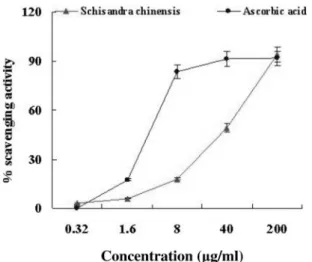
The mechanism for antioxidants to remove free radical involves donating hydrogen to a free radical and hence its reduction to an unreactive species through removing the odd electron feature which is responsible for radical reactivity (Wang et al., 2008). In this assay, the DPPH radical absorptions at 515 nm were inhibited in the presence of the varying concen- trations of the extracts from Schisandra Chinensis. This reduc- tion in absorbance is related to the anti-radical efficiency of the extracts. The DPPH scavenging efficiency values of the extracts are presented in Fig. 1. DPPH free radical scavenging activities of the extracts were 3% at 0.32 μg/ml, 6% at 1.6 μg/ml, 18%
at 8 μg/ml, 49% at 40 μg/ml and 94% at 200 μg/ml.
Hydroxyl radical scavenging activity of Schisandra Chinensis
Among the oxygen radicals, hydroxyl radical is the most reactive and induces severe damage to adjacent biomolecules (Sakanaka et al., 2005). Hydroxyl radical scavenging is an im- portant antioxidant activity because of very high reactivity of hydroxyl radical which enables it to react with a wide range of molecules found in living cells such as sugars, amino acids, lipids and nucleotides (Stohs and Bagchi, 1995). In this assay, the extracts removed hydroxyl radical by 8% at 0.32 μg/ml, 15% at 1.6 μg/ml, 41% at 8 μg/ml, 78% at 40 μg/ml and 94%
at 200 μg/ml, respectively (Fig. 2).
Hydrogen peroxide scavenging activity of Schisandra Chinensis
Fig. 1. DPPH free radical scavenging activities of the extracts from Schisandra chinensis. Reaction mixture containing 40 μl of test samples (4 mg/ml dissolved in DMSO) and 760 μl of 300 μM DPPH ethanol solution were incubated at 37℃ for 30 min and absorbance was measured at 515 nm. The absorbance values were converted to scavenging effects (%) and data plotted as the means of replicate scavenging effect (%) values ± 5 S.D. (n=3) against extract concentration in μg extract per ml reaction volume.
Ascorbic acid was used for the positive control.
Fig. 2. Hydroxyl radical scavenging activities of the extracts from Schisandra chinensis. The reaction mixture (800 μl) contained 250 μl of FeSO4 (1.5 mM), 175 μl of H2O2 (6 mM), 300 μl of sodium salicylate (20 mM) and varying concentrations of the extracts. After a reaction for 30 min at 37 oC, the absorbance of the hydroxylated salicylate complex was measured at 562 nm.
The absorbance values were converted to scavenging effects (%) and data plotted as the means of replicate scavenging effect (%) values ± 5 S.D. (n=3) against extract concentration in μg extract per ml reaction volume. Ascorbic acid was used for the positive control.
Fig. 3. Hydrogen peroxide scavenging activities of the extracts from Schisandra chinensis. One hundred micro liter of 0.1 M phosphate buffer (pH 5.0), 40 μl of the extracts and 60 μl of 1 mM hydrogen peroxide were mixed, and then incubated for 5 min at 37℃. After 5 min, 400 μl of 1.25 mM ABTS and 400 μl of peroxidase (1unit/ml) were added to the mixture, and then incubated for 10 min at 37℃. After 10 min, the absorbance was read at 405 nm. The absorbance values were converted to scavenging effects (%) and data plotted as the means of replicate scavenging effect (%) values ± 5 S.D. (n=3) against extract concentration in μg extract per ml reaction volume.
Ascorbic acid was used for the positive control.
Hydrogen peroxide can attack many cellular energy-producing systems (Hyslop et al., 1998). Hydrogen peroxide itself is not very reactive, but it can sometimes be toxic to cells, since it may give rise to hydroxyl radicals inside the cell (Halliwell and Aruoma, 1991). In this assay (Fig. 3), the extracts removed hy- drogen peroxide by 1% at 0.32 μg/ml, 12% at 1.6 μg/ml, 38%
at 8 μg/ml, 78% at 40 μg/ml and 96% at 200 μg/ml, respectively.
Inhibitory effect of Schisandra Chinensis on oxidative DNA damage induced by hydroxyl radical
The inhibitory effects of the extracts from Schisandra Chinensis on oxidative DNA damage caused by hydroxyl radical were investigated through in vitro DNA double strand break assay using φX-174 RF I plasmid DNA and DNA migration assay in the cellular system. Excessive ROS (e.g. hydrogen peroxide) can lead to DNA oxidation, causing cell damage to all cellular constituents. Irreparable DNA damage is involved in carcino- genesis, aging and other degenerative diseases (Cozzi et al., 1997). Oxidative DNA damage can lead to mutations and is suspected to be a major cause of cancer (Schwarz et al., 1984).
(a) (b)
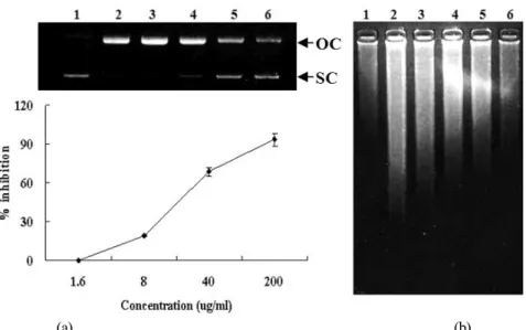
Fig. 4. Inhibitory effect of the extracts from Schisandra chinensis on hydroxyl radical-induced DNA damage. (A) Oxidative damage of φ X-174 RF I plasmid DNA caused by hydroxyl radical. The plot shows % inhibition of the extracts against the conversion of SC into OC induced by hydroxyl radical. % inhibition was calculated by the density using the software Un-SCAN-IT gel Version 5.1 (Silk Scientific, Inc.). (B) Intracellular DNA damage assay of NIH 3T3 cells caused by hydroxyl radical. Lane 1 is treated with nothing, and lane 2 is treated with 1 mM FeSO4 and 1 mM H2O2 without extracts. Lane 3~5 were treated with varying concentrations of the extract (1.6, 8, 40 and 200 μg/ml) in presence of 1 mM FeSO4 and 1 mM H2O2. Hydroxyl radical was generated from Fenton reaction between FeSO4 and H2O2.
The extracts inhibited the conversion of supercoiled form (SC) to open-circular form (OC) induced by hydroxyl radical in φ X-174 RF I plasmid DNA by 19% at 8 μg/ml, 69% at 40 μg/ml and 93% at 200 μg/ml (Fig. 4A). In addition, it inhibited DNA migration induced by ROS in a does-dependent manner (Fig.
4B). DNA migration assay is a sensitive biomarker of the DNA damage.
Inhibitory effect of Schisandra Chinensis on oxidative cell damage induced by hydroxyl radical
Inhibitory effect of the extracts from Schisandra Chinensis on oxidative cell damage induced by ROS was examined using MTT assay and lipid peroxidation assay. ROS damage involves injury to cellular membranes. Measurement of lipid peroxidation is used as an indicator of membrane damage in mammalian cells. Lipid peroxidation can cause a cascade effect of lipid- derived radicals, thereby causing additional membrane damage.
The products of lipid peroxidation, malondialdehyde and other group of aldehyde products may also cause DNA damage (Vaca et al., 1988). It has also been proposed that free radicals derived from lipid peroxidation may function as tumor initiator (Esterbauer, 1982). Our result showed that the extracts inhibit
the lipid peroxidation by 22% at 1.6 μg/ml, 42% at 8 μg/ml, 69% at 40 μg/ml and 88% at 200 μg/ml (Fig. 5A). In MTT assay, the treatment of hydrogen peroxide induced the cell death by about 49% while the extracts prevented the cells from the death by 12% at 1.6 μg/ml, 44% at 8 μg/ml, 63% at 40 μg/ml and 90%
at 200 μg/ml (Fig. 5B). The treatment of the extracts from Schisandra Chinensis without H2O2 had no effect on lipid peroxidation and cell viability (not shown).
Discussion
The results from this study demonstrate that Schisandra Chinensis inhibits oxidative DNA and cell damage induced by hydroxyl radical via its antioxidant activities. In non-cellular systems, the extracts from Schisandra Chinensis effectively scavenged DPPH radical, hydrogen peroxide and hydroxyl radical, and prevented DNA damage induced by hydroxyl radical. In a cell system, the extracts also effectively inhibited intracellular DNA damage, lipid peroxidation and cell death induced by hydroxyl radical.
ROS have been associated with pathogenic processes including carcinogenesis through direct effects on DNA directly and by
Fig. 5. Inhibitory effect of the extracts from Schisandra chinensis on oxidative cell damage induced by hydroxyl radical. (A) The inhibitory effect of the extracts on the inhibition of lipid peroxidation was evaluated by measuring the amount of TBARS formation using NIH 3T3 cells. (B) The viability of NIH 3T3 cells was evaluated by a MTT assay. Hydroxyl radical used in lipid peroxidation assay and MTT assay was generated from Fenton reaction between 1 mM H2O2 and 1 mM FeSO4.The treatment of the extracts from Schisandra chinensis without H2O2 had no effect on lipid peroxidation and cell viability (not shown).
acting as a tumor promoter (Wiseman and Halliwell, 1996).
ROS damage can be affected by two factors: (1) scavenging of radicals formed during reactions and (2) inhibiting the radi- cal generation (Leonard et al., 2006). Cellular systems generate a variety of ROS such as superoxide, hydrogen peroxide and hydroxyl radical. Hydroxyl radical is an extremely reactive species, reacting with virtually all known bio-molecules at diffusion-limited rates of reactions (~107-1010 M/s). Moreover, this radical has been shown to per-oxidize lipids, oxidize protein, and promote DNA strand scission (Grisham, 1992). This DNA damage by hydroxyl radical has been shown to play a key role in the carcinogenesis (Barreto et al., 2005). Although hydrogen peroxide is a more powerful oxidant than is superoxide, it is relatively unreactive toward most biologic substrates. However, the Fenton reaction between Fe2+ and H2O2 generates Fe3+
and hydroxyl radical (Halliwell and Gutteridge, 1992). The scavenging of hydroxyl radical by the extracts from Schisandra Chinensis demonstrates its effectiveness against biologically generated radicals. Moreover, the scavenging of H2O2 by the extracts from Schisandra Chinensis demonstrates its effec- tiveness for inhibiting the radical generation. The present investigation also examined the ability of the extracts from Schisandra Chinensis to inhibit DNA damage in phi X-174 RF I plasmid DNA cleavage and intracellular DNA migration from exposure to hydroxyl radical. Hydroxyl radical was gen- erated from the Fenton reaction between Fe2+ and H2O2. The
result of the present study indicates that the extracts from Schisandra Chinensis can inhibit DNA damage caused by hydroxyl radical. Carcinogens such as chromium, asbestos and nickel exert their carcinogenic effect, in part, through production of ROS (Leonard et al., 2004). Another aspect of ROS damage involves injury to cellular membranes. Measure- ment of lipid peroxidation is used as an indicator of membrane damage (Leonard et al., 2006). Lipid peroxidation can cause a cascade effect of lipid-derived radicals, thereby causing addi- tional membrane damage. The products of lipid peroxidation, malondialdehyde and other groups of aldehyde products may also cause DNA damage (Vaca et al., 1988). It has also been proposed that free radicals derived from lipid peroxidation function as tumor initiators (Esterbauer, 1982). Our result showed that the extracts from Schisandra Chinensis were effective in inhibiting lipid peroxidation of cellular membranes.
The results of the present investigation indicate the followings:
(1) the extracts from Schisandra Chinensis scavenged DPPH radical, hydrogen peroxide and hydroxyl radical; (2) DNA damage from hydroxyl radical produced by the Fenton reaction was inhibited by the extracts from Schisandra Chinensis; (3) cell death and lipid peroxidation in cell membranes caused by ex- posure to hydroxyl radical were inhibited by the extracts from Schisandra Chinensis;. Together, these data indicate that Schi- sandra Chinensis possesses a spectrum of antioxidant and DNA-protective properties.
Acknowledgements
This work was supported by a support work Human Re- sources Development and GB-Regional Innovation Research Program (2008), Korea
Literature Cited
Anonymous. 1992. Pharmacopoeia of the People’s Republic of China, English Edition, Guangdong Science and Technology Press, Guangzhou. 82–83.
Barreto, R., S. Kawakita, J. Tsuchiya, E. Minelli, K. Pavasuthi- pasit, A. Helmy and F. Marotta. 2005. Metal-induced oxidative damage in cultured hepatocytes and hepatic lysosomal fraction:
beneficial effect of a curcumin/absinthium compound. Chi- nese J. Digest. Dis.6: 31–36.
Barzilai, A. and K.I. Yamamoto. 2004. DNA damage responses to oxidative stress. DNA Repair. 3: 1109-1115.
Cho, E.S., K.W. Lee and H.J. Lee. 2008. Cocoa procyanidins pro- tect PC12 cells from hydrogen-peroxide-induced apoptosis by inhibiting activation of p38 MAPK and JNK. Mutat. Res.
640: 123-130.
Cozzi, R., R. Ricordy, T. Aglitti, V. Gatta, P. Perticone and R.
De Salvia. 1997. Ascorbic acid and beta-carotene as modula- tors of oxidative damage. Carcinogenesis 18: 223-228.
Esterbauer, H. 1982. In: McBrien, D.C.H., Slater, T.F. (Eds), Free Radicals, Lipid Peroxidation and Cancer. Academic Press, New York, 101-128.
Grisham, M.B. 1992. Reactive oxygen metabolism. In: Grisham M.B., ed. Reactive metabolites of oxygen and nitrogen in biology and medicine. Austin: RG Landers Company, 39.
Ghosal, D., M.V. Omelchenko, E.K. Gaidamakova, V.Y. Matro- sova, A. Vasilenko, A. Venkateswaran, M. Zhai, H.M. Kosta- ndarithes, H. Brim, K.S. Makarova, L.P. Wackett, J.K. Fred- rickson and M.J. Daly. 2005. How radiation kills cells: survival of Deinococcus radiodurans and Shewanella oneidensis under oxidative stress. FEMS Microbiol. Rev. 29: 361–375.
Halliwell, B. and O.L. Aruoma. 1991. The biological toxicity of free radicals and other reactive species. Free radicals and food additives. 41.
Halliwell, B. and J.M.C. Gutteridge. 1992. Biologically relevant metal ion-dependent OH generation. An update. FEBS Lett.
307: 108.
Hus, B., I.M. Coupar and K. Ng. 2006. Antioxidant activity of
hot water extract from the fruit of the Doum palm. Hyphaene thebaica. Food Chem. 98: 317-328.
Hyslop, P.A., D.B. Hinshaw, W.A. Halsey Jr, I.U. Schraufstatter, R.D. Sauerheber and R.G. Spragg. 1998. Mechanisms of oxidant-mediated cell injury. The glycolytic and mitochondrial pathways of ADP phosphorylation are major intracellular tar- gets inactivated by hydrogen peroxide. J. Biol. Chem. 263:
1665–1675.
Jung, Y. and Y. Surh. 2001. Oxidative DNA damage and cytot- oxicity unduced by copper-stimulated redox cycling of sal- solinol, a neurotoxic tetrahydroisoquinoline alkaloid. Free Radic. Biol. Med. 30: 1407-1417.
Kang, K.A., K.H. Lee, S.W. Chae, R. Zhang, M.S. Jung, Y.K.
Lee, S.Y. Kim, H.S. Kim, H.G. Joo, J.W. Park, Y.M. Ham, L.
H. Lee and J.W. Hyun. 2005. Eckol isolated from Ecklonia cava attenuates oxidative stress induced cell damage in lung fibroblast cells. FEBS Lett. 579: 6295-6304.
Kang, K.A., R. Zhang, M.J. Piao, D.O. Ko, Z.H. Wang, B.J. Kim, J.W. Park, H.S. Kim, D.H. Kim and J.W. Hyun. 2008. Protec- tive effect of irisolidone, a metabolite of kakkalide, against hydrogen peroxide induced cell damage via antioxidant effect.
Bioorgan. Med. Chem. 16: 1133-1141.
Leonard, S.S., D. Keil, T. Mehlman, S. Proper, X. Shi and G.K.
Harris. 2006. Essiac Tea: Scavenging of reactive oxygen species and effects on DNA damage. J. Ethnopharmacol. 103, 288- 296.
Leonard, S.S., J.R. Roberts, J.M. Antonini, V. Castranova and X. Shi. 2004. PbCrO4 mediates cellular responses via reactive oxygen species. Mol. Cell. Biochem. 255: 171–179.
Lin, J.K., Y.C. Liang and S.Y. Lin Shiau. 1999. Cancer chemo- prevention by tea polyphenols through mitotic signal transduc- tion blockade. Biochem. Pharmacol. 6: 911-915.
Longwen, C., P.E. Bowen, D. Berzy, F. Aryee and R.E. Riley.
1999. Diet modification affects DNA oxidative damage in healthy humans. Free Radic. Biol. Med. 26: 695-703.
Pao, T.T., K.T. Liu, K.F. Hsu and C.Y. Sung. 1975. Studies on Fructus Schizandrae I: effects on increased SGPT levels in animals caused by hepatotoxic chemical agents. J. Chinese Med. 54: 275–277.
Pick, E. and Y. Keisari. 1980. A simple colorimetric method for the measurement of hydrogen peroxide produced by cells in culture. J. Immunol. Methods 38: 161–170.
Sakanaka, S., Y. Tachibana and Y. Okada. 2005. Preparation and antioxidant properties of extracts of Japanese persimmon leaf tea (kakinoha-cha). Food Chem. 89: 569-575.
Seifried, H.E., D.E. Anderson, E.I. Fisher and J.A. Milner. 2007.
A review of the interaction among dietary antioxidants and reactive oxygen species. J. Nutri. Biochem. 18: 567-579.
Schwarz, S.M., G. Peres, W. Kunz, G. Furstenberger, W. Kittstein and F. Marks. 1984. On the role of superoxide anion radicals in skin tumour promotion. Carcinogenesis. 5: 1663-1670.
Smirnoff, N. and Q.J. Cumbes. 1989. Hydroxyl radical scavenging activity of compatible solutes. Phytochemistry. 28: 1057-1060.
Stohs, S.J. and D. Bagchi. 1995. Oxidative mechanism in the toxicity of metal ions. Free Radic. Biol. Med. 18: 321-336.
Vaca, C.E., J. Wilhelm and M. Harms-Ringdahl. 1988. Interaction of lipid peroxidation products with DNA. A review. Mutat.
Res. 195: 137-149.
Wang, T., L.X. Chen, Y. Long, W.M. Wu and Wang. 2008. DNA damage induced by caffeic acid phenyl ester in the presence of Cu(II) ions: Potential mechanism of its anticancer properties.
Cancer Lett. 263: 77-88.
Wiseman, H. and B. Halliwell. 1996. Damage to DNA by reactive oxygen and nitrogen species: role in inflammatory disease and progression to cancer. J. Biochem. 313: 17–29.
(Received 29 October 2008 ; Accepted 16 March 2009)