• 검색 결과가 없습니다.

Biomechanical Differences of Lower Extremity Joints at the Frontal Plane during Sidestep Cutting in Male and Female Judo Athletes

N/A
N/A
Protected

Academic year: 2021

Share "Biomechanical Differences of Lower Extremity Joints at the Frontal Plane during Sidestep Cutting in Male and Female Judo Athletes"

Copied!
7
0
0

로드 중.... (전체 텍스트 보기)

전체 글

(1)

http://dx.doi.org/10.12925/jkocs.2018.35.1.55

Biomechanical Differences of Lower Extremity Joints at the Frontal Plane during Sidestep Cutting in Male

and Female Judo Athletes

Hyun Yun

Department of Judo, College of Martial Arts, Yongin University, 134 Yongindaehak-ro, Cheoin-gu, Yongin-si, Gyeonggi-do 170-92, Korea

(Received January 17, 2018; Revised February 12, 2018; Accepted February 28, 2018)

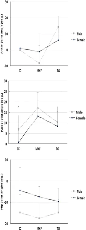
Abstract : The purpose of this study was to analyze the biomechanical differences of lower extremity joints of the frontal plane during sidestep cutting in male and female Judo athletes. In the knee and hip joint, the female group showed a smaller angle than the male group at the time of IC(initial contact). But peak knee joint adduction moment of female group was greater than male group( p <.05). Therefore, female Judo athletes were more likely to injure their knees at the point where their initial foot contacted the ground than male athletes during sidestep cutting.

Keywords : Lower extremity joints, Biomechanics, Frontal plane, Judo athletes, Sidestep cutting

1. Introduction

Acute injuries of the lower extremities frequently occur in athletes performing sports activities [1]. Several previous studies on joint injuries have been reported to be relatively more frequent in female athletes than in men [2-4]. Female athletes have higher risk of contact anterior cruciate ligament injuries than male athletes, which may be due to body structure differences such as lower limb strength and Q angle [5, 6].

Judo, the official sport of the Olympic Games, has recently reduced the size of the stadium, banned lower body attacks, and imposed strict penalties on defensive players to



Corresponding author (E-mail: [email protected])

induce more aggressive games. Judo players perform their movements quickly by attacking and defending their feet. In addition, the attack technique that supports the body weight with one foot increases the risk of injury because it increases the load on the leg joint of the foot during the defense of the opponent [7]. Induction athletes perform physical rotation exercises centering on the footsteps when attacking techniques are used to obtain the opponent's score. At this time, the lower extremity joints are performed to a combination of adduction/abduction in the kinematic chain aspect.

Knee joint injuries to Judo players account

for about 20% of total injury, and the joint

injury rate of the lower limb following the

arm joints that come to contact with the

opponent among the injury types of Judo

(2)

players is the second [8]. In specially, differences in the motion of the lower limb joints occurring on the frontal plane during the sudden directional change affect the risk of injury to the knee joint [9]. The alignment of the lower limb joints is recognized as an important index to determine the risk of joint injuries, because sudden load occurs in the lower limb joint after foot to ground contact.

Therefore, the purpose of this study was to analyze the biomechanical differences of lower extremity joints of the frontal plane during sidestep cutting in male and female Judo athletes.

2. Methods

2.1. Subjects

The subjects of this study were 15 male (age = 22.17 ± 1.41 yrs, body weight = 75.62 ± 4.55 kg, height = 1.76 ± 0.03 m, career = 7.26 ± 1.30 yrs) and female (age = 20.92 ± 1.16 yrs, weight = 64.03 ± 6.19 kg, height = 1.64 ± 0.06 m, career = 7.80 ± 1.50 yrs) Judo athletes, who had not experienced orthopedic history and neurological history were selected. All the subjects were fully explained about the purpose of the experiment, and the experiment agreement was made.

2.2. Data collection

Height and weight were measured in all participants. Moreover, an anthropometer (SM-324) was used to measure body segment length and circumference that were required for applying the scaling method for body segment parameters. The subjects were wearing shorts and t - shirts in black spandex and participated in the experiment with their personal running shoes. Subjects were instructed to perform a directional change using a left foot at distance of 4.5m from the starting point of the pre-displayed starting point. In the direction of the subject's motion,

and the direction change performed by supporting is performed on the right side. At this time, information on the direction was recognized on the ground using a blue tape.

After sufficient stretching to prevent injury to the subjects, which could occur due to the sudden change of direction, the direction change operation was performed for 10 minutes. In order to analyze the motion of the human body in three-dimensional analysis, 35 15-mm reflective markers suitable for a plug-in-gait model were attached on the participants for 3D motion analysis during the sidestep cutting. A three-dimensional coordinate acquisition area of the cameras was created using a T-shaped wand composed of four reflective markers, and then the NLT(nonlinear transformation) to calculate the positional coordinates of the reflection markers on the human body in the 3D motion analysis. And the global coordinate system in which the forward and backward directions of the movement progression direction are defined as the Y-axis, the medial and lateral directions are defined as the X-axis, and the vertical direction is defined as the Z-axis. We defined the local coordinate system of the head, body, pelvis, femur, lower leg, foot, upper arm, forearm, and hand segment using reflection markers attached to the human body.

The visual information device LED (Light Emitting Diode, Visol, Korea) was installed at 8m ahead of the starting line to prevent the subjects from performing pre-bodily movements before the change of direction.

Two electronic sensors (Photoelectric sensor,

Viso, Korea) were installed at the points

before redirection at intervals of 2.5 m to

prevent the differences in the speed of motion

in collecting the directional movement of

subjects. LEDs were used to visualize the

direction of the subjects. The subjects started

to operate according to the first light signal of

the LED. When they passed the two electronic

sensors, they collected information about the

(3)

Ground reaction forces factors

Gravity

Body segment inertial parameters

Biomechanical model

Joint angle in frontal plane

Peak adduction/abduction lower extremity joints moment

Motion analysis

Fig. 2. Schematic diagram for computing lower extremity joints angles and moments.

speed and transmitted the information about the redirection through the LED signal. The subjects performed a linear motion. And all electronic signal equipments are A/D Snyc.

box (VSAD-102-32C, Visol, Korea).

Biomechanical data of subjects were collected at 100 Hz using 8 Vicon cameras (T10S, Vicon, UK) and at 1000 Hz using force plate (OR6-7-1000, AMTI, Inc, Watertown, MA , USA, sampling rate).

2.3. Data analyze

In this study, to analyze the kinematic factors of the lower limb during the sidestep cutting, the point where the left foot contacts the ground and the vertical repulsion force is 10N or higher is referred to as event 1 (initial contact), event 2 was max knee flexion (MKF) while the left foot was in contact with the ground, event 3 was toe off (TO).

The 3 dimensional coordinate data of the reflection markers attached to the human body were filtered by a second-order low-pass butterworth filter (6 Hz) and the data collected by the ground reaction force were filtered by a second-order butterworth filter (50 Hz).

Z

G Y X

Fig. 1. Coordinate system of lower body segment.

The subjects' data were modeled and data processed using Kwon3d XP software (Visol Inc, Korea). The cardan orientation method calculates the relative orientation angle of the distal segmental coordinate system to the proximal segmental coordinate system after setting the local coordinate system (X axis:

medial/lateral direction, Y axis:

anterior/posterior direction, Z axis: vertical

direction). And the angle of the human joint was

calculated. the kinematical data and the ground

reaction force data calculated by the experiment

(4)

are substituted into the joint equilibrium equation which is calculated by the inverse dynamics method as biomechanical model, and the body inertia values ​​of the subjects are calculated using the measured data joint reaction force and joint moment were calculated (fig. 2).

The ankle, knee, hip joint motion was defined as the following: positive (+) and negative (-) values in the AP axis as adduction and abduction, respectively.

2.4. Statistics

In this study, we compared the adduction and abduction of the lower extremity joints on the frontal plane during the change of

direction of the male and female Judo athletes. In order to verify the difference of analysis factors, SPSS ver. (SPSS Inc., Chicago, IL, USA) Statistical analysis was performed using independent t-test. The significance level of data processing was set to α = .05.

3. Results and Discussion (Table 1) and (Fig. 3) showed the results of the analysis of the kinematic factors in the frontal plane during the direction change of the Judo players participating in this study.

Table 1. Biomechanical lower extremity joints angle on frontal plane between male and female

Joint Event Male Female t-value p-value

Ankle

Initial Contact -1.22 (10.15)

-0.96

(9.65) 0.590 .560

Max Knee Flexion -7.24 (10.99)

-1.12

(11.70) 1.453 .158

Toe Off 8.18

(6.77)

6.11

(8.14) -.746 .462

Knee

Initial Contact 5.94 (6.59)

0.76

(3.93) -2.547 .017

*

Max Knee Flexion 17.70 (7.24)

13.21

(0.06) -1.739 .093

Toe Off 10.67

(7.11) 8.39

(9.50) -.733 .470

Hip

Initial Contact -10.46 (8.59)

-4.39

(6.76) 2.105 .045

*

Max Knee Flexion -10.25 (0.04)

-7.35

(4.75) 1.270 .215

Toe Off -5.14

(5.89)

-9.62

(6.00) -2.026 .053

Note.

*

p<.05

(5)

Fig. 3. Lower extremity joints angle value at the IC, MKF, TO between male and female.

Fig. 4. Peak add/abduction moment of lower extremity joints between male and female.

There was no statistically significant difference between the male and female groups at the IC (t=0.590, p=.560), MKF (t=1.453, p=.158), TO (t=-0.746, p=.462) in the ankle joint. In the knee joint, the female group showed a smaller angle than the male group at the time of IC (t=-2.547, p=.017). MKF (t=-1.739, p=.093) and TO (t=-0.733, p=.470) showed no statistical difference between the two groups.

In the hip joint, the female group showed a smaller angle than the male group at the time of IC (t=2.105, p=.045). MKF (t=1.270, p=.215) and TO (t=-2.026, p=.053) showed no statistical difference between the two groups.

(Fig. 4) shows the results of analysis of the

peak add/abduction moments of the left lower

extremity joints of the male and female group

in the sidestep cutting. There was no

statistically significant difference between the

male and female groups in the factors of peak

add/abduction moment of the ankle (t=0.290,

p=.774) and hip (t=1.042, p=.307) joints in

the turning direction. Only the peak

add/abduction moment of the knee (t=2.807,

p=.009) joint showed statistical differences

between the two groups.

(6)

The level of the repulsive force of the ground that occurs during the body deceleration exercise increases the risk of non-contact injury of the knee joint [10].

During the body decelerating movement, a large load is generated in the knee joint with the contact of the foot and the ground, and the anterior movement of the femur due to the movement of the ankle and the knee joint can be improved. And it is reported that this increases the risk of injury to anterior cruciate ligament [6, 11, 12].

It has been reported that the adduction moment generated by the add/abduction of the supporting foot and the increase of the extension moment due to the extensor contraction of the quadriceps muscle increase the compressive force and shear force of the knee joint [13, 14].

In this study, the female group was smaller than the male group in the add/abduction angle of the supporting foot of the knee and hip joint at the initial contact with the ground. Movement of the knee joint at the time of changing direction of the body movement affects the maximum add/abduction moment of the knee joint [15, 16]. This increase in the moment of the knee joint increases the compressive force and shear force of the knee joint with the increase of the extension moment due to the stretch contraction of the quadriceps muscle [4, 17].

The result of this study showed that the knee joint adduction moment of female Judo group in induction was higher than that of male Judo group [16, 18].

4. Conclusions

The purpose of this study was to analyze the biomechanical differences of lower extremity joints of the frontal plane during sidestep cutting in male and female Judo athletes. In female group, the angle of knee and hip joint was smaller than that of male

Judo player at the initial contact point. The peak adduction/abduction moment of the knee joints of female Judo players group were higher than those of male during the sidestep cutting performance. Therefore, we found that the risk of injuries to knee joints in female Judo athletes was relatively higher than that of male Judo athletes during a sudden exercise in the side direction.

Acknowledgements

This paper was supported by Yongin University research funding in 2016.

References

1. B. Hang, “Acute sports-related lower extremity injuries”, Clin. Pediatr Emerg.

Med, Vol.14, No.4 pp. 304-317, (2013).

2. T. E. Hewett, G. D. Myer, K. R. Ford, R.

S. Heidt, A. J. Colosimo, S. G. McLean, A. J. van den Bogert, M. V. Paterno, P.

Succop, “Biomechanical measures of neuromuscular control and valgus loading of the knee predict anterior cruciate ligament injury risk in female athletes: a prospective study” Am. J. Sports Med, Vol.33, No.4 pp. 492-501, (2005).

3. C. R. James, P. S. Sizer, D. W. Starch, T.

E. Lockhart, J. Slauterbeck, “Gender differences among sagittal plane knee kinematic and ground reaction force characteristics during a rapid sprint and cut maneuver” Res. Q. Exerc. Sport, Vol.75, No.1 pp. 31-38, (2004).

4. K. H. Kim, J. H. Cho “The influence of cutting direction on risk factors of anterior cruciate ligament injury” J. Sports Leisure Studies, Vol.48, No.2 pp. 795-802, (2012).

5. J. D. Chappell, B. Yu, D. T. Kirkendall,

W. E. Garrett, “A comparison of knee

kinetics between male and female

(7)

recreational athletes in stop-jump tasks”

Am. J. Sports Med, Vol.30, No.2 pp.

261-267, (2002).

6. B. Yu, W. E. Garrett, “Mechanisms of non-contact ACL injuries” Br. J. Sports Med, Vol.41, No.1 pp. 47-51, (2007) 7. S. Koshida, T. Deguchi, K. Miyashita, K.

Iwai, Y. Urabe, “The common mechanisms of anterior cruciate ligament injuries in judo: a retrospective analysis” Br. J. Sports Med, Vol.44, No.12 pp. 856-861, (2010).

8. U. M. Kujala, S. Taimela, I. Antti-Poika, S. Orava, R. Tuominen, P. Myllynen,

“Acute injuries in soccer, ice hockey, volleyball, basketball, judo, and karate:

analysis of national registry data” Br.

Med. J, Vol.311, No.2 pp. 1465-1468, (1995).

9. J. R. Houck, A. Duncan, K. E. De Haven, “Comparison of frontal plane trunk kinematics and hip and knee moments during anticipated and unanticipated walking and side step cutting tasks” Gait Posture , Vol.24, No.3 pp.

314-322, (2006).

10. T. E. Hewett, G. D. Myer, K. R. Ford,

“Anterior cruciate ligament injuries in female athletes: Part 1, mechanisms and risk factors” Am. J. Sports Med, Vol.34, No.2 pp. 299-311, (2006).

11. I. Estevan, C. Falco, J. F. Silvernail, D.

Jandacka, “Comparison of Lower Limb Segments Kinematics in a Taekwondo Kick. An Approach to the Proximal to Distal Motion” J. Hum. Kinet, Vol.47, No.1 pp. 41-49, (2015).

12. S. W. Arms, M. H. Pope, R. J. Johnson, R. A. Fischer, I. Arvidsson, E. Eriksson,

“The biomechanics of anterior cruciate ligament rehabilitation and reconstruction”

Am. J. Sports Med, Vol.12, No.1 pp.

8-18, (1984).

13. H. Yun, M. S. Kwon “A comparison of the lower extremity kinetics between male and female university student majoring in judo during the randomly cued cutting performance” Kor. J. Sports Sci , Vol.26, No.3 pp. 1367-1376, (2017).

14. C. D. Pollard, I. M. Davis, J. Hamill,

“Influence of gender on hip and knee mechanics during a randomly cued cutting maneuver” Clin. Biomech, Vol.19, No.10 pp. 1022-1031, (2004).

15. S. G. McLean, X. Huang, Van Den Bogert, J. Antonie, “Association between lower extremity posture at contact and peak knee valgus moment during sidestepping: implications for ACL injury”

Clin. Biomech, Vol.20, No.8 pp. 863-870, (2005).

16. K. R. Ford, G. D. Myer, H. E. Toms, T.

E. Hewett, “Gender differences in the kinematics of unanticipated cutting in young athletes” Med. Sci. Sports Exerc, Vol.37, No.1 pp. 124-129, (2005).

17. I. K. Choi, J. I. Kim, W. K. Sin, D. W.

Choi, “Kinematic Analysis of Knee Joint during Pre/Post Decision Cutting Maneuver” J. Sports Leisure Studies, Vol.47, No2. pp. 845-854, (2012).

18. M. J. Decker, M. R. Torry, D. J. Wyland, W. I. Sterett, J. R. Steadman, “Gender differences in lower extremity kinematics, kinetics and energy absorption during landing” Clin. Biomech, Vol.18, No.7 pp.

662-669, (2003).

수치

Fig.  2.  Schematic  diagram  for  computing  lower  extremity  joints  angles  and  moments.
Fig.  4.  Peak  add/abduction  moment  of  lower  extremity  joints  between  male  and  female.

참조

관련 문서

The study photgraphs of male dentist dressed in formal attire (A) white coat1. The study photgraphs of female dentist dressed in formal attire (A)

The purpose of this study was to investigate the lower extremity muscle strength and student health of male middle school students. Twenty middle school

A crystal plane at an angle θ to the close-packed plane will contain broken bonds in excess of the close-packed plane due to the atoms at the steps..

Third, the analysis of the body-centered vertical speed between male.. and female students showed that male students appeared faster than female students at

The purpose of this study was to analyze the impaction pattern of the impacted mandibular third molar and the relationship with the inferior alveolar nerve

This study attempts to analyze the differences of pre-service secondary teachers’ mathematical noticing and confirm the factors that cause the differences

Among the sub-factors of self-esteem, positive, male was higher than female, and on the contrary, negative In this study, females were higher than males,

1) Because of the maximum plunge force area, the heat generation in FSW of dissimilar joints was maximum at Al6061-T6 side which lead to brittle fracture of the weld