• 검색 결과가 없습니다.

성인 남성에서 단안에 발생한 임균성 결막염 1례

N/A
N/A
Protected

Academic year: 2021

Share "성인 남성에서 단안에 발생한 임균성 결막염 1례"

Copied!
5
0
0

로드 중.... (전체 텍스트 보기)

전체 글

(1)

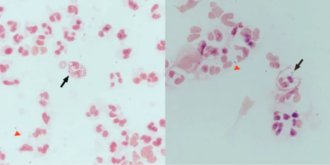
Gonococcal conjunctivitis is rare in adults and, if not treated properly, can cause corneal perforation. Gonococcal conjunctivitis typically presents with a severe mucopurulent discharge, similar to that associated with viral conjunctivitis. Here, we describe a case of monocular gonococcal conjunctivitis, including its clinical characteristics and slit-lamp images, which was initially misdiagnosed as epidemic conjunctivitis. A 20-year-old man was referred to our hospital with no improvement in monocular infection and purulent ocular discharge after 2-wk treatment using antibiotic and 0.1% fluorometholone eye drops at the local ophthalmic clinic. Initially, 0.5% loteprednol eye drops were used since we suspected viral conjunctivitis. Following this treatment, conjunctival infection worsened and a yellow-white ocular discharge covered the conjunctiva and cornea surface. Additional history taking revealed that the patient had sexual contact with a prostitute 1 wk prior to symptom presentation and, after the encounter, he took antibiotics for genital discharge at the local urology clinic, but self-discontinued treatment. A Gram staining showed gram-negative diplococci and culture of collected ocular discharge from the palpebral conjunctiva revealed growth of Neisseria gonorrhoeae , confirming gonococcal conjunctivitis. Following this, the patient was systemically treated with 3rd generation cephalosporin antibiotics. After 3-d treatment, conjunctival infection and purulent ocular discharge had significantly improved. When clinical symptoms are aggravated following steroid eye drop treatment for suspected monocular viral conjunctivitis, gonococcal conjunctivitis must be considered as a differential diagnosis Keywords: Cephalosporin, Gonococcal conjunctivitis, Neisseria gonorrheae

Received: September 30, 2018 Revised: November 20, 2018 Accepted: December 28, 2018

Corresponding Author: Jong Hwa Jun, M.D.

Department of Ophthalmology, Keimyung University School of Medicine, 56 Dalseong-ro, Jung-gu, Daegu 41931, Korea Tel: +82-53-250-7708

E-mail: [email protected]

• The authors report no conflict of interest in this work.

Department of Ophthalmology, Laboratory Medicine

1

, Keimyung University School of Medicine, Daegu, Korea

You Hyun Lee, M.D., Nam Hee Ryoo

1

, M.D., Jong Hwa Jun, M.D.

A Case of Monocular Gonococcal Conjunctivitis in an Adult Male

계명대학교 의과대학 안과학교실, 진단검사의학교실1

이유현 · 류남희

1

· 전종화

성인 남성에서 단안에 발생한 임균성 결막염 1례

© Copyright

Keimyung University School of Medicine 2018

(2)

서 론

임균에 의한 결막염은 신생아 이외에는 드물다고 알려져 있으나 남성에서는 감염된 요로 생식계 분비물의 자가접종, 여성에서는 콘돔을 사용하지 않은 구강 성교의 증가로 인해 성인에서의 발병률이 증가하고 있다[1-3].

임균성 결막염은 1,000안 당 0.19-0.60안의 유병률을 가지고 있으며 젊은 남성이 여성보다 더 흔한 것으로 알려져 있다[4,5]. 임상 양상은 양안 또는 단안에서 초급성으로 발생하는 심한 결막 충혈과 부종, 많은 양의 화농성 분비물이 특징적인 소견이며 이 외에도 안구 불편감 또는 통증, 귓바퀴 앞 임파선염, 안검 부종, 각막 상피 및 실질의 염증을 관찰할 수 있으며 치료가 늦어지면 각막 천공으로 영구적 시력손실을 초래할 수 있어 빠른 진단과 치료가 중요하다[6-8]. 임균성 결막염은 그람 염색과 세균 배양검사에서 90% 이상 진단되며 치료에 대해서는 여러 보고가 있지만, 점안 항생제에 대해서는 효과가 불분명하며 1 g ceftriaxone의 전신적 투여가 중요하다는데 의견이 일치한다[9-11]. 국내외에서 임균성 각결막염을 3세대 cephalosporin을 전신적으로 사용하여 효과적으로 치료한 보고가 있으나 세극등현미경 사진을 함께 첨부한 경우는 드물며 있더라도 사진 해상도가 좋지 않아 진료에 참고하기에 어려움이 있다[12,13]. 이에 저자들은 20세 성인 남자환자에서 초기에 유행성 결막염으로 오진한 단안에 발생한 임균성 결막염 1례를 진단하고 치료한 경험을 그 임상 양상 및 세극등현미경 사진과 함께 보고하고자 한다.

증 례

20세 남자 환자가 3주 전부터 지속된 우안의 안검 부종 및 결막 충혈, 부종, 화농성 분비물 및 간헐적인 통증을 주소로 내원하였다. 환자는 증상 발현 후 개인 안과에서 바이러스 결막염 의심하에 0.5% 레보플록사신(Cravit

, Santen pharmaceutical Co., Osaka, Japan), 0.1%

플루오로메토론 점안액(Ocumetholone

, Samil, Seoul, K o r e a)을 세 시간마다 점안하였고 오플록사신 안연고(Ocuflox

, Samil, Seoul, Korea)를 자기 전에 점안하여 2주 동안 치료하였으나 호전을 보이지 않아

본원으로 전원 되었다.

본원 내원 당시 세극등현미경검사에서 우안 상하안검의 부종, 결막의 심한 충혈 및 부종이 있었고 중등도의 분비물이 관찰되었다. 반면 좌안에는 특별한 이상이 관찰되지 않았다. 우안의 나안시력은 20/50, 교정시력은 20/25이었다. 초기 진료 시 아데노 바이러스에 의한 결막염을 우선적으로 고려하여 우안 주위의 부종 및 염증을 감소시킬 목적으로 0.1% 플루오로메토론 점안액을 0.5% 로테프레드놀 점안액(Lotemax

, Bausch &

Lomb, Tampa, FL, USA)로 바꾸고 하루 5회 점안하도록 하고 경과 관찰 하였다. 4일 뒤 시행한 세극등현미경검사에서 우안의 결막 충혈은 급격히 악화되었으며 화농성 분비물의 양 또한 증가하여 상하안검 결막과, 구결막 및 각막을 모두 뒤덮을 정도로 극히 심해진 양상이 관찰되었다(Fig. 1).

좌안에 증상 및 징후가 없는 점과 추가적인 자세한 병력

청취에서 최초 안증상 발현 일주일 전 유흥업소에

종사하는 여성과 성적접촉을 하였음을 확인하였다. 또한

성적 접촉 3~4일 후 배뇨통으로 요도염 의심 하에 개인

비뇨기과 의원에서 치료 도중 증상이 약간 호전되자

자의적으로 치료를 중단한 적이 있었다는 것을

확인하였다. 당시 소변검사를 시행하였으나 정확한

진단은 환자가 기억하지 못하였다. 이에 세균성 결막염을

의심하여 무균 면봉으로 우안 화농성 분비물을 채취해

슬라이드에 도말하여 그람 염색(Gram staining)을 시행하였고

세균배양 검사를 위해 검체를 혈액한천배지(blood agar

plate)에 접종하였다. 그람 염색에서는 intra- and extracelluar

그람음성쌍구균이 관찰되었으며 세균배양검사에서는

Neisseria gonorrheae가 동정되어 임균성 결막염을

확진하였다(Fig. 2). 즉각적으로 로테프레드놀 점안제의

사용을 중지하였고 치료를 위해 감염내과에 협진

의뢰하여 1 g ceftriaxone (Rocephin

, Roche Diagnostics

Corp., USA)을 1회 정주하였으며 3세대 cephalosporin계

경구 항생제(Suprax

, Dong-A Pharmaceutical Co., Seoul,

Korea)를 하루에 두 번씩 5일간 복용하도록 하였다. 치료

시작 후 3일째 우안 나안시력은 20/20으로 회복되었으며

세극등현미경 검사에서 결막충혈과 황백색의 화농성

분비물은 급격히 호전되었다(Fig. 3). 이후 환자는

경과관찰 중 임의로 내원을 중단하였다.

(3)

고 찰

임균에 의한 안감염은 일반적으로 안검 부종, 화농성 분비물, 충혈, 결막 부종 등의 증상을 동반하며 적절히 치료하지 않으면 감염이 결막에서 각막으로 이동하여 주변부 각막 궤양을 일으키며 빠르게 진행하여 심하면 각막궤양 및 천공을 유발할 수 있다[6,14]. 임균의 잠복 기간은 3일에서 19일까지 다양하고 성적접촉 병력의 누락으로 임균성 결막염 진단이 지연되는 경우가 많다[15]. 본 증례에서도 7일간의 잠복기간이 있었으며 성적 접촉 과거력이 뒤늦게 확인되어 진단이 늦어졌다.

McAnena 등[4]은 성인에서 발생하는 임균성 결막염은 남성과 단안에서 호발한다 하였는데 본 증례에서도 환자는 20세 남자였으며 우안에서만 병변이 관찰되었다.

또한, 소아에서와는 다르게 성인에서 발생하는 임균성

결막염의 감염경로는 대부분 감염된 소변이나 생식기 분비물에 접촉하거나 오염된 손에 의한 균의 직접 접종에 의한 것으로 알려져 있는데 환자의 경우도 증상 발현 1주일 전 성적 접촉 및 이후 요도염 증상의 발현이 있어 이에 의한 발생을 의심할 수 있었다[16]. 본 증례의 경우 안 증상과 배뇨통이 동반되어 비뇨기과에서 요도염으로 치료를 받은 적이 있었지만 Peters 등[11]은 요도염에 치료되는 항생제의 농도가 안구 병변을 치료하기에는 부족하다고 보고하였으며 환자가 치료를 자의로 중단하여 안 증상은 지속되었던 것으로 여겨지며 전신 항생제의 사용에 의해 플루오로메토론 등의 스테로이드 점안제의 사용 중에도 각막 침범 등의 양상은 거의 없었던 것으로 생각된다. 다만, 환자가 비뇨기과 치료를 자의로 중단하여 요도염 및 결막염의 완전한 치료가 이루어 지지 않고 본원 내원 당시 초기 0.5% 로테프레드놀 점안액을 사용하고 안구 Fig. 1. Photographs at 4th day after the 0.5% loteprednol eye drop was used. (A) Cornea surface covered with yellow-white thick purulent ocular discharge (arrow). (B-D) Severe chemosis and conjunctival injection with yellow-white purulent ocular discharge at bulbar conjunctiva (B, arrow head), and palpebral conjunctiva (D, arrow head).

A

C

B

D

(4)

병변이 악화된 것은 적절한 항생제 치료가 이루어지기 전에 점안 스테로이드제를 사용하여 잠재적으로 질병의 경과를 연장함과 동시에 세균의 감염력을 증가시켰기 때문으로 생각된다.

임균성 결막염의 효과적인 치료에 대한 연구 중 국외에서는 Haimovici 등[9]이 ceftriaxone을 1회 근주하여 13명의 환자를 효과적으로 치료하였고 국내에서는 Kim

등[12]이 ceftriaxone을 하루 1회 5일 동안 근주함과 동시에 3세대 cephalosporin계 점안제를 사용하여 1명의 환자를 성공적으로 치료한 보고가 있다. 본 증례에서는 ceftriaxone (Rocephin

)을 1회 근주하고 5일간의 경구약 복용으로 임상 증상의 급격한 호전을 경험하였는데 이는 임균성 결막염의 치료에는 전신적 항생제 투여가 중요함을 보여준다.

Fig. 2. Gram staining of the yellow-white purulent ocular discharge smear showed many polymorphonuclear cells with intra- (arrow) and extracelluar (arrow head) gram negative reniform diplococcic (x1000, oil immersion).

Fig. 3. Photographs at 3rd day after the 3rd-generation cephalosporin antibiotic was used systemically. (A) Disappearance of yellow-white purulent ocular discharge on cornea. (B) Dramatically improved yellow-white purulent ocular discharge in lower palpebral conjunctiva.

A B

(5)

임균성 결막염은 치료 시작 후 12시간 이내에 화농성 분비물이 호전되고 6시간 이내에 세균배양 검사에서 Neisseria gonorrheae의 음성이 확인된다고 알려져 있다[17]. 본 증례에서는 치료 후 세균배양 검사를 시행하지 않았지만, 치료 시작 후 3일째 화농성 분비물이 급속히 호전됨을 확인하였다.

요 약

결론적으로 젊은 성인 남성에서 단안에 아데노 바이러스에 의한 결막염을 의심하여 점안 스테로이드제를 사용 후 임상 양상이 악화되어 황백색의 화농성 분비물이 결막 및 각막에서 관찰되고 심한 충혈과 결막 부종이 관찰된다면 임균성 결막염을 감별 진단의 하나로 고려하여야 한다.

참 고 문 헌

1. Quirke M, Cullinane A. Recent trends in chlamydial and gonococcal conjunctivitis among neonates and adults in an Irish hospital. Int J Infect Dis 2008;12:371-3.

2. Lee JS, Choi HY, Lee JE, Lee SH, Oum BS.

Gonococcal keratoconjunctivitis in adults. Eye (Lond) 2002;16:646-9.

3. Copen CE, Chandra A, Martinez G. Prevalence and timing of oral sex with opposite-sex partners among females and males aged 15-24 years: United States, 2007-2010. Natl Health Stat Report 2012;16:1-14.

4. McAnena L, Knowles SJ, Curry A, Cassidy L.

Prevalence of gonococcal conjunctivitis in adults and neonates. Eye (Lond) 2015;29:875-80.

5. Lahra MM. Australian Gonococcal Surveillance Programme annual report, 2012. Commun Dis Intell Q Rep 2013;37:E233-9.

6. Wan WL, Farkas GC, May WN, Robin JB. The clinical

characteristics and course of adult gonococcal conjunctivitis. Am J Ophthalmol 1986;102:575-83.

7. Schwab L, Tizazu T. Destructive epidemic Neisseria gonorrheae keratoconjunctivitis in African adults. Br J Ophthalmol 1985;69:525-8.

8. Kawashima M, Kawakita T, Den S, Tomita M, Shimazaki J. Surgical management of corneal perforation secondary to gonococcal keratoconjunctivitis. Eye (Lond) 2009;23:339-44.

9. Haimovici R, Roussel TJ. Treatment of gonococcal conjunctivitis with single-dose intramuscular ceftriaxone. Am J Ophthalmol 1989;107:511-4.

10. Azari AA, Barney NP. Conjunctivitis: a systematic review of diagnosis and treatment. JAMA 2013;310:1721-9.

11. Peters RP, Verweij SP, McIntyre JA, Schaftenaar E.

Gonococcal Conjunctivitis Despite Successful Treatment of Male Urethritis Syndrome. Sex Transm Dis 2016;43:120-1.

12. Kim HK, Lee JS, Lee JR. Gonococcal Keratoconjunctivitis in Adult. J Korean Ophthalmol Soc 2002;43:786-90.

13. Bodurtha Smith AJ, Holzman SB, Manesh RS, Perl TM. Gonococcal Conjunctivitis: A Case Report of an Unusual Mode of Transmission. J Pediatr Adolesc Gynecol 2017;30:501-2.

14. McElnea E, Stapleton P, Khan S, Stokes J, Higgins G.

Challenges in the management of Neisseria gonorrhoeae keratitis. Int Ophthalmol 2015;35:135-40.

15. Bruins SC, Tight RR. Laboratory-acquired gonococcal conjunctivitis. JAMA 1979;241:274.

16. Diena BB, Wallace R, Ashton FE, Johnson W, Platenaude B. Gonococcal conjunctivitis: accidental infection. Can Med Assoc J 1976;115:609, 612.

17. Fraunfelder FT, Fraunfelder FW, Roy FH. Roy and

Fraunfelder's current ocular therapy. Philadelphia,

Pa: Elsevier Saunders; 2008.

수치

Fig. 2. Gram staining of the yellow-white purulent ocular discharge smear showed many polymorphonuclear cells  with intra- (arrow) and extracelluar (arrow head) gram negative reniform diplococcic (x1000, oil immersion).

참조

관련 문서

the material balance for a reaction component must be made for a differential element of volume dV.. For constant-volume reaction.. The integral can be evaluated in any

• In a attraction-dominant condition, when a gas expands, some kinetic energy of them must be converted to potential energy to reach greater

2.World Health Organization: Clinical management of severe acute respiratory infection when Novel coronavirus (nCoV) infection is suspected: Interim Guidance.

(1.1 points) Which of the following is the best example of the great damage in (a) The damage they do is particularly great when they are relied upon as guide in the process

(Background) The standard triple therapy used as the first-line treatment for Helicobacter pylori infection are a combination of proton pump inhibitor(PPI),

We suggest that it will be helpful for diagnosis, treatment and prevention of adverse drug reaction through known causative drug and clinical

Several methods are considered for permanent disposal of spent nuclear fuel, which is a high-level radioactive waste, such as disposal of the deep layer, disposal of

According to above, liquid based cytology is considered to be adequate for routine oral examination or diagnostic tool for oral dysplasia and carcinoma