• 검색 결과가 없습니다.

Quantification of Microstructures in Mice Alveolar Bone using Micro-computed tomography (${\mu}CT$)

N/A
N/A
Protected

Academic year: 2021

Share "Quantification of Microstructures in Mice Alveolar Bone using Micro-computed tomography (${\mu}CT$)"

Copied!
6
0
0

로드 중.... (전체 텍스트 보기)

전체 글

(1)

http://dx.doi.org/10.11620/IJOB.2013.38.3.087

*Correspondence to: Hae-Ryoung Park, Division of Pre-service Teacher Training Course and General Studies, Kwangju Women's University, 201, Yeodai-gil, Gwangsan-gu, Gwangju 506-713, Republic of Korea, Tel: +82-62-950-3751,

Fax: +82-62-950-3617, e-mail: hrpark@kwu.ac.kr

This is an Open-Access article distributed under the terms of the Creative Commons Attribution Non-Commercial License(http://creati- vecommons.org/licenses/by-nc/3.0) which permits unrestricted non- commercial use, distribution, and reproduction in any medium, pro- vided the original work is properly cited.

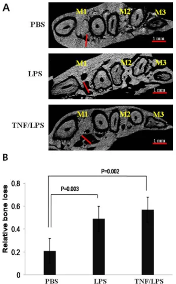
Quantification of Microstructures in Mice Alveolar Bone using Micro-computed tomography ( µCT)

Hae-Ryoung Park

1

*, Hyun-Jin Kim

2

, and Byung-Ju Park

3

1

Division of Pre-service Teacher Training Course and General Studies, Kwangju Women's University

2

Department of Oral Anatomy, School of Dentistry, Wonkwang University

3

Department of Biochemistry, School of Dentistry, Chonnam National University

(received May 13, 2013 ; revised September 6, 2013 ; accepted September 9, 2013)

Periodontal inflammation increases the risk of tooth loss, particularly in cases where there is an associated loss of alveolar bone and periodontal ligament (PDL). Histological and morphometric evaluation of periodontal inflammation is difficult. Especially, the lengths of the periodontal ligament and interdental alveolar bone space have not been quan- tified. A quantitative imaging procedure applicable to an animal model would be an important clinical study. The purpose of this study was to quantify the loss of alveolar bone and perio- dontal ligament by evaluation with micro-computed tomo- graphy (micro-CT). Another purpose was to investigate diffe- rences in infections with systemic E. coli LPS and TNF-α on E. coli lipopolysaccharide (LPS) in loss of alveolar bone and periodontal ligament model on mice. This study showed that linear measurements of alveolar bone loss were repre- sented with an increasing trend of the periodontal ligament length and interdental alveolar process space. The effects of systemic E. coli LPS and TNF-α on an E. coli LPS-induced periodontitis mice model were investigated in this research.

Loss of periodontal ligament and alveolar bone were eval-

uated by micro-computed tomography (micro-CT) and cal- culated by the two- and three dimensional microstructure morphometric parameters. Also, there was a significantly increasing trend of the interdental alveolar process space in E. coli LPS and TNF-α on E. coli LPS compared to PBS. And E. coli LPS and TNF-α on E. coli LPS had a slightly increa- sing trend of the periodontal ligament length. The increa- sing trend of TNF- α on the LPS-induced mice model in this experiment supports the previous studies on the contribu- tion of periodontal diseases in the pathogenesis of systemic diseases. Also, our findings offer a unique model for the study of the role of LPS-induced TNF- α in systemic and chronic local inflammatory processes and inflammatory diseases. In this study, we performed rapidly quantification of the perio- dontal inflammatory processes and periodontal bone loss using micro-computed tomography (micro-CT) in mice.

Key words: Micro-CT, alveolar bone loss, periodontal ligament, TNF- α, E. coli LPS

Introduction

A common inflammatory oral disease involves perio- dontal disease and periodontal tissue linked with a systemic disorder. Bacteria are important causes of bone pathology in common conditions such as periodontitis and dental cysts [1]. Periodontal disease increases the risk of tooth loss, particularly in cases with an associated loss of alveolar bone

87

(2)

and periodontal ligament (PDL) [2]. Periodontal inflamma- tion is represented by the loss of supporting connective tissue and alveolar bone around the teeth [3]. Bacterial infections produce destruction of the periodontal tissue caused by the inflammatory response to the pathogenic bacteria. Immune mediators such as interleukin-1 (IL-1), tumor necrosis factor alpha (TNF- α), and IL-6 have been found to be abundantly expressed in periodontal disease [4]. Periodontal diseases are a group of infections that lead to inflammation of the gingiva, destruction of the periodontal tissues, and, in severe cases, loss of alveolar bone with eventual exfoliation of the teeth [5]. The inflammation induces detachment of the perio- dontal ligament, with loosening of the connection between the gingiva and alveolar bone, allowing the extension of the oral bacterial infection [6]. Periodontal disease has been sus- pected as a trigger of systemic disorders. Penetration of bac- terial products, such as lipopolysaccharide (LPS) may reach into deeper periodontal tissues. This may affect the systemic blood circulation and cytokine production [7]. Interleukin-1 β (IL-1 β) shows a significantly increased production of low dose LPS groups but TNF- α does not show a significant inc- rease in production [8]. The cytokine dysregulation asso- ciated with prolonged TNF- α expression represents a mecha- nism through which bacteria may induce a more damaging inflammatory disease [9]. Lipopolysaccharides (LPS) of gram- negative bacteria were potent inducers of pro-inflammatory mediators and could initiate numerous host-mediated destruc- tive processes [10,11].

Micro-computed tomograpy (Micro-CT) offers signifi- cant potential for identifying microstructures [12,13]. Micro- CT for preclinical applications provides higher spatial reso- lution images than medical or dental CT for clinical assess- ment.

To achieve three-dimensional evaluation of alveolar bone loss in periodontitis animal models, micro-computed tomog- raphy (micro-CT) has been used in recent studies [14,15].

This study was investigated the effects of alveolar bone loss in E. coli LPS and TNF-α on E. coli LPS-induced mice evaluated by micro-CT. And also this method suggested that the periodontal ligament and interdental alveolar process space length could be quantified using the micro-CT. Our findings offer a TNF- α on LPS-induced mice model to study the role of inflammatory processes and the degree as quantified by micro-CT. This study was done to assess periodontal tissue breakdown after E. coli LPS and TNF-α on LPS-induced

experimental periodontitis in mice and the use of micro-CT as a rapid quantification method for alveolar bone and PDL loss assessment.

Materials & Methods

Animals model preparation

BALB/c 8-week-old female mice were randomly divided into 3 groups (n = 10 per each groups). All animals were housed at standard temperature and humidity with a 12-h light/dark cycle, and their body weights were recorded before the test procedures. The mice were fed with standard rodent chow and water ad libitum. The experimental protocols were approved by the Chonnam National University Institutional Animal Care and Use Committee. The mice (20 g per each) had experimental periodontitis induced in the interdental region between the mandible first (M1), second (M2) and third molars (M3).

Inflammatory Bone Loss Model

The bone loss model was generated as previously described [16]. To initiate alveolar bone loss, micro-injections of 10 mg/kg E. coli LPS (20 μg per injection in 2 μl PBS total volume, Sigma, MO, USA) were made directly into the pa- latal interproximal gingiva between the first and second mo- lars on the left side. The same volume of PBS was injected into the same region on the right side [17]. E. coli LPS and PBS injections were repeated 3 times each week over 10 weeks (denoted by LPS and PBS). The E. coli LPS treated side was injected with TNF- α (PeproTech, seoul, Korea) (deno- ted by TNF/LPS). Oral injections of 100 μg/kg of TNF-α were repeated two times per week over a 10 week period.

Micro-CT and Bone Volume Fraction (BVF) Analysis

Micro-CT analysis was performed as previously described

[18]. Briefly, after euthanasia, the molar with intact surrounding

tissue from each animal was dissected and fixed in freshly

prepared 4% paraformaldehyde (pH 7.2) at 4°C overnight

for scanning by Micro-CT. Specimens were scanned by Sky-

scan 1172 Desk Top micro-CT. The system used was Scan-

ner of Skyscan1172, Instrument S/N=054, Hardware version

of A, software of Version 1. 5. Analysis with GE microview

software (Microview Software Technologies, USA) was done

for quantification of the bone volume fraction (BVF). Loss

of bone volume was assessed by means of 3-D displays as

(3)

previously described [19]. Briefly, given a series of pro- jection images, a stack of 2D sections was reconstructed for each specimen (the number of sections depended on the desired height) and stored in the bmp format condition. In each scan, a standardized tridimensional region of interest (ROI) was defined by the following landmarks: first molar (M1), second molar (M2) and third molar (M3).

Statistics

Data were expressed as the mean±SD from three indepen- dent triplicate experiments. Statistical analysis was perfor- med by one-way analysis of variance (ANOVA) followed by the Student’s t-test. For all analyses, p<0.05 was considered statistically significant.

Results

The effects of oral administration of E. coli LPS and TNF-

Fig. 1. The daily food uptake and body weight of mice. A daily food intake (g/day) was checked in PBS, LPS and TNF/ LPS mice (A). Body weight changes were monitored in PBS, LPS and TNF/LPS mice until 20 weeks (B). Numbers of animals studied were 10 in each group.

α on food intake and body weight was assessed. The mice were given an oral injection with 10 mg/kg of E. coli LPS repeated 3 times each week over 10 weeks. The mice were orally injected with 10 mg/kg of E. coli LPS with the ad- dition of 10 μg/kg of TNF-α repeated two times per week over a 10 week period. The control mice were treated with PBS over the same period. The effects of E. coli LPS and TNF- α/E. coli LPS on food intake and body weight were checked time dependently for 20 weeks. The food intake of the TNF/LPS mice was decreased compared with that of the PBS and LPS mice in a time dependent manner (Fig. 1A). The body weight of the LPS and TNF/LPS mice was decreased more

Fig. 2. Effects of LPS and TNF/LPS on periodontal ligament length. The 2D displayed periodontal ligament length at PBS, LPS and TNF/LPS mice (A). Comparison of the relative bone loss in- dicated that the periodontal ligament length in LPS and TNF/

LPS mice was significantly greater than that in PBS mice (B).

The indicated white bar was 0.5 mm. The white bar was the

distance of the periodontal ligament length. *indicates a signi-

ficant difference, p < 0.05.

(4)

than the body weight of the PBS mice (Fig. 1B).

Two-dimensional (2D) serial sections showed all the mandibular molar teeth extracted from the mice. Alveolar bone loss was estimated by measuring the length of the periodontal ligament and interdental alveolar process space in the 2D reconstruction images. Transverse serial sections (0.5 mm) images with the corresponding micro-CT marked off the first molar (M1), second molar (M2) and third molar (M3). The length of the periodontal ligament in the LPS and TNF/LPS showed an increase compared with PBS (Fig. 2A).

Quantification of the periodontal ligament length revealed

Fig. 3. Effects of LPS and TNF/LPS on the interdental alveolar process space. The 2D displayed the length of the alveolar process space loss at PBS, LPS and TNF/LPS mice (A).

Comparison of the relative bone loss indicated that the alveolar process space length in LPS and TNF/LPS mice was significantly greater than that in PBS mice (B). The indicated red bar was 1 mm. The red bar was the distance of the alveolar process space. *indicates a significant difference, p < 0.05.

statistically significant increases in LPS and TNF/LPS compared with PBS (Fig. 2B). The interdental alveolar process space length in the LPS and TNF/LPS was greater than in PBS (Fig. 3A). Quantification of the alveolar process space length revealed statistically significant increases in LPS and TNF/LPS compared with PBS (Fig. 3B). There was no difference between LPS and TNF/LPS in the length of the periodontal ligament and interdental alveolar process space. There was no difference in alveolar bone loss bet- ween TNF- α and TNF-α in the E. coli LPS mice. The alveolar bone loss was represented as the length of the pe- riodontal ligament and interdental alveolar process space using 2D- and 3D measurements. The treatment with 10 mg/kg of E. coli LPS and 100 μg/kg of TNF-α caused destruction of the periodontal ligament and alveolar process space. The leng- ths of the periodontal ligament and alveolar process space were more affected by 100 μg/kg of TNF-α (TNF/ LPS) in 10 mg/kg of E. coli LPS-induced on mice.

Fig. 4. LPS and TNF/LPS treated mice demonstrated signi- ficantly decreased alveolar bone volume fraction (BVF) in com- parison with PBS. The 3D representative specimens displayed the periodontal ligament and alveolar process space. TNF or TNF/LPS mice demonstrated a decreased alveolar bone vo- lume fraction (BVF) in comparirson with PBS mice. The vo- lume of interest (VOI) was established in the micro-CT recon- struction. VOI was established with a three dimensional recons- truction displaying the periodontal ligament and alveolar bone loss in PBS, LPS and TNF/LPS mice. M1; first molar, M2;

second molar and M3; third molar.

(5)

Volumetric micro-CT measurements were three dimen- sional (3D) structures in the periodontal ligament and alveo- lar process space. 3D reconstruction of the mandibular bone showed the lingual surface at the alveolar bone. Analysis of the alveolar bone including all three molars can be done by using the method for creating 3D ROIs. TNF/ LPS showed reduction of the alveolar bone process (Fig. 4). 3D micro- CT measurements showed the alveolar bone loss in LPS and TNF/ LPS compared with PBS.

Discussion

In this study, E. coli LPS or TNF-α on E. coli LPS in- duced inflammation was successfully used to induce experi- mental periodontitis in mice. TNF- α can be induced by bacterial products and is considered as an early event in the cascade leading to inflammation [20]. Intragingival injec- tion of 10 mg/kg of E. coli LPS has been accepted as a useful experimental model of periodontitis with alveolar bone loss. 10 weeks after, LPS injection resulted in significant gin- gival and periodontal inflammation with inflammatory infil- trate interdental bone loss. It indicated that intragingival injec- tion of LPS in the rat provides an easily induced reprodu- cible experimental model of periodontal inflammation [21]. Our results showed site-specific, alveolar bone loss, and inflam- mation in this model. Quantification of the periodontal liga- ment length and interdental alveolar process space was done using micro-CT methods. We found that the methods yielded comparable results for detecting alveolar bone loss. More- over, the osteoclast activity observed in this study was con- sistent with the progression of inflammation in a monkey model [22]. Periodontal disease is the most frequent cause of tooth loss, including osteoporosis [23]. This finding was similar to the research done by Fokkema, et al. [24]. Micro- CT is a recent technology available in bone laboratories. Micro- CT has been widly used in the study of bone metabolism.

Previous reports showed that micro-CT is an effective me- thod for the histomorphometrical analysis of long bone ino- variectomized rats and gene-deficient mice [25]. A 3D finite element model of a tooth with different levels of bone height was constructed to estimate the reduction [26]. The selected landmarks for quantification of alveolar bone (ROF, the most distal end of molar roots, and tangent line between roots on the coronal plane for landmark detection of ROI)

provided critical criteria and reproducibility to measure vo- lumetric osseous parameters. These approaches for the quan- titative assessment of periodontal osseous structures demon- strated the reliability and reproducibility of 3-D micro-CT measurements of alveolar bone loss.

In this study, the roles of E. coli LPS or TNF-α in E. coli LPS-induced alveolar bone loss mice model were studied.

But no difference between E. coli LPS and TNF-α on E. coli LPS-induced alveolar bone loss were found using micro- CT. The destroyed periodontal ligaments and alveolar process space showed the same results of alveolar bone loss. This experiment, once again, proved the contribution of perio- dontal disease in the pathogenesis of systemic diseases.

Further study needs to be done to investigate the close relationship between periodontal disease and systemic dis- ease using in vivo micro-CT imaging approaches for real- time assessments of alveolar bone changes that could lead to dynamic, rather than static, measures of osseous change.

Acknowledgement

This study was supported by a grant (CRI13011-1) of the Chonnam University Hospital Research Institute of Clinical Medicine.

References

1. Nair SP, Meghji S, Wilson M, Reddi K, White P, Hen- derson B. Bacterially induced bone destruction: mecha- nisms and misconceptions. Infect Immun. 1996;64(7):

2371-2380.

2. Cochran DL. Inflammation and bone loss in periodontal disease. J Periodontol. 2008;79(8 Suppl):1569-1576.

3. Pihlstrom BL, Michalowicz BS, Johnson NW. Periodontal diseases. The Lancet.2005;366(9499):1809-1820.

4. Boch JA, Wara-aswapati N, Auron PE. Interleukin 1 Signal Transduction-Current Concepts and Relevance to Periodon- titis. J Dent Res. 2001; 80(2):400-407.

5. Haffajee AD, Socransky SS. Microbial etiological agents of destructive periodontal diseases. Periodontol 2000;994:

5:78-111.

6. Baker PJ. The role of immune responses in bone loss during periodontal disease. Microbes Infect. 2000; 2(10):

1181-1192.

7. Henderson B, Poole S, Wilson M. Bacterial modulins: a novel class of virulence factors which cause host tissue pathology by inducing cytokine synthesis. Microbiol Rev.

1996;60:316-341.

(6)

8. Li X, Kolltveit KM, Tronstad L, Olsen I. Systemic diseases Caused by Oral Infection. Clin. Microbiol. Rev. 2000;13 (4):547-558.

9. Jonarta AL, Asmara W, Astuti I, Tandelilin RTC. 1 Sys- temic IL-1 β and TNF-α Productions of E. coli Lipo- polysaccharide-Induced Periodontitis Model on Rats. The Indonesian J Dent Res. 2010;1(1):49-54.

10. Kim SR, Jeong SK, Kim WS, Jeon HJ, Park HJ, Kim MK, Jang HO, Yun I, Bae SK, Bae MK. The Lipopolysaccha- ride from Porphyromonas gingivalis Induces Vascular Per- meability. Int J Oral Biol. 2011;36(1):23-29

11. Jain S, Darveau RP. Contribution of Porphyromonas gingi- valis lipopolysachharide to periodontitis. Periodontol 2000.

2010; 54(1): 53-70.

12. Park CH, Abramson ZR, Taba M, Jr. Jin Q, Chang J, Kreider JM, Giannobile WV. Three-dimensional micro-com- puted tomographic imaging of alveolar bone in experi- mental bone loss or repair. J Periodontol. 2007;78(2):

273-281.

13. Moon HS, Won YY, Kim KD, Lee SW, Baek MH, Kook HK, Chung MK. The three dimensional analysis of micros- tructure of Mandibular trabecular bone. Implantology. 2003;

10:2312-24.

14. Naito T, Hosokawa R, Yokota M. Three-dimensional al- veolar bone morphology analysis using computed tomo- graphy. J Periodontol 1998;69:584-589.

15. Wilensky A, Gabet Y, Yumoto H, Houri-Haddad Y, Sha- pira L. Three-dimensional quantification of alveolar bone loss in Porphyromonas gingivalis-infected mice using micro- computed tomography. J Periodontol. 2005;76:1282-1286.

16. Sartori R, Li F, and Kirkwood KL. MAP Kinase Phosp- hatase-1 Protects against Inflammatory Bone Loss. J Dent Res. 2009;88(12):1125-1130.

17. Rogers JE, Li F, Coatney DD, Rossa C, Bronson P, Krieder JM. Actinobacillus actinomycetemcomitans lipopolysaccharide- mediated experimental bone loss model for aggressive pe- riodontitis. J Periodontol. 2007;78:550-558.

18. Park HR , Park BJ. Micro-CT analysis of LPS-induced Al- veolar Bone Loss in Diabetic Mice. Int J Oral Biol. 2012;

37(3): 85-90.

19. Kirkwood KL, Li F, Rogers JE, Otremba J, Coatney DD, Kreider JM. A p38alpha selective mitogen-activated protein kinase inhibitor prevents periodontal bone loss. J Phar- macol Exp Ther. 2007;320:56-63.

20. Teng YTA, Nguyen H, Gao X, Kong XX, Gorczynski RM, Singh B, Ellen RP, Penninger JM. Functional human T-cell immunity and osteoprotegerin ligand control alveolar bone destruction in periodontal infection. J.Clin.Invest. 2000;106 (6):R59-67.

21. Miyauchi M, Sato S, Kitagawa S, Hiraoka M, KudoY, Ogawa I, Zhao M, Takata T. Cytokine expression in rat molar gingival periodontal tissues after topical application of lipopolysaccharide. Histochem Cell Biol. 2001;116: 57-62.

22. Graves DT, Delima AJ, Assuma R, Amar S, Oates T, Co- chran D. Interleukin-1 and tumor necrosis factor antago- nists inhibit the progression of inflammatory cell infilt- ration toward alveolar bone in experimental periodontitis. J Periodontol. 1998;69:1419-1425.

23. Marques MR, Silva MAD, Barros SP. Periodontal disease and osteoporosis association and mechanisms: A review of the literature. Braz J Oral Sci. 2003;2(4):137-140.

24. Fokkema S, Loos B, Slegte C, Van Der Velden U. A type 2 response in lipopolysaccharide (LPS)-stimulated whole blood cell cultures from periodontitis patients. Clin Exp Immu- nol. 2002;127:374-378.

25. Peterkofsky A, Wang G, Garrett DS, Lee BR, Seok YJ, Clore GM. Three-dimensional structures of protein-protein complexes in the E. coli PTS. J Mol Microbiol Biotechnol.

2001; 3(3):347-354.

26. Ishijima MTK, Rittling SR, Yamashita T, Kurosawa H, Den-

hardt DT, Nifuji A, Noda M. Resistance to unloading-

induced three-dimensional bone loss in osteopontin-defi-

cient mice. J Bone Miner Res. 2002;17:661-667.

수치

Fig. 2. Effects of LPS and TNF/LPS on periodontal ligament  length. The 2D displayed periodontal ligament length at PBS, LPS  and TNF/LPS mice (A)
Fig. 3. Effects of LPS and TNF/LPS on the interdental alveolar  process space. The 2D displayed the length of the alveolar  process space loss at PBS, LPS and TNF/LPS mice (A)

참조

관련 문서

In other words, pomegranate extract has an effect of inhibiting the progression of alveolar bone loss due to periodontal inflammation by reducing the expression of COX-1 and

Periodontal ligament cells were embedded in acellular dermal matrix(Alloderm Ⓡ ). Roots of unextracted teeth were classified into the positive control group, in

success rates of dental implants placed at the time of or after alveolar ridge augmentation with an autogenous mandibular bone graft and titanium mesh: a 3-to

 In the present study, as a preliminary study, a LACC-based Compton CT was developed to estimate the activity of the spot inside the radioactive waste drum. To

The rationale for augmentation of the residual alveolar socket at the time of tooth removal such as socket reservation, socket augmentation and ridge

Biochemical markers of bone turnover can be classified according to the process that underlie in markers of bone formation, products of the osteoblast

Higher power view of the box area of the left figure showed poor new-bone formation around the implant (asterisk) and low BIC. Villanueva

The purpose of this study was to analyze the impaction pattern of the impacted mandibular third molar and the relationship with the inferior alveolar nerve