파골세포의 골 흡수에 미치는 녹용의 억제효과
김윤경․최윤홍1․송정훈2․장성조3․김현정4․이창훈4․안선호4․이지은4․김정중1․최민규1*
원광대학교 약학대학 한약학과, 1 : 의과대학 해부학교실, 2 : 성형외과학교실, 3 : 신경외과학교실, 4 : 내과학교실
Inhibitory Effect of Deer Antler on Osteoclastic Bone Resorption
Yun Kyung Kim, Yun Hong Choi1, Jeong Hoon Song2, Sung Jo Jang3, Hyun Jung Kim4, Chang Hoon Lee4, Ho Seon Ahn4, Ji Eun Lee4, Jeong Joong Kim1, Min Kyu Choi1*
Department of Oriental Pharmacy, School of Pharmacy,
1 : Department of Anatomy, 2 : Department of Plastic Surgery, 3 : Department of Neurosurgery, 4 : Department of Internal Medicine, School of Medicine, Wonkwang University
We have previously shown that water extract of deer antler (WEDA) inhibited RANKL-mediated osteoclast differentiation from bone marrow macrophages by suppressing c-Fos and NFATc1 expression. Thus, we examined the effect of WEDA in inflammation-induced bone loss in vivo. Here we found that WEDA inhibited osteoblast-supported osteoclast differentiation induced by lipopolysaccharide (LPS). However, WEDA did not suppress the expression of receptor activator of NF-κB ligand (RANKL) in response to LPS in osteoblasts. WEDA also inhibited the bone resorptive activity of mature osteoclasts. To examine the effect of WEDA on bone loss, when LPS injected subcutaneously in mice, bone loss was greatly increased, but WEDA treatment inhibited LPS-mediated bone loss.
Taken together, we conclude that WEDA inhibited osteoclast differentiation and bone resorption in vitro and in vivo.
Thus WEDA may be useful in the treatment of bone-related disorders.
Key words : osteoclast, deer antler, lipopolysaccharide
* 교신저자 : 최민규, 익산시 신용동 344-2, 원광대학교 의과대학 해부학교실
․E-mail : [email protected], ․Tel : 063-850-6761
․접수 : 2009/11/16 ․수정 : 2009/11/30 ․채택 : 2009/12/11
서 론
골 항상성(Bone homeostasis)은 골 형성과 골 흡수 사이의 균형에 의해 유지된다 1) . 이러한 균형은 폐경기 여성들의 에스트 로젠 결핍과 만성 염증에 의해 쉽게 파괴되는데 골다공증과 류 마티스 관절염과 같은 골 손실은 골을 형성하는 조골세포 (osteoblast)의 골 형성 억제 보다는 파골세포(osteoclast)의 과도 한 활성에 의해 유도된다 2,3) . 또한 골석화증은 파골세포의 형성 및 기능의 장애로 비정상적인 뼈가 형성되는 질환으로 이 모든 질병은 파골세포와 깊은 관련이 있다 4) .
파골세포는 조혈모세포에서 유래된 세포로 골을 흡수하는 유일한 세포이며 단핵구/대식세포 계열의 세포가 융합되어 파골 세포로 분화된다 5) . 단핵구/대식세포는 파골세포의 전구세포로 receptor activator of NF-κB (RANK) 수용체를 발현하고 조골세 포에서 발현하는 RANK ligand (RANKL)와 macrophage-colony
stimulating factor (M-CSF)의 자극에 의해서 분화된다 1,6) .
RANKL은 tumor necrosis factor (TNF)-related
activation-induced cytokine (TRANCE), osteoprotegerin (OPG)
ligand라고도 불리며 TNF 계열의 사이토카인으로 활성화된 T
세포에서 발현되는 사이토카인으로 처음 밝혀졌다 1) . RANKL는
파골세포의 형성에 중요한 사이토카인으로 RANKL이 결핍된 생
쥐에서 파골세포의 형성이 억제되었고 심각한 골석화증이 유도
되었다 7) . RANKL은 대부분 조골세포에서 발현되며 interleukin
1 (IL-1), prostaglandin E2 (PGE2)와 1α, 25-dihydroxyvitamin
D3 (VitD3)등의 자극에 의해 발현이 유도된다 8-10) . 전구세포에서
발현되는 RANK와 RANKL의 결합은 파골세포의 분화에 중요한
여러 신호 전달물질을 활성화시킨다. 특히 mitogen-activated
protein kinase (MAPK)인 p38과 JNK 그리고 전사인자인 NF-κB
등은 파골세포 분화에 필수적인 전사인자 c-Fos와 nuclear factor
of activated T cells (NFAT)c1의 발현을 촉진 한다 11,12) . 또한 전
사인자 Mi transcription factor (MITF)와 PU.1이 파골세포의 분
화에 중요하다고 알려져있다. 이 전사인자들은 파골세포의 지표
인 tartrate resistant acid phosphatase (TRAP), calcitonin
receptor, osteoclast-associated receptor (OSCAR)등의 발현을 유 도한다 11,13) .
현재 사용되고 있는 골다공증 치료제 및 골 질환 치료제인 selective estrogen receptor modulator(SERM)와 bisphosphonate 는 합성 약품으로 많은 부작용이 대두되고 있다 14,15) . 최근 단삼 (丹蔘)에서 추출한 tanshinone IIA, 노각나무 추출물, 녹용의 chloroform 추출물이 파골세포의 분화 및 기능에 탁월한 효과를 나타낸다고 보고되었다 16-18) . 저자는 최근 녹용, 커큐민, 두충, 토 사자등이 파골세포의 분화 및 기능을 억제한다고 보고하였다
19-22)
. 특히 이들은 고대부터 현대까지 널리 섭취되고 있으며 부작 용이 없어 골다공증 치료제로 이용될 수 있을 것이다. 녹용은 면 역 질환 치료제로 사용되고 관절염 치료에 좋은 효과를 나타낸 다고 보고되었다 23) . 따라서 본 연구에서 저자는 생체 내 골 질환 모델에서 녹용의 효과를 검증하고자 하였다. 생체 내 파골세포는 조골세포에서 발현하는 RANKL과 염증 부위의 T 세포에서 발현 되는 RANKL에 의해 파골세포 형성이 유도되어 골 흡수가 증가 된다. 저자는 강력한 염증 유도제인 lipopolysaccharide (LPS)를 이용하여 조골세포에 의존적인 파골세포 분화에 미치는 녹용의 효과를 규명하고자 하였으며 골 질환 생쥐 모델에서 녹용의 효 과를 검증하고자 하였다.
재료 및 방법
1. 시료
녹용(옴니허브, 한국)은 예전에 발표한 논문에서와 같이 추 출하여 시료를 얻었다 19) . VitD3, PGE2, LPS, TRAP 용액은 Sigma Aldrich(St. Louis, MO, U.S.A)사에서 구입하였다.
Hydroxyapatite-coated plate는 오스코텍(천안, 한국)사의 제품을 사용하였다.
2. 파골세포 분화에 미치는 녹용의 효과
골수세포를 얻기 위해 ICR 5주령 생쥐의 대퇴골과 경골을 분리하고 골 속질 공간을 1cc 주사기로 수세하여 골수세포를 얻 었다. 또한 생후 1일령 생쥐의 두개골을 적출하고 0.1%
collagenase와 0.2% dispase를 이용하여 두개골에서 조골세포를 추출하였다. 조골세포 (2 X 10 4 /well)와 골수세포 (2 X 10 5 /well) 는 48-well plate에 첨가하고 LPS와 녹용 추출물을 투여하여 6일 간 배양 하였다. 6일 배양 후, 배양한 세포는 TRAP 용액으로 염 색하고 붉은색으로 염색된 세포를 파골세포로 간주하였다.
3. RANKL 발현에 대한 녹용의 효과
녹용 추출물을 전처리한 실험군과 대조군 조골세포에 LPS 를 각각 1일과 2일 처리하였다. 실험에 사용한 세포는 TRIzol (Invitrogen) 용액으로 용해하여 제조사의 방법에 따라 RNA를 분리했다. 분리한 RNA 1 ㎍은 oligo dT primer, dNTP, buffer, dithiothreitol, RNase inhibitor와 Superscript II reverse transcriptase를 이용해 cDNA를 합성했다. 합성된 cDNA는 다음 과 같은 primer를 이용하여 PCR 증폭을 했다. RANKL sense,
5'-CAGGTTTGCAGGACTCGAC-3' ; RANKL antisense 5'-AGCAGGGAAGGGTTGGACA-3 '; GAPDH sense, 5'-ACCACAGTCCATGCCATCAC-3' ; GAPDH antisense, 5'-TCCACCACCCTGTTGCTGTA-3' ; PCR 후, PCR 산물은 1%
agarose gel에서 전기영동하고 Et-Br로 염색하여 U.V.상에서 관찰 했다.
4. 골 흡수에 대한 녹용의 효과
성숙 파골세포를 얻기 위해 collagen을 90-mm 배양접시에 코팅하고 골수세포와 조골세포를 첨가하여 VitD3와 PGE2를 넣 고 배양하였다. 6일 후, collagenase로 성숙 파골세포를 떼어내고 hydroxyapatite-coated 48-well plate에 첨가하였다. 골 흡수에 녹 용의 효과를 관찰하기 위해 녹용 추출물을 농도별로 처리하고 24시간 배양하였다. 배양 후 세포는 제거하고 골 흡수 정도를 분 석하였다.
5. 생체 내 골 흡수에 미치는 녹용의 효과
ICR 6주령 생쥐는 마취하고 녹용 추출물(1 mg/생쥐)과 LPS(30 μg/생쥐)를 머리에 피하 주사하였다. 생쥐는 SPF(specifie pathogen free)조건에서 5일간 생육하였다. 5 일 후 생쥐는 경추 탈골법 으로 희생하고 두개골을 얻었다. 두개골은 4%
paraformaldehyde로 1일간 고정하고 12% EDTA로 탈회하여 paraffin 포매 후 조직절편은 hematoxylin & eosin (H&E)으로 염색하였다.
6. 조골세포의 분화에 대한 녹용의 효과
조골세포는(2 X 10 4 /well) 48-well plate에 첨가하고 ascorbic acid (50 μg/ml)와 β-glycerophosphate (10 mM)를 처리하고 녹 용 추출물을 투여하여 7일간 배양하였다. 7일간 배양 후 alkaline phosphatase (ALP) 용액으로 염색하고 파란색으로 염색된 세포 를 조골세포로 간주하였다.
7. 통계분석
정량적인 결과는 평균값과 표준편차로 표시하였다. 통계적 인 차이는 Student's t-test를 이용하여 분석하였고 p 값이 0.05 이하인 경우 통계적으로 유의한 것으로 간주하여 별표(*)로 표시 하였다.
결 과
1. 파골세포의 분화에 미치는 녹용의 효과
저자는 최근에 파골세포의 분화에 녹용의 효과를 보고하였
다 19) . 그러나 생체 내에서 파골세포의 분화와 골 흡수에 녹용의
효과를 검증하기 위해 생체 내에서 파골세포의 분화에 중요한
역할을 하는 조골세포와 파골세포의 분화에 녹용의 효과를 규명
하고자 하였다. 조골세포와 골수세포를 공배양하고 파골세포의
분화를 유도하여 골 흡수를 촉진하는 LPS와 녹용 추출물을 처리
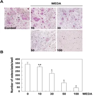
하였다. 녹용 추출물 10 μg/ml로 처리한 실험군에서는 억제 효
과가 없었지만 녹용 추출물 농도의 증가에 따라 파골세포의 수 가 유의하게 억제되었다(Fig. 1A). 또한 파골세포의 수가 농도 증 가에 따라 감소되는 것을 확인하였다(Fig. 1B). 이 결과로 녹용 추출물이 조골세포에 의해 촉진되는 파골세포의 분화를 억제한 다고 할 수 있다.
Fig. 1. Effect of water extract of deer antler (WEDA) on LPS-induced osteoclast differentiation.
(A) Bone marrow cells and osteoblasts were cocultured for 6 days with LPS (1 μg/ml) in the presence of increasing concentrations of WEDA (μg/ml). Cells were fixed in 3.7% formalin, permeabilized in 0.1% Triton X-100, and stained for TRAP. (B) TRAP-positive cells were counted as osteoclasts. Significant difference from control (no treatment) is marked by an asterisk. n.s., not significant.Fig. 2. Effect of WEDA on the expression of RANKL induced by LPS.
(A) Osteoblasts were pretreated with or without WEDA (100 μg/ml) for 1 hour and then treated with LPS (1 μg/ml) for the indicated time. Total RNA was isolated from the cells and the expression of RANKL and GAPDH mRNA was analyzed by RT-PCR. (B) Relative levels of RANKL mRNA was quantified using Image Pro-plus program version. 4.0 and normalized to GAPDH.2. 조골세포에서 파골세포 유도인자 발현에 미치는 녹용의 효과 비록 조골세포는 골 형성에 중요한 세포이지만, 생체 내에서 파골세포의 분화에 중요한 RANKL를 발현하여 파골세포의 형성 을 조절한다 1) . 녹용 추출물이 조골세포에 의한 파골세포의 분화 를 억제하였기 때문에 조골세포에 의한 RANKL발현에 녹용 추 출물의 효과를 검증하였다. 조골세포에 녹용 추출물을 처리하고 LPS를 처리하였다. LPS는 조골세포에서 RANKL의 발현을 촉진 하였고 녹용 추출물을 전처리한 실험군 에서도 LPS는 RANKL 의 발현을 촉진하였다(Fig. 2). 이 결과로 녹용 추출물은 조골세 포의 기능을 억제하지 않고 파골세포에 직접적으로 작용한다고 할 수 있다.
3. 파골세포의 골 흡수에 대한 녹용의 효과
다음으로 파골세포의 골 흡수에 녹용 추출물의 효과를 연구 하였다. 골수세포와 조골세포의 공배양으로 얻은 성숙 파골세포 를 hydroxyapatite plate에 첨가하고 녹용 추출물을 농도별로 처 리하였다. 대조군에서는 파골세포에 의해 hydroxyapatite가 흡수 되었지만 녹용 추출물을 처리한 실험군에서는 농도 증감에 따라 hydroxyapatite 흡수가 억제되는 것을 확인하였다(Fig. 3). 이 결 과는 녹용 추출물이 파골세포의 형성뿐만 아니라 골 흡수도 억 제한다고 할 수 있다.
Fig. 3. Effect of WEDA on osteoclastic bone resorption. (
A) Bone marrow cells and osteoblasts were co-cultured for 6 days in the presence of VitD3 and PGE2. Mature osteoclasts were seeded on hydroxyapatite-coated 48-well plates and then treated for 24 hours in the presence of the WEDA concentrations. (B) Pit area (indicated by the arrows) was quantified using Image Pro-plus program version. 4.0. *P<0.05, significant differences from no WEDA treatment.4. 생체 내 골 흡수에 미치는 녹용의 효과
녹용 추출물이 골수세포에서 파골세포로의 분화와 골 흡수
를 억제하였기 때문에 생쥐를 이용하여 생체 내 골 흡수에 녹용
추출물의 효과를 검증하였다. LPS는 강력한 염증 인자로 생체
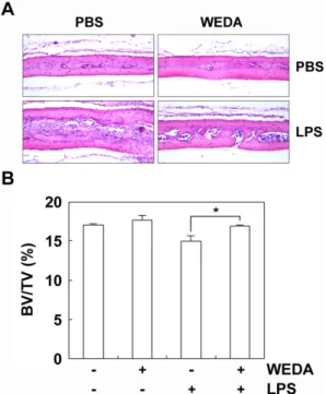
내 검증하였다. LP촉진하여 골 흡수를 유도한다. 따라서 LPS를 생쥐에 투여하고 녹용 추출물을 투여한 생쥐에서 골 흡수 였다젮도를 확인하였다. LPS는 골 흡수를 촉진하였지만 녹용 추 출물을 같이 투여한 생쥐에서는 골 흡수가 억제되는 것을 확인 할 수 있다(Fig. 4).
Fig. 4. Effect of WEDA on LPS-induced bone loss.
(A) 6-week-old ICR mice were injected subcutaneously at the calvaria with WEDA (1 mg/mouse) and/or LPS (30 μg/mouse). Mice were sacrificed 5 days. Calvarial bones were isolated, fixed in 4% paraformaldehyde for 1 day, decalcified with 12% EDTA, and embedded in paraffin. Sections were stained with H&E. (B) Bone volume/tissue volume (BV/TV) was analyzed using the histomorphometric results. Significant difference between the indicated groups are marked by an asterisk.Fig. 5. Effect of WEDA on osteoblast differentiation.
Osteoblasts were cultured for 7 days with ascorbic acid (50 μg/ml) and β-glycerophosphate (10 mM) in the presence of increasing concentrations of WEDA (μg/ml). Cells were fixed in 3.7% formalin and stained for alkaline phosphatase.5. 조골세포 분화에 대한 녹용의 효과
녹용 추출물이 생체 내에서 골 파괴를 억제하는 효과를 관 찰하였다. 골은 골의 흡수와 형성에 의해서 양질의 골이 유지되 는데 녹용 추출물이 조골세포의 분화에는 어떤 효과를 나타내는 지 확인하였다. 조골세포는 ascorbic acid와 β-glycerophosphate 를 첨가하고 녹용 추출물을 처리하였다. 7일 후 ALP 염색으로
조골세포의 분화를 관찰하였지만 녹용 추출물은 조골세포의 분 화에 효과가 없었다(Fig. 5). 이 결과로 녹용 추출물은 파골세포 의 분화와 기능에만 관여한다는 것을 알 수 있다.
고 찰
조골세포는 골 형성에 중요한 세포이지만 RANKL의 발현을 통해 파골세포의 형성과 기능에 깊이 관여하는 세포이다 3) . 본 연 구에서 녹용 추출물은 조골세포에 의해 유도되는 파골세포 분화 를 억제하였다(Fig. 1). LPS는 강력한 염증 유도 인자로 수용체인 Toll-like receptor 4와 결합하여 myeloid differentiation protein 88 (MyD88)을 활성화 시키고 여러 신호 전달물질의 활성화 유도 로 조골세포에서 RANKL의 발현을 촉진하여 파골세포의 분화를 유도한다고 보고되었다 8,24) . 그러나 LPS를 처리한 조골세포에서 발현되는 RANKL은 녹용 추출물에 의해 억제되지 않았다(Fig.
2). 저자는 녹용 추출물은 RANKL에 의해 유도되는 전사인자 c-Fos와 NFATc1의 발현을 억제하여 파골세포 분화를 억제한다 고 보고하였다 19) . 이들의 결과로 녹용 추출물은 파골세포의 분화 를 조절하는 조골세포에는 영향 없이 골수세포에서 파골세포로 의 분화에 직접적으로 관여 한다고 할 수 있다.
파골세포는 유일하게 골을 흡수하는 세포로 여러 신호전달 경로를 통해 골 흡수가 이루어진다. 골을 흡수하는 파골세포의 가장 큰 특징은 세포막이 주름지고 clear zone이 형성된다. 특히 파골세포가 골에 부착하기 위해서 clear zone에 integrin을 발현 하는데 αvβ3 integrin이 골 표면에 RGD (arginine-glycine- aspartic acid) sequence를 인지하여 골에 부착된다. 골에 부착된 파골세포는 골의 흡착 부위에 H+, cathepsin K, TRAP등을 분비 하여 골을 흡수한다 2) . 최근 calcitonin이 파골세포의 액틴 구조를 파괴하여 골 흡수를 억제한다고 보고되었다 25) . 이들의 결과로 골 에 파골세포의 부착이 중요하다 할 수 있다. 본 연구에서 녹용 추출물은 분화와 골 흡수를 억제한다는 것을 확인하였다(Fig. 1, 3). 비록 파골세포의 부착과 H+, cathepsin K, TRAP의 분비에 대한 녹용 추출물의 효과를 검증하지 못하였지만 녹용 추출물은 골수세포에서 파골세포의 분화를 억제할 뿐만 아니라 골을 흡수 하는 성숙 파골세포의 기능도 억제한다고 할 수 있다.
골 항상성은 조골세포와 파골세포의 활성 조절에 따라 유지
되지만 조골세포와 파골세포는 수많은 사이토카인과 호르몬에
의해 조절된다. 생체 내 IL-1, TNF-α등의 증가는 파골세포의 분
화를 촉진하여 골다공증을 유도 하지만 interferon-γ와 에스트로
젠 등은 파골세포의 분화를 억제한다 11) . 비록 녹용 추출물은 조
골세포에서 발현되는 RANKL에 의한 파골세포 분화에 억제 효
과가 나타났지만 생체 내 골의 손실에 대한 녹용 추출물의 효과
는 알 수 없었다. 따라서 저자는 LPS를 투여한 생쥐에서 녹용
추출물의 효과를 검증하였다. 녹용 추출물은 LPS에 의해 유도되
는 골 손실을 억제하였다(Fig. 4). 골 손실의 억제는 파골세포의
형성 및 활성의 증가로 발생되지만 최근 조골세포의 활성을 유
도하여 골 손실을 억제하는 연구가 보고되고 있다 26) . 따라서 저
자는 조골세포의 분화에 대한 녹용 추출물의 효과를 연구하였지
만 조골세포의 분화에 녹용 추출물은 효과가 없었다(Fig. 5). 이 들의 결과로 녹용 추출물은 조골세포의 골 형성에는 효과 없이 파골세포의 골 흡수를 억제하여 생체 내 골 손실을 억제한다고 할 수 있다.
결 론
본 연구에서 생체 내 골 손실에 미치는 녹용의 효과를 검증 하고자 하였다.
먼저 조골세포와 골수세포를 공배양하고 LPS를 처리하여 파골세포의 분화에 녹용 추출물의 효과를 확인하였다. 녹용 추 출물은 LPS를 투여한 파골세포의 분화를 억제하였다. 그러나 LPS에 의한 RANKL 발현 억제에는 효과가 없었다. 이 결과는 녹용 추출물이 파골세포의 분화에 직접적으로 관여한다고 할 수 있다. 또한 녹용 추출물은 파골세포의 골 흡수를 억제하였다.
LPS를 생쥐에 투여했을 때 골 손실이 나타났지만 녹용 추출물 을 같이 투여한 생쥐에서는 골 손실이 억제되었으며 조골세포의 분화에는 효과가 없었다.
결론으로 녹용 추출물은 조골세포의 기능에는 관여하지 않 고 파골세포에 직접적으로 관여하여 골다공증과 같은 질환에 녹 용이 치료제로 이용될 수 있으리라 기대한다.
감사의글
이 논문은 2007년도 원광대학교 교비 지원에 의해 수행되었 습니다.
참고문헌