Lymphotoxin β Receptor Stimulation Is Linked to MLCK Activity and Suppresses Stress Fiber Formation in Agonistic Anti-LTβR Antibody-stimulated Fibroblastic Reticular Cells
Min Hwan Kim3 and Jong-Hwan Lee1,2,3*
1Biomedical Engineering and Biotechnology Major, Division of Applied Bioengineering, College of Engineering, Dong-eui University, Busan 614-714, Korea
2Department of Biotechnology and Bioengineering, College of Engineering, Dong-eui University, Busan 614-714, Korea
3Department of Smart-Biohealth, College of Engineering, Dong-eui University, Busan 614-714, Korea Received July 23, 2017 /Revised October 7, 2017 /Accepted October 19, 2017
The lymphotoxin β receptor (LTβR), a member of the tumor necrosis factor receptor family, plays an important role in lymphoid tissue’s architecture and organogenesis. We found that LTβR stimulation induced changes in stress fibers (SFs) in fibroblastic reticular cells (FRCs). MLCK and ROCK play crit- ical roles in the regulation of SF formation in cells. The present study was performed to investigate the antifibrotic effects on SF regulation of LTβR signaling, with a focus on MLCK inhibition. The ef- fect of LTβR on the SF change was analyzed using immunoblot and fluorescence assays and agonistic anti-LTβR antibody-treated FRCs. In addition, we checked the level of Rho-guanosine diphosphate (GDP)/guanosine triphosphate (GTP) exchange activity with FRC lysate. Phospho-ezrin proteins acting as membrane-cytoskeleton linkers completely de-phosphorylated in agonistic anti-LTβR antibody-treated FRCs. The actin bundles rearranged into SFs, where phospho-myosin light chain (p-MLC) co-localized in FRCs. ML7-treated FRCs completely blocked SFs and showed retraction and shrinkage processes comparable to those observed in agonistic anti-LTβR antibody-treated cells. Inhibition of ROCK activ- ity induced changes in the actin cytoskeleton organization; however, some SFs remained in the cells, while they were completely disrupted by MLCK inhibition with ML7. We showed that the phosphor- ylation of MLC was completely abolished with LTβR stimulation in FRCs. When LTβR was stimu- lated with the agonistic anti-LTβR antibody, the Rho-GDP/GTP exchange activity was reduced, how- ever, the activity was not completely abolished. Collectively, the results illustrated that MLCK was potently responsible for the SF regulation triggered via LTβR signaling in FRCs.
Key words : FRC (fibroblastic reticular cell), MLCK, LTβR (lymphotoxin β receptor), ROCK, SF (stress fiber)
*Corresponding author
*Tel : +82-51-890-2280, Fax : +82-505-182-6897
*E-mail : [email protected]
This is an Open-Access article distributed under the terms of the Creative Commons Attribution Non-Commercial License (http://creativecommons.org/licenses/by-nc/3.0) which permits unrestricted non-commercial use, distribution, and reproduction in any medium, provided the original work is properly cited.
Journal of Life Science 2017 Vol. 27. No. 10. 1199~1206 DOI : https://doi.org/10.5352/JLS.2017.27.10.1199
Introduction
One of the critical hallmarks of immune responses is the rapid and extensive expansion of lymph nodes (LNs).
During this process, the 3 dimensional internal architecture of LN is maintained revealing the existence of mechanisms able to balance LN integrity with structural flexibility.
Fibroblastic reticular cells (FRC) are LN-resident mesen- chymal cells that secrete and remodel extracellular matrix to construct a complicated reticular mesh that filter draining
lymph [21]. LNs are meeting spaces for T lymphocytes and antigen presenting cells (APC) such as DC and macrophage.
FRCs represents various soluble factors and membrane bounded molecules to interact with microenviroment im- mune cells. For instance, FRC produces IL-7 in the T cell region and exist in close contact with T cells and DCs [6, 19]. FRC also expresses the homeostatic chemokines CCL19/
CCL21 [4] and ICAM-1, which together act as the guiding cue and driving force for the intranodal migration of lym- phocytes and DCs [7]. The interaction between tumor ne- crosis factor (TNF) and TNF receptor super family (TNFRSF) plays important roles in cell differentiation, survival, and death, which further orchestrates lymphoid organogenesis, activation, and homeostasis of immune cells [3, 13, 24].
Lymphotoxin-β receptor (LTβR) is one of the TNFRSFs [16].
LTβR ligands (LTα1β2 and LIGHT) displaying on the sur- face of immune cells such as DC and T cells were critical
mediators between FRC and leucocytes [16].
Mammalian cells can generate traction forces against solid extra cellular supports by assembling contractile stress fibers (SFs), which are bundles of filamentous actin (F-actin), ac- tin-binding proteins, and nonmuscle myosin II (NMMII) [18]. These traction forces are crucial to a variety of funda- mental cellular properties and behaviors, including motility, mechanosensing, shape stability, polarity, and fate determi- nation [25]. There are two kinases playing a critical role on SF formation in mammalian cells; Rho GTPase kinase (ROCK) and myosin light-chain kinse (MLCK) [2, 5, 17].
Inhibition of actomyosin contractility using inhibitors of the ROCK and myosin light-chain kinase (MLCK) pathway sup- presses SF formation in the central and peripheral regions, respectively [10]. However, there are no reports which one is a more important function on SF alteration in FRC. So far, many immunologic parameters have investigated in im- mune interaction, but little attention has focused on the SF alteration aspects of FRC triggered via LTβR. To understand these tasks, FRC was stimulated via LTβR with agonistic anti- LTβR antibody [6, 12]. Here, in order to investigate SF regulation between ROCK and MLCK in FRCs when FRC treated with agonistic anti-LTβR antibody, the cytoskeleton alteration, with particular emphasis on SF, studied in FRCs.
Materials and Methods
Cell culture
FRC was established as described previously [12]. FRC was maintained in 10% fetal calf serum–dulbeco's modified eagle's medium (DMEM) supplemented with streptomycin and penicillin.
Reagent and antibodies
The following antibodies or fluorescent probes were used in this study: Inhibitor Y27632 (Sigma-Aldrich, St. Louis, MO), rhodamine phalloidin (Cytoskeleton, Denver, CO), SuperSignal West Pico Chemiluminescent Substrate for de- tecting signal in Western blot (Thermo Scientific, Waltham, MA), pMLC (phospho Thr18/Ser19) primary antibody (Cell Signaling, Danvers, MA), anti-myosin light chains (anti- MLCs) antibody (Cell Signaling, Danvers, MA), anti-ezrin (Upstate Biotechnology, Lake Placid, NY), anti–p-ERM (Cell Signaling, Danvers, MA), anti-rabbit and anti-goat per- oxidase-conjugated secondary antibody for Western blot (Cell Signaling, Danvers, MA). RhoA activation assay kit
was from Cytoskeleton, Inc. (Denver, CO). Polyclonal goat antibody against LTβR extracellular domain and control IgG for cell stimulation purchased from R&D Systems.
Immunoblot
FRC was pretreated with agonistic anti-LTβR antibody (100 ng/ml) and were lysed in a 5× sodium dodecyl sulfate (SDS) sample buffer. After the samples were boiled, equal amounts of total lysates were separated by SDS-PAGE and transferred onto polyvinylidene difluoride membranes. The membranes were soaked in a blocking solution (5% skim milk and 0.2% Tween 20-PBS) for 1 hr, and then incubated with primary antibodies for 1 hr. After being washed with Tween 20-PBS, membranes were incubated with appropriate horse radish peroxidase (HRP)-conjugated secondary anti- bodies for 1 hr. Specific bands were visualized by an en- hanced chemiluminescence (ECL) method (ECL+; Amersham Biosciences, Piscataway, NJ, USA).
Immunofluorescence microscopy
FRCs were treated with agonistic anti-LTβR antibody (100 ng/ml), as described above, were gently rinsed, fixed in 4%
formaldehyde in PBS for 5 min, washed with PBS, and stained with rhodamine-labeled phalloidin (5 μg/ml) for 45 min. For ROCK and MLCK inhibition, cells were pretreated with 5 μM Y-27632 or 10 μM ML-7 (Sigma, St. Louis, MO), respectively, for 1 hr before imaging. For immunofluor- escence staining, cells were fixed with 3% PFA in PBS and permeabilized with 0.2% Triton X-100/PBS or 0.5% sap- onin/PBS. To detect the intracellular localization of p-ezrin we performed a TCA fixation method, which inactivates phosphatases and maintains the level of p-ERM proteins during sample processing [11], with a slight modification.
In brief, cells were fixed in suspension with 10% TCA sol- ution for 15 min at room temperature, and were rinsed three times with PBS. After being blocked with 5% BSA-PBS, cells were stained with antibodies for 30~60 min. The nucleus sained with 0.1 μg/ml DAPI. Stained cells were mounted with Permafluor aqueous mounting medium (Immunotech, Monrovia, CA) and examined with a Zeiss photomicroscope equipped for fluorescence microscopy. Digital images proc- essed with Adobe Photoshop software.
Rho A pull-down assay
More than 3×10⁷ cells were lysed with a 2-ml RIPA buffer.
The amount of Rho-GTP in the reaction solution measured
A B
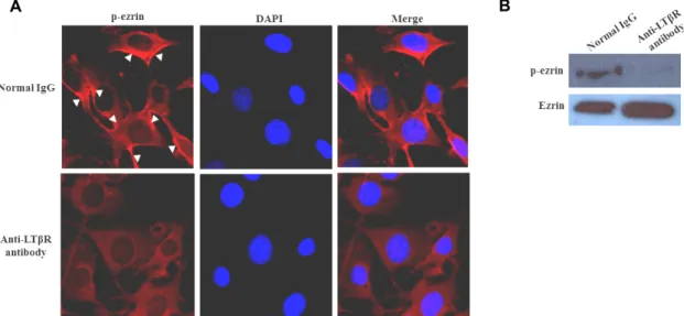
Fig. 1. Effects of LTβR signaling on p-ezrin distribution and expression in FRCs. (A) FRCs were treated with 100 ng/ml anti-LTβR antibody for 24 hr and were exhibited with a marked signal intensity against p-ezrin at peripheral FRC membrane. (B) FRC (5×10⁶ cells) was grown on 10 mm dish plate. FRC was incubated with anti-LTβR antibody for 24 hr. After incubation, cell was washed with PBS. Cell was lyzed with RIPA buffer and protein concentration of FRC lysate was measured by BCA method. The expression degree of p-ezrin and ezrin was detected by Western blot.
by a pull-down method based on the specific binding to Rhotekin-RBD followed by Western blotting using an an- ti-Rho antibody (Rho-activation assay biochem kit; BK306;
Cytoskeleton, Denver, CO, USA). The relative amount of ac- tive Rho compared with that in the control calculated by measuring the band density of Rho and normalized total RhoA density.
Results and Discussion
The localization of p-ezrin is regulated by LTβR signalling in FRCs
We examined localization and phosphorylation of ezrin proteins in FRCs. To address the details of p-ezrin local- ization in the FRC, we stained Thr 567 p-ezrin with anti- p-ezrin antibody into FRCs. In this experiment, we used an agonistic anti LT-bR antibody which is a commercially avail- able polyclonal antibody against the extracellular part of LT βR to stimulate signal cascades [6, 12]. In normal IgG treated cells, p-ezrin was detectable at the plasma membrane (Fig.
1A, arrowhead). In contrast to IgG treated cells, p-ezrin sig- nal was disappear in agonistic anti-LTβR antibody treated cells (Fig. 1A). In accordance with this observation, agonistic anti-LTβR antibody completely abolished the phosphor- ylation of ezrin proteins (Fig. 1B). This suggests that LTβR associates with ezrin and LTβR signaling is related to ezrin dephosphorylation in FRC. Ezrin belongs to the family of
closely related proteins, ezrin, radixin and moesin (ERM), which tether the actin cytoskeleton to the plasma membrane [1]. CT domain of ezrin undergoes phosphorylation at Thr 567 site and ezrin protein functions as membrane–cytoske- leton linkers by binding to the membrane proteins at their NT domains and to F-actin at its CT domain [9, 11]. Thus, this result suggests that ezrin play a role as a cross-linker between membrane and cytoskeleton in FRC and ezrin func- tions appear to be under the control of these cellular processes.
Co-localization of actin filaments and pMLC in SF structure of FRC
Ezrin is targeting to cytoskeleton, in other words, SF, within the cells. SFs are composed of actin bundle held to- gether by α-actinin, fascin, espin, filamin, and myosin II bun- dle [12]. They generate contractile force and play a central role in cell structure, adhesion, motility, morphogenesis, and cell-to-cell interaction of many specialized eukaryotic cells [21]. Consistent with previous findings with FRCs, the local- ization of pMLC and actin filaments to observe SF structure examined in FRC. Fig. 2 illustrated the stress fiber dis- tribution in a control monolayer as viewed by fluorescence microscopy. SFs formed thick cables. SF connected to the nuclear periphery with the processes of these flat and spread FRCs (Fig. 2). Moreover, in normal FRC, pMLC and actin filaments showed a SF pattern, which was co-localized with
A B
Fig. 2. Agonistic anti-LTβR antibody disrupted the formation of SF in FRC. FRCs were treated with agonistic anti-LTβR antibody (100 ng/ml) for 24 hr and examined for changes in F-actin distribution (phalloidin staining) and p-MLC. (A) Agonistic anti-LTβ R antibody suppressed formation of actin and p-MLC SFs compared with that shown in normal IgG-treated control cells.
pMLC and actin co-localized in normal FRC. (B) FRC (5×10⁶ cells) was grown on 10 mm dish plate. FRC was incubated with anti-LTβR antibody for 24 hr. After incubation, cell was lyzed with RIPA buffer and protein concentration of FRC lysate was measured by BCA method. The expression degree of β-actin and GAPDH was detected by Western blot. Actin filaments abolished completely in agonistic anti-LTβR treated FRC. GAPDH was used as a loading control.
each other (Fig. 2). However, LTβR activation by agonistic anti-LTβR antibody induced the abrogation of SF connecting the processes of the FRC (Fig. 2). In addition, when FRCs were treated with an agonistic anti-LRβR antibody, actin was almost abolished (Fig. 2). In addition, pMLC scarcely detected in the presence of agonistic anti-LTβR antibody stimulation (Fig. 2). Furthermore, upon agonistic anti-LTβR antibody stimulation, less intense co-staining of pMLC and actin filament in a SF pattern observed (Fig. 2). These results indicate that actin and p-MLC bundles integrated in SF and LTβR signal regulates SF formation in FRC. In cultured mammalian cells, actomyosin SFs are perhaps the most sig- nificant and widely studied [2, 15, 23]. These structures, which are composed of antiparallel arrays of F-actin stabi- lized by actin-binding proteins and interleaved with NMMII, contribute to cytoskeletal prestress by anchoring into cell-ECM adhesions and permitting the cell to generate trac- tion against the extracellular matrix (ECM) [20]. It is becom- ing increasingly appreciated that the mechanical balance be- tween tensile prestress in the cellular cytoskeleton and the elastic resistance of the ECM can strongly regulate a wide variety of fundamental cellular properties, including shape, polarity, motility, fate decisions, and tissue structural integrity. Our results imply that FRC also contains the basic structure to communicate with intercellular counterparts.
Effects of a MLCK inhibitor, ML7, on SF disruption in FRCs.
To evaluate the involvement of the MLCK pathway on
FRC morphology alteration and SF formation in FRC, we treated a specific inhibitor of ML7 in FRCs. After stim- ulation, cells fixed and stained with rhodamine-conjugated phalloidin to visualize F-actin. The SF formation found in the control FRCs. In contrast, cellular alterations were read- ily apparent after 1 hr of treatment with ML7. With re- traction and shrink of processes similar to those observed in agonistic anti-LTβR antibody-treated cells, SFs in ML7-treated FRCs were blocked completely and were not seen (Fig. 3). Indeed, both of the central and peripheral SFs disrupted after addition of the MLCK inhibitors and simulta- neously morphological changed occurred in FRC (Fig. 3).
This result suggests that LTβR stimulation linked to MLCK inhibition for SF disruption in FRC. One potential ex- planation for the above data is that peripheral fibers may be wider than central fibers and thus make stronger con- tributions toward maintaining SF independently of their subcellular location. Intriguingly, these results are consistent with Katoh and colleagues’ [8] earlier studies showing that stimulation of MLCK activity in isolated SF preparations yielded more extensive contractions than stimulation of ROCK activity, and that ML7-mediated disruption of periph- eral SFs was accompanied by a loss of the cells’ spread mor- phology [20].
Role of MLCK in SF formation and reorientation We observed that SFs were less prevalent in agonistic an- ti-LTβR antibody treated FRC in Fig. 2. ROCK and MLCK regulate SF populations in several cells [2, 20]. We reasoned
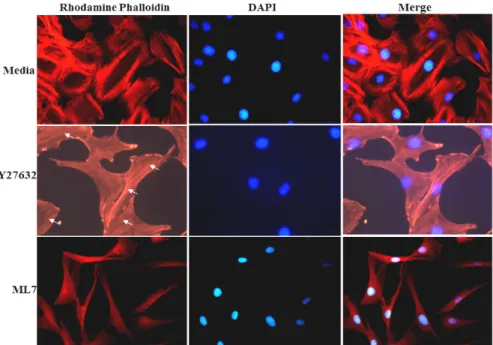
Fig. 3. LTβR was linked to myosin on SF formation in FRC. FRC on chamber slides were stimulated with 100 ng/ml agonistic anti-LTβR antibody and stained for actin SF. FRC were treated with 5 μM ML7 for 1 hr and exhibited a marked decrease in actin SF. However, similar morphology between ML7 treated cells and agonistic anti-LTβR antibody treated cells was observed.
Fig. 4. Roles of MLCK and ROCK on SF alignment in FRCs. Representative images of FRCs adhered on slide chamber subjected for 1 hr after treatment with 5 μM ML7 or 10 μM Y27632. A part of actin SF remained in Y27632 treated FRC (arrow), while actin SF completely abrogated in ML7 treated FRC.
that LTβR signal and MLCK or ROCK contribution to cell SF might be involved in as well. Thus, to assess the involve- ment of MLCK and ROCK pathways on FRCs, we treated the FRCs with inhibitors of either 5 μM MLCK (ML7) or 10 μM ROCK (Y27632) for 30 min and used immuno-
fluorescence to examine the effect on the spatial distribution of SFs. In cells treated with ML7, SFs were completely atte- nuated (Fig. 4). In contrast, there was some reduction in the number of SFs in cells treated with Y27632 (Fig. 4). We ob- served that ML7 treatment led to complete attenuation of
A B
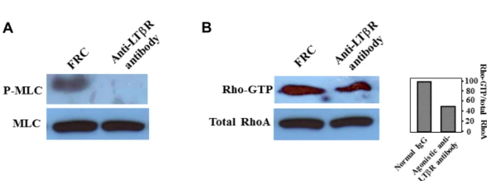
Fig. 5. Effect of LTβR signal on MLC phosphorylation and RhoA activation. (A) Normal IgG increased MLC phosphorylation (p-MLC) over the control, which was abolished by 100 ng/ml agonistic anti-LTβR antibody. (B) LTβR stimulation induces a significant decrease in active RhoA compared with control. MLC and total RhoA was used as a loading control.
SFs, while some actin bundles were observed in cells treated with Y27632 (Fig. 4). Our results indicate that agonistic an- ti-LTβR antibody-induced SF disruption is dependent on MLCK rather than ROCK. ROCK and MLCK regulates dif- ferent populations of SFs: peripheral SFs are sensitive to MLCK inhibition, while central SFs are sensitive to ROCK inhibition [10]. As described in previous report [20], SFs lo- cated at the cellular periphery and center are predominantly activated by MLCK and ROCK, respectively. Our ob- servations of the present study suggest that there is also dis- tinct subcellular localization of SF in FRC. Overall, these ef- fects are inconsistent with previous results obtained in fibro- blasts [20], implying that different regional SF control mech- anisms operate in FRCs, i.e., MLCK preferentially regulates both peripheral and central SF assembly in FRC.
Rho GTPase activity after agonistic anti-LTβR antibody
We examined the activation of the small GTPase RhoA in cultured FRCs treated with agonistic anti-LTβR antibody.
Agonistic anti-LTβR antibody caused about a half-fold de- crease in GTP-bound RhoA, however, RhoA-GTP levels re- mained not completely diminished from agonistic anti-LTβR antibody treated FRC (Fig. 5). However, anti-LTβR antibody completely abolished the phosphorylation of MLC (Fig. 5).
MLC itself is phosphorylated at multiple sites. Among them, T18 and S19 are the phosphorylation sites associated with an increase in myosin ATPase activity, the formation of actin filaments such as SFs. MLCK is the first kinase that was identified to phosphorylate T18 and S19 [14]. MLCK phos- phorylates MLC with preference for S19 over T18; therefore, the phosphorylation of S19 and T18 takes place in a sequen- tial manner [14]. Later, other kinases including Rho-kinase, Zipper-interacting kinase and integrin-linked kinase were al-
so identified to phosphorylate MLC with no preference be- tween T18 and S19 [14]. Thus, our results indicate that LTβR signal linked to closer MLCK pathway than ROCK signal pathway for SF alteration in FRC. However, whether MLCK and ROCK play any differential roles in SF regulation re- mains to be investigated in detail.
Acknowledgment
This research was supported by the Basic Science Research Program through the National Research Foundation of Korea (NRF) funded by the Ministry of Education, Science and Technology (Grant #: 2017R1D1A1B03028537).
References
1. Biri-Kovács, B., Kiss, B., Vadászi, H., Gógl, G., Pálfy, G., Török, G., Homolya, L., Bodor, A. and Nyitray, L. 2017.
Ezrin interacts with S100A4 via both its N- and C-terminal domains. PLoS One 12, e0177489.
2. Chang, C. W. and Kumar, S. 2015. Differential contributions of nonmuscle myosin II isoforms and functional domains to stress fiber mechanics. Sci. Rep. 5, 13736.
3. Chen, L. and Flies, D. B. 2013. Molecular mechanisms of T cell co-stimulation and co-inhibition. Nat. Rev. Immunol.
13, 227-242.
4. Comerford, I., Harata-Lee, Y., Bunting, M. D., Gregor, C., Kara, E. E. and McColl, S. R. 2013. A myriad of functions and complex regulation of the CCR7/CCL19/CCL21 che- mokine axis in the adaptive immune system. Cytokine Growth Factor Rev. 24, 269-283.
5. Connell, L. E. and Helfman, D. M. 2006. Myosin light chain kinase plays a role in the regulation of epithelial cell survival. J. Cell Sci. 119, 2269-2281.
6. Katakai, T., Hara, T., Sugai, M., Gonda, H. and Shimizu, A. 2004. Lymph node fibroblastic reticular cells construct the stromal reticulum via contact with lymphocytes. J. Exp.
Med. 200, 783-795.
7. Katakai, T. and Kinashi, T. 2016. Microenvironmental con- trol of high-speed interstitial T cell migration in the lymph node. Front Immunol. 7, 194.
8. Katoh, K., Kano, Y., Amano, M., Kaibuchi, K. and Fujiwara, K. 2001. Stress fiber organization regulated by MLCK and Rho-kinase in cultured human fibroblasts. Am. J. Physiol.
Cell Physiol. 280, 1669-1679.
9. Kovacs-Kasa, A., Gorshkov, B. A., Kim, K. M., Kumar, S., Black, S. M., Fulton, D. J., Dimitropoulou, C., Catravas, J.
D. and Verin, A. D. 2016. The protective role of MLCP-medi- ated ERM dephosphorylation in endotoxin-induced lung in- jury in vitro and in vivo. Sci. Rep. 6, 39018.
10. Lee, C. F., Haase, C., Deguchi, S. and Kaunas, R. 2010. Cyclic stretch-induced stress fiber dynamics - dependence on strain rate, Rho-kinase and MLCK. Biochem. Biophys. Res. Commun.
401, 344-349.
11. Lee, J. H., Katakai, T., Hara, T., Gonda, H., Sugai, M. and Shimizu, A. 2004. Roles of p-ERM and Rho-ROCK signaling in lymphocyte polarity and uropod formation. J. Cell Biol.
167, 327-337.
12. Lee, J. S., Kim, Y. H. and Lee, J. H. 2013. Involvement of RhoA/ROCK signaling for alteration of stress fiber via lym- photoxin β receptor stimulation in fibroblastic reticular cell isolated from lymph node. Anim. Cells Syst. 6, 421-428.
13. Locksley, R. M., Killeen, N. and Lenardo, M. J. 2001. The TNF and TNF receptor superfamilies: integrating mamma- lian biology. Cell 104, 487-501.
14. Loirand, G. and Pacaud, P. 2014. Involvement of Rho GTPases and their regulators in the pathogenesis of hyper- tension. Small GTPases 5, 1-10.
15. Lu, L., Oswald, S. J., Ngu, H. and Yin, F. C. 2008. Mechanical properties of actin stress fibers in living cells. Biophys. J. 95, 6060-6071.
16. Lu, T. T. and Browning, J. L. 2014. Role of the Lymphotoxin/
LIGHT system in the development and maintenance of reticular networks and vasculature in lymphoid tissues.
Front Immunol. 5, 47.
17. McKenzie, J. A. and Ridley, A. J. 2007. Roles of Rho/ROCK and MLCK in TNF-alpha-induced changes in endothelial morphology and permeability. J. Cell Physiol. 213, 221-228.
18. Oakes, P. W., Wagner, E., Brand, C. A., Probst, D., Linke, M., Schwarz, U. S., Glotzer, M. and Gardel, M. L. 2017.
Optogenetic control of RhoA reveals zyxin-mediated elas- ticity of stress fibres. Nat. Commun. 8, 15817.
19. Onder, L., Narang, P., Scandella, E., Chai, Q., Iolyeva, M., Hoorweg, K., Halin, C., Richie, E., Kaye, P., Westermann, J., Cupedo, T., Coles, M. and Ludewig, B. 2012. IL-7-produc- ing stromal cells are critical for lymph node remodeling.
Blood 120, 4675-4683.
20. Tanner, K., Boudreau, A., Bissell, M. J. and Kumar, S. 2010.
Dissecting regional variations in stress fiber mechanics in living cells with laser nanosurgery. Biophys. J. 99, 2775-2783.
21. Thompson, H. L., Smithey, M. J., Surh, C. D. and Nikolich- Žugich, J. 2017. Functional and homeostatic impact of age-related changes in lymph node stroma. Front Immunol.
8, 706.
22. Tojkander, S., Gateva, G., Husain, A., Krishnan, R. and Lappalainen, P. 2015. Generation of contractile actomyosin bundles depends on mechanosensitive actin filament assem- bly and disassembly. Elife 4, e06126.
23. Wu, Z., Plotnikov, S. V., Moalim, A. Y., Waterman, C. M.
and Liu, J. 2017. Two distinct actin networks mediate trac- tion oscillations to confer focal adhesion mechanosensing.
Biophys. J. 112, 780-794.
24. Zhao, L., Chen J., Liu, L., Gao, J., Guo B. and Zhu, B. 2015.
Essential role of TNF-alpha in development of spleen fibro- blastic reticular cells. Cell Immunol. 293, 130-136.
초록:FRC에서 agonistic anti-LTβR antibody의 LTβR 자극은 MLCK 연관성 및 stress fiber 형성에 대한 강력한 억제 작용
김민환3․이종환1,2,3*
(1동의대학교 바이오응용공학부 의생명공학전공, 2동의대학교 생명공학과, 3동의대학교 스마트바이오헬스학과)
종양괴사인자 수용체 일종인 Lymphotoxin β receptor (LTβR)은 림프 구조와 기관 형성에 중요한 역학을 한다.
우리는 fibroblastic reticular cell (FRC)에서 agonistic anti-LTβR antibody로 LTβR을 자극하면 stress fiber (SF)에 변화가 생긴다는 것을 알았다. MLCK와 ROCK는 세포에서 SF 형성 기여에 중요한 역할을 한다. 본 연구는 MLCK 저해에 초점을 맞추어 LTβR 신호 전달은 SF 조절로 항섬유화 효과에 대하여 조사하였다. SF 변화에 대한 LTβR 의 기능 조사를 위해 agonistic anti-LTβR antibody로 처리된 FRC와 세포 추출물을 이용하여 immunoblot, fluo- rescence assay와 Rho-guanosine diphosphate (GDP)/guanosine triphosphate (GTP) exchange 활성 분석법으로 분석하였다. 세포막과 세포골격 연결자 ezrin은 agonistic anti-LTβR antibody 처리된 FRC에서 완전히 탈인사화 가 유도되었다. Actomysoisn에 의한 SF를 확인하였고 인산화 myosin light chain (p-MLC)인 함께 co-localization 되는 것도 확인하였다. ML7 처리된 FRC에서 agonistic anti-LTβR antibody 처리된 세포에서 관찰되는 유사한 현 상인 SF분해, 세포막 응축과 쇠퇴 현상이 나타났다. ROCK 활성저해는 액틴 골격 변화는 유도하였으나 부분적으 로 SF가 세포에 남아 있었다. 반면, ML7에 의한 MLCK저해는 SF를 완전히 분해하였다. 또한, LTβR 자극은 MLC 인산화를 완전히 억제하였지만, Rho-GDP/GTP exchange 활성변화에서는 감소는 되었으나 활성이 완전히 없어지 지는 않았다. 결론적으로 이런 결과들은 FRC에서 LTβR신호전달을 통해 유도되는 SF 조절에는 MLCK가 보다 더 강력한 역할을 한다는 것을 제시하고 있다.