Regulation of Inflammatory Cytokine Production by Bee Venom in Rat Chondrocytes
Eun Jung Kim, Gye Yeop Kim*
Department of Physical Therapy, College of Health and Wellfare, Dongshin University
Bee venom acupuncture (BVA), as a kind of herbal acupuncture, involved injecting diluted bee venom into acupoints and is used for pain, osteoarthritis and rheumatoid arthritis patients. BVA is growing in popularity, especially in Korea, and is used primarily for pain relief in many kinds of diseases. However, the effect of bee venom anti-inflammatory related action in lipopolysaccharide (LPS) induced chondrocyte stress have not been reported yet.
The aim of this study was to investigate the effect of bee venom of cell viability and inflammatory cytokine in rat articular chondrocyte cultures stimulated with lipopolysaccharide. Inflammation was induced in rat chondrocytes by treatment with 10 ㎍/㎖ LPS. The change of cell viability were decreased in chondrocytes after treatment with lipopolysaccharide. The cell viability revealed that BV exerted no significant cytotoxicity in the rat chondrocyte. Bee venom inhibited decreased cell viability in the presence of lipopolysaccharide (10 ㎍/㎖) in a dose dependent manner(0.1, 0.5, 1.0 and 5.0 ㎍/㎖) at bee venom(p<0.05). Tumor necrosis factor (TNF)-α production in the presence of lipopolysaccharide(1 ㎍/㎖) was also inhibited in a dose dependent manner (p<0.05 from bee venom 0.1 ㎍/㎖).
Interleukin (IL)-6 production in the presence of lipopolysaccharide (10 ㎍/㎖) was inhibited as well (p<0.05 at bee venom 0.1, 0.5, 1.0 and 5.0 ㎍/㎖, respectively). Our results demonstrate that bee venom was a anti-inflammatory agent of chondrocytes. Bee venom may exert its anti inflammatory effects through inhibition of TNF-α and IL-6 synthesis, and may then pain relief and reduce the articular destruction.
Key words : Bee venom, Chondrocyte, Inflammatory cytokine
* To whom correspondence should be addressed at : Gye Yeop Kim, Department of Physical Therapy, College of Health & Wellfare, Dongshin University, Naju, Jeonnam
․E-mail : [email protected], ․Tel : 061-330-3391
․Received : 2010/11/08 ․Revised : 2010/11/28 ․Accepted : 2010/12/14
Introduction
Osteoarthritis (OA) is a degenerative joint disease, considered to be one of the major public health problems
1). Its prevalence after age 65 is increasing in incidence as a chronic condition widespread, and it is likely to rise further with the obesity epidemic
2,3). Women are affected more than men
4), and The most common site in hands, knees, hips, and spine
4,5). Patients predominant symptoms are joint pain, stiffness and swelling in the early
6,7). Joint pain typically worsened with weight bearing and functional activity.
Osteoarthritis involved degeneration of articular cartilage as well as erosion of articular cartilage, variable degrees of synovitis, and osteophytes
4,5). The histological pathology reflects the result of joint inflammation and cartilage erosion.
Since the initial stages of OA involve increased cell
proliferation, proteinases, inflammation cytokines, and other inflammatory mediators by chondrocytes
8). The relationship between the increased levels of catabolic enzymes and inflammatory mediators such as prostaglandins, nitric oxide, interleukin-1β (IL-1β), and tumor necrosis factor α (TNF-α) in OA synovial fluids and joint tissue
8,9).
Current treatment options used to manage OA are fail to reverse the degenerative process of OA. Among the commonly used pharmacologic agents are nonsteroidal anti-inflammatory drugs (NSAIDs), corticosteroids, and hyaluronan prepa- rations
10). NSAIDs are widely used but their prolonged consumption is associated with serious adverse side effects such as gastrointestinal ulcerations
11). The need for effective treatment modalities with fewer side effects has prompted OA patients to consider complementary approaches to control pain as well as to improve function and quality of life.
Among the inflammatory mediators associated with inflammatory and degenerative joint diseases, tumor necrosis factor alpha (TNF-α) and interleukin 6 (IL-6) is well established as a key mediator in the progression of cartilage degeneration.
Also there were reported to be upregulated in cartilage of
experimental OA patients and in vivo model
9,12). High levels of TNF-α are detected in the synovial lining of rheumatic joints and in chondrocytes of osteoarthritic joints
13). TNF-α promotes further expression of cytokines by synovial cells and chondrocytes, thereby sustaining a renewal of local inflammatory mediators
14).
Bee venom (BV) has been used as treatment against wide variety of ailments including inflammatory diseases for centuries in some Asian countries including Korea
15,16). It is a complex mixture of substances that are known to induce allergic responses in humans. Nevertheless, BV has been used to treat arthritis, pain, and rheumatoid diseases
17,18). Bee venom relieve pain and inflammation in osteoarthritis patients or rheumatoid arthritis and stop its progression, but not yet studies have so far failed to prove convincingly that it anti-inflammatory related works in lipopolysaccharide (LPS) induced chondrocyte stress. So our research has focused on the chondrocyte as the cellular inflammation mediator of OA pathogenesis.
This study investigated the effect of bee venom in inflammatory cytokine such as TNF-α and IL-6, which are implicated in osteoarthritis by using rat primary cultured chondrocyte exposed to an LPS-induced stress.
Materials & Methods
1. Preparation of bee venom
A working solution of bee venom was prepared by diluting 1,000 ㎎ of dried bee venom solid (Daihan Pharm, Co.
Lted, Korea). Wherein 0.1 g of BV was added to 99.9 ㎖ of distilled water yielding a bee venom solution of 1 ㎎/㎖. The BV solution was further diluted to 0.1 ㎍/㎖ to 100 ㎍/㎖ for use in the experiments.
2. Chondrocyte isolation and culture
Cartilage of normal appearance was aseptically prepared from the femoral and tibial articular surfaces of knee joints of 300 g Sprague-Dawley rats. Rats were used after our college ethics committee approved that our protocol fulfilled the guide of animal experience established by the Korean academy of medical sciences. Briefly, EDTA-CMFT(calcium magnesium free tyrode)(0.1%), trypsin (0.15%) and collagenase(2 ㎎/㎖) were treated for total 3 hr. After washing and centrifugation, cells were resuspended in Ham's F12/Dulbecco's modified Eagle's medium with 10% fetal calf serum and antibiotics (penicillin 100 U + streptomycin 50 ㎍/㎖). Cells were then washed and seeded in culture flasks or 96-well plate at a density of 10
4cells/㎠. The cells were grown at 37℃ with 5% CO
2in air. The
cells from a single animal were obtained for each experiment.
The medium was changed every 1 days. The first passage cells were used in all experiments
19).
3. Cell viability
Cell viability was analyzed using a MTT assay.
Tetrazolium salts such as MTT are metabolic by mitochondroal dehydrogenases to form a blue formazen dye and are there fore useful for the measurement of cell viability. The cell were gently washed with Hanks' balanced salt solution(HBSS, Sigma Co., USA), and exposed to rat primary chondrocyte. After washing the cells, cultures medium containing 0.5 ㎎/㎖ of MTT was added to each well. The cells were incubated for 2 hr at 37℃, the supernatant was removed and the formed formazen crystals in viable cells were solubilized with 110 ㎕ of dimethl sulfoxide. A 100 ㎕ aliquot of each sample was then translated to 96 well plates and the absorbance of each well was measured at 550 ㎚ with ELISA reader (Bio-Rad instrument, USA). Data were expressed as a percentage of control measure in the absence of rat primary chondrocyte20).
4. ELISA assays for TNF-α and IL-6 measure
TNF-α and IL-6 levels in culture media were determined by the quantikine human immunoassay (R&D system Inc., USA). Which employ the quantitative sandwich enzyme linked immunoassay (ELISA) technique. The sensitivity limit of both assays was about 20 pg/㎖, the linear assay range was between 100 and 2,000 pg/㎖. Aliquots of 100 ㎕ from control or LPS-treated cultures were analysed according to the manufactures instruction.
5. Statistical analysis
Results were expressed as means ± standard deviation(S.D.) of least three separate experiments. The difference between two mean values was analyzed by student t-test. Values of p<0.05 were considered as significantly different.
Results
1. Effect of LPS in rat chondrocyte viability
Fig. 1 showed that cell viability was also assessed using
the MTT assay (Fig. 1). The effect of cell viability on LPS in
the rat chondrocyte was examined as variable LPS time and
dose. The dependence of the inhibitory effect of LPS on the
LPS concentration (ranging from 0 to 100 ㎍/㎖) was
examined. Fig. 1A shows that LPS at 0.5 ㎍/㎖ (24hr) did not
inhibit cell viability. However when added at levels over 1 ㎍/
㎖, LPS caused significant inhibition of cell viability (86.51 ± 12.84 % decrease vs. control; p<0.05). As the effect of LPS on cell viability may possibly be influenced by oxidative stress (Fig. 1A). Fig. 1B shows that cell viability occurred at a significantly reduced rate after a 2 hr incubation period with LPS 10 ㎍/㎖ (85.44 ± 9.57 % decrease vs. control; p<0.05).
This level of inhibition was maintained over 48 hr incubation period. The dependence of the inhibitory effect of LPS on the LPS time (ranging from 0 to 48 hr) was examined (Fig. 1B).
Thus, 24 hr incubation period and 10 ㎍/㎖ was used (Fig. 1).
0 20 40 60 80 100 120
0
C el l V ia b ili ty ( % o f co n tr o l)
Lipopolysaccharide(LPS) 24hr 5
1 3 10
0.5 50 100
*
***
***
***
(ug/ml)
**
***
1A
C el l V ia bi lit y (% o f c on tr ol )
0 20 40 60 80 100 120
**
***
*
***
Lipopolysaccharide(LPS) 10 ug/ml
0.5 (hr)
1B
0 1 2 5 12 24 48
*
Fig. 1. Dose-and time-dependent effects of LPS on cell viability in rat chondrocyte. (Values are means±S.D. of three independent experiments.
*P
< 0.05,
**P < 0.01,
***P < 0.001 compared to the control)
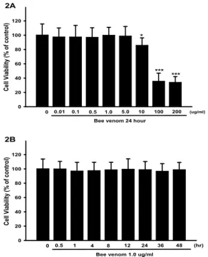
2. Effect of bee venom treatment in rat chondrocyte viability The effect of variable concentration and time bee venom on cell viability by the rat chondrocyte was examined as a function of dose and time. Fig. 2A shows that cell viability incubated with BV at 0 ㎍/㎖, 0.01 ㎍/㎖, 0.1 ㎍/㎖, 0.5 ㎍/㎖, 1.0 ㎍/㎖, 5.0 ㎍/㎖ for 24 hour were 97.34 ± 12.74 %, 97.26 ± 16.43 %, 96.86 ± 13.51 %, 99.74 ± 11.28 %, 98.58 ± 13.57 % of the control value, respectively. The cell viability revealed that BV exerted no significant cytotoxicity in the rat chondrocyte.
However when added at levels over 10 ㎍/㎖, BV caused significant inhibition of cell viability. The inhibitory effect of BV was 85.64 ± 10.58 %, 35.46 ± 11.47 %, 33.85 ± 8.32 % decreased at 10, 100 and 200 ㎍/㎖ (Fig. 2A). As the effect of BV on cell viability may possibly be significantly cytotoxicity influenced by high concentration. The cell viability revealed that 1.0 ㎍/㎖ BV related incubation time exerted no
significant inhibition effect in rat chondrocyte (Fig. 2B).
0 20 40 60 80 100 120
0 0.1
*
1.0
0.5 5.0
0.01
Bee venom 24 hour
100 200
C el l V ia b ili ty ( % o f co n tr o l)
10 (ug/ml)
*** ***
2A
0 20 40 60 80 100 120
0 0.5 1 4 8 12
Bee venom 1.0 ug/ml
36 48
C el l V ia b ili ty ( % o f co n tr o l)
24 (hr)
2B
Fig. 2. Dose-and time-dependent effects of BV on cell viability in rat chondrocyte. (Values are means±S.D. of three independent experiments.
*P <
0.05,
***P < 0.001 compared to the control)
3. Effect of LPS and bee venom treatment on cell viability in rat chondrocyte proliferation
The effect of bee venom and LPS on cell viability by the rat chondrocyte was examined as a function of dose. Fig. 3 shows that cell proliferation occurred at a significantly increased rate after a 24 hr incubation period with 0.1 and 0.5
㎍/㎖ BV. This level of inhibition was maintained over 48 hr incubation period. BV caused significant cell proliferation when added over 24 hr. Thus, a 24 hr incubation period was used (Fig. 3).
0 20 40 60 80 100 120
LPS 10 ug/ml -
- 0.1
Bee venom (ug/ml)
+ + +
0.5 -
4 hr
*
C el l V ia b ili ty ( % o f co n tr o l)
# #
# #
0.1
+ +
0.5 24 hr
0.1
+ +
0.5 48 hr
Fig. 3. Effect of LPS and BV treatment on cell viability in rat chondrocyte. (Values are means±S.D. of three independent experiments.
*P <
0.05 compared to the control.
#P < 0.05 compared to the LPS-treated group)
4. Effect of bee venom on TNF-α synthesis
This results showed that the TNF-α levels by BV in the rat chondrocyte. The level of TNF-α was markedly increased to 238.50 ± 18.32 pg/㎖ following treatment with 10 ㎍/㎖ LPS for 24 h, while decreased to 180.54 ± 13.51 pg/㎖, 124. 48 ± 11.28 pg/㎖, 90.35 ± 13.57 pg/㎖, and 82.34 ± 10.58 pg/㎖ in cells treated with BV at 0.01 ㎍/㎖, 0.1 ㎍/㎖, 0.5 ㎍/㎖, 1.0 ㎍ /㎖, 5.0 ㎍/㎖, respectively(Fig. 4).
0 50 100 150 200 250
TNF-αconcentration (pg/ml)
LPS 10 ug/ml -
- 0.1
*
#
Bee venom (ug/ml)
+ + + + + +
1.0
0.5 5.0
- 0.01
##
### ###
LPS and BV 24hr incuvation
Fig. 4. Effect of bee venom dose dependent on TNF-α production in rat chondrocyte. (Values are means±S.D. of three independent experiments.
*
P < 0.05 compared to the control.
#P < 0.05,
##P < 0.01,
###P < 0.001 compared to the LPS-treated group)
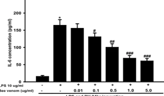
5. Effect of bee venom on IL-6 synthesis
This results showed that the IL-6 levels by BV in the rat chondrocyte. The level of TNF-α was markedly increased to 238.50 ± 18.32 pg/㎖ following treatment with 10 ㎍/㎖ LPS for 24 h, while decreased to 180.54 ± 13.51 pg/㎖, 124. 48 ± 11.28 pg/㎖, 90.35 ± 13.57 pg/㎖, and 82.34 ± 10.58 pg/㎖ in cells treated with BV at 0.01 ㎍/㎖, 0.1 ㎍/㎖, 0.5 ㎍/㎖, 1.0 ㎍ /㎖, 5.0 ㎍/㎖, respectively (Fig. 5).
0 50 100 150 200
IL-6 concentration (pg/ml)
LPS 10 ug/ml -
- 0.1
Bee venom (ug/ml)
+ + + + + +
1.0
0.5 5.0
- 0.01
LPS and BV 24hr incuvation
*
#
##
### ###