천식 모델 마우스에서 골쇄보의 항천식 효과
김승택1, 이장천2, 이영철1*
1:상지대학교 한의과대학 본초학교실, 2:부산대학교 한의학전문대학원
The therapeutic effect of Drynariae Rhizoma in a mouse model of allergic asthma
Seung-Taik Kim
1, Jang-Cheon Lee
2, Young-Cheol Lee
1,*1:Department of Herbology, College of Oriental Medicine, Sangji University, Wonju 220-702, Republic of Korea 2:Division of Pharmacology and Basic Korean Medicine, School of Korean Medicine, Pusan National University,
Pusan 609-735, South Korea
ABSTRACT
Objective:Allergic asthma is a chronic airway disease that affects millions of people in the developed world.
The disease is characterized by concurring airway inflammation, Th2 cytokine production, increased mucus secretion, airway hyperresponsiveness (AHR) to inhaled antigen, and pulmonary fibrosis. To investigate the therapeutic and anti-asthmatic effects of Drynariae Rhizoma (DR), we examined the influence of DR on the development of pulmonary eosinophilic inflammation and airway hyperresponsiveness in a mouse model of allergic asthma.
Methods:In this study, BALB/c mice were systemically sensitized to ovalbumin (OVA) followed intratracheally, intraperitoneally, and by aerosol allergen challenges. We investigated the effect of DR on airway hyperresponsiveness, pulmonary eosinophilic infiltration, various immune cell phenotypes, Th2 cytokine production and OVA specific IgE production in a mouse model of asthma.
Results:In asthmatic mice, we found that DR–treated groups had suppressed eosinophil infiltration, allergic airway inflammation and AHR by suppressing the production of IL-5, IL-13 and OVA specific IgE.
Conclusions:Our data suggest that the therapeutic mechanism by which DR effectively treats asthma is based on reductions of Th2 cytokines (IL-5), eotaxin, OVA-specific IgE production and eosinophil infiltration.
Key words:Drynariae Rhizoma, asthma, eosinophil, IL-5, CCR3, eotaxin.
1. Introduction
Allergic asthma is driven by inappropriate Th2 immune responses against otherwise innocuous environmental allergens. This results in the infiltration of inflammatory leukocytes into the lung tissue, airway hyperreactivity (AHR). Allergic asthma is a chronic infammatory disease of the airways characterized by airway eosinophilia and goblet cell hyperplasia with mucus hypersecretion to inhaled allergens and nonspecific stimuli
1,2). Eosinophilic inflammation is regulated to a major extent by activated
T lymphocytes in the airways that secrete the T helper-2 (Th2) cytokine interleukin (IL)-5. This cytokine is a central mediator in the regulation of eosinophilic inflammation through effects on the proliferation, differentiation, and activation of eosinophils
3). Eosinophilic inflammation has been considered to be the hallmark of airway inflammation in asthma
4).The inflammatory process in allergic asthma is dominated by Th2 cells, which produce IL-4, IL-5, and IL-13
5). In particular, IL-4, IL-5, and IL-13, which are produced by Th2 cells, are all related to inflammatory changes in the airway via the
*교신저자:이영철. 강원도 원주시 우산동 상지대학교 한의과대학 본초학교실.
․ Tel:033-730-0672. ․ Fax:033-730-0653. ․ E-mail:[email protected].
․ 접수:2011년 11월 9일 ․ 수정:2011년 11월 27일 ․ 채택:2011년 12월 16일
activation of eosinophils and the production of immuno-globulin E (IgE) by B cells
6). IgE is the effector antibody in atopic reactions and plays a central role in the pathogenesis of allergic asthma.
Moreover, it is well established that there is a strong interaction between eosinophils and Th2 cells in the asthmatic airways and that Th2 cell-derived cytokines, namely IL-4, IL-5, and IL-13, play critical roles in orchestrating and amplifying allergic inflammation in asthma
7).
Drynariae Rhizoma ( Drynaria fortunei (kunze) J.
Sm) is a fern plant widely used in South Korea.
Gol-Se-Bo in Korean extract(DR) is one of candidates known to be effective for the treatment of inflammation, hyperlipemia, arteriosclerosis, and gynecological diseases such as osteophoresis and bone resorption according to the ancient Chinese and Korean medicinal and herbal literature
8). Park et al.
also reported that DR was effective in the protection of streptomycin induced cytotoxicity on cultured mouse fibroblasts
9). Previous phytochemical studies reported triterpenes, flavonoids, and phenylpropanoids in this plant
10-12). However, we have not seen any reports on the anti-asthmatic and anti-inflammatory activity of DR has been reported in vivo.
The aim of this study was to evaluate the control activity of DR extract on Th1- and Th2-type cytokines, the development of allergic airway inflammation and airway hyperresponsiveness.
Therefore, we decided to investigate anti-inflammatory and anti-asthmatic effects of DR extract in a mouse model of allergic asthma.
2. Materials and methods
2.1. Plant material and preparation of extracts
The sample of DR were purchased from wonju Jaeil Korean Herbs Co. Ltd. (Wonju, Korea) in April, 2009 and identified by Professor Young-Cheol Lee, College of Oriental Medicine, Sangji University in Wonju, Korea, and a voucher specimens (DR) are deposited in our laboratory (Department of Herbology, College of Oriental Medicine, Sangji University Wonju 220-702, Republic of Korea). Plant material (200 g) was extracted three times with H
2O. Then, the extract was filtered and evaporated on a rotatory evaporator (Rotary evaporator, BUCHIB-480, Switzerland) and finally dried by a freeze drier (Freezedryer, EYELAFDU-540, Japan) to yield the extract(25.0g) of DR. The yield(w/w) of the extract was about 12.5%.
2.2. Animals
Five-week-old female BALB/c mice were obtained from Daehan Biolink Co. LTD. (Eumsung, Republic of Korea). Our study was approved by the committee for animal welfare at the institution Daejeon University.
Moreover, all animal procedures were conducted in accordance with the guidelines of the Institutional Animal Care and Use Committee of the Korea Research Institute of Bioscience and Biotechnology (Daejeon, Republic of Korea).
2.3. Ovalbumin (OVA) sensitization and inhalation
As per the modified protocol previously described
13), OVA (500 ㎍/㎖) in PBS was mixed with equal volumes of 10%(w/v) aluminum potassium sulfate(alum; Sigma, M.O., USA) in distilled water, then incubated for 60 min at RT after adjustment to pH 6.5 using 10 N NaOH, and centrifuged at 750×g for 5 minutes. The OVA/alum pellet was resuspened to the original volume in distilled water. All mice were immunized on three different days (first day of 2, 3, 4 weeks before inhalational exposure) by intraperitoneal (i.p.) injections of 0.2 ml alum-precipitated antigen containing 100 ㎍ of OVA (Sigma-Aldrich Korea, Korea) bound to 4 mg of aluminum hydroxide (Sigma-Aldrich Korea. Korea) in PBS. Seven days after the second sensitization intratracheally injected with 250㎍ of OVA (on day 21) on the back of the tongue, mice were exposed to aerosolized OVA for 30 min/day, 3 days/week for 5 weeks (at a flow rate of 250 L/min, 1% OVA in normal saline for first 4 weeks and 2% OVA in normal saline for last 1 week). DR (50 and 200 mg/kg) were orally administered 3 times a week for the last 5 weeks. One day after the last OVA exposures (2% OVA inhalation), airway hyperresponsiveness was determined and samples (bronchoalveolar lavage fluid, lung cells, and serum) were collected for further molecular analyses.
2.4. BALF (Bronchoalveolar lavage fluid)
Immediately following assessment of AHR, mice
were sacrificed with an i.p. injection of sodium
pentoparbitone (100 mg/kg). The trachea was
cannulated and BAL obtained by washing the airway
lumina. Briefly, cells in the lungs were recovered by
flushing 1 ml of BAL fluid (1 mM EDTA, 10% FBS,
PBS) into the lungs via the trachea. Total cell counts
were determined and 100 μl of fluid was cytospun
onto glass slides using a Cytospin centrifuge (Cellspin, Hanil, Korea) (400g for 4 minutes). Differential cell counts were performed after staining with a Diff-Quik StainSet(Baxter Healthcare Corp., Miami, Florida, USA).The supernatant of BALF was stored at -25℃
for determination of cytokine levels.
2.5. Digestion of pulmonary tissue and cells preparations
Single cell suspensions from lung tissues and BALF were isolated by mechanical disruption in RPMI 1640 medium supplemented with 2 mM L-glutamine, 100 U/ml penicillin, 100 µg/ml streptomycin, 50 µM 2-mercaptoethanol, 20 mM HEPES, and 2%
heat-inactivated fetal bovine serum (FBS, GIBCO, Grand Island, NY). Briefly, the lungs were removed from thoracic cavity. After mincing using sterile scalpels, the tissue was incubated in PBS containing 1 mg/ml Collgenase IV and 2 mg/ml Dispase for 40 min at 37°C in a sterile polypropylene tube. After incubation, lung tissue was vigorously pipetted up and down to further dissolve remaining tissue clumps and then filtered using a 70 mm cell-strainer (Falcon, Le Pont de Claix, France).
2.6. Determination airway hyperresponsiveness (AHR)
Airway hyperresponsiveness in mice was estimated using a previously described method with modifications
13). A Buxco system (Biosystem XA;
Buxco Electronics Inc, Troy, Conn) was used to evaluate the extent of airway constriction in different groups of mice following the protocol described previously.
Penh is equal to Pause × PEF/PIF, where Pause = (Te-Tr)/Tr (PIF, peak inspiratory flow; PEF: peak expiratory flow; Te, expiratory time;Tr, relaxation time). In this experiment mice were aerosolized with OVA for 30 min/day, 3 days/week for 5 weeks. At 24 hours after the final inhalation, mice were given aerosolized normal saline, followed by 3.125, 6.25, 12.5, 25, 50 mg/mL methacholine (Sigma-Aldrich, St.
Louis, MO) serially. Airway reactivity was then monitored for 30 minutes. Differences of Penh value between groups were evaluated using a unpaired Student’s t-test.
2.7. Hematoxylin-eosin (H&E), Masson- trichrome (M-T) and Periodic acid-Schiff (PAS) staining
BALB/c mice were injected, inhaled and sprayed with OVA for 5 weeks (three times a week) to induce asthma. Two experimental groups were treated with different concentrations of DR for the later 5 weeks (5 times/week). At the end of the experiment, the lungs were removed and analyzed histologically using a modified protocol previously described
13).
Briefly, the lung tissue was embedded in paraffin, then cut into 3 ㎛ thickness sections, stained with H&E solution or M-T solution. The tissue was subsequently mounted and cover-slipped with Dako-mounting medium (Dakocytomation; Denmark Carpinteria CA). The degree of airway inflammatory cell infiltration was scored in a double-blind screen by two independent. The degree of peribronchiole and perivascular inflammation was evaluated by a subjective scale of 0–2, as modified protocol described previously
13). Periodic acid-Schiff (PAS) stain was performed to identify mucus secretion in lung tissue.
Frozen sections (30 mm in thickness) of each tissue were made. The each sample section was mounted on the gelatin-coated slide, stained with PAS reagents, dehydrated and coverslipped with the permount. The PAS-positive goblet cells were counted manually and normalized to the length of the bronchial epithelial perimeter on the basal side, and expressed as the number of PAS-positive cells per mm of basement membrane.
2.8. Antibodies and flow cytometric analysis
All antibodies (such as anti-CD3, CCR3, CD11b, Gr-1 etc.) for flow cytometric analysis were purchased from Becton Dickinson (BD) PharMingen (SanDiego,CA). Cells from lung tissues and BALF were stained with the indicated antibodies in staining buffer (PBS containing 1% FBS and 0.01% NaN3)for 10 min on ice, and analyzed by two color flow cytometry on a FACSCalibur using Cell Quest software(BD Biosciences, MountainView, CA) for data expression.
2.9. Enzyme-Linked Immunosorbent Assay (ELISA)
Interleukin(IFN-γ, IL-4, IL-5, IL-13 etc.)
production in BALF and anti-OVA IgE in serum of
the indicated mice (n=5) was measured by ELISA
according to the manufacturer's instructions with a
monoclonal antibody-based mouse interleukin ELISA
kit (R&D system). OVA-specific IL-4 and IFN-γ
production were investigated from spleen cells
suspended in RPMI 1640 medium supplemented with 2
mM L-glutamine, and 5% fetal bovine serum. The
spleen cells were then cultured for 48 hrs at a concentration of 1 X 10
5cells/well in 96-well culture plates(Corning Inc, Cambridge, Mass) with or without 1 ug/ml of OVA in a humidified atmosphere of 5%
CO
2in air at 37℃. The culture supernatants were collected and assayed for IFN-γ and IL-4 antibodies induced by OVA using ELISA.
2.10. Statistical Analysis
Data were analyzed by one-way analysis of variance (ANOVA) or unpaired Student's t-test followed by Dunnett's multiple comparison test (SPSS version 14.0 statistic software). The difference between the normal group and the control group (OVA+vehicle) was clearly distinguished, and for this reason, statistical significance between the normal group and the control group was not shown in the figures and tables to put an emphasis on the statistical differences between the experimental groups and the control group. Results (presented as mean ± standard error of mean) were considered statistically significant if P values were <0.05 (*), <0.01 (**), or
<0.001 (***).
3. Results
3.1. Inhibitory effect of DR on airway hyperresponsiveness (AHR)
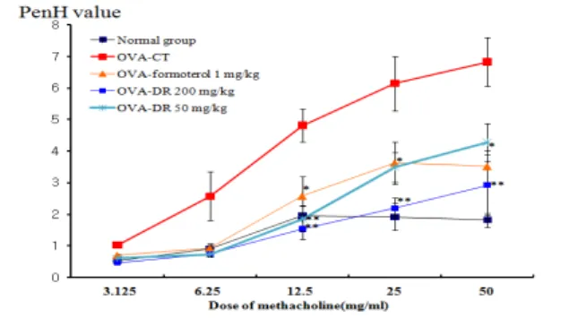
In a mouse model of allergic asthma, we compared the effects of DR, delivered by means of nebulization only or in association with oral administration. Both DR treatments were equally efficient at reducing AHR to methacholine, as measured on the basis of whole-body plethysmography (Fig. 1).
PenH was measured using a Buxco system on day 1 after final inhalation and samples (lung, BALF, serum, etc.) were immediately collected. Methacholine treatment is useful to exhibit the distinct effect of drugs on Penh value by way of inducing AHR. In OVA-sensitized and -challenged mice, the dose–
response curve of Penh value was shifted to the upper of the image compared with that of normal mice (Fig.
1). As shown in Figure 1, relative to animals sensitized with OVA (Control group), AHR to methachoine was reduced in DR (200, 50 mg/kg) treated mice (p<0.01, p<0.05).
Figure 1. Effect of DR on airway hyperresponsiveness in OVA-challenged mice.
Mice were sensitized and challenged by OVA as described in the Materials and methods. Airway responsiveness to aerosolized methacholine was measured with a Buxco box, as described in Materials and Methods. Mice were placed into the main chamber and were nebulized first with PBS, then with increasing doses (3.125 to 50 mg/ml) of methacholine for 3 min for each nebulization. Data represent the means ± SEM from five independent experiments. * P<0.05, ** P<0.01 versus control goup.
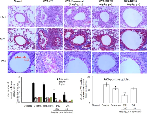
3.2. Histological analysis of lung sections
Next we studied the tissue inflammation in the lungs. As shown in Fig. 2, the inflammation degree of peribronchial and perivascular regions in mice with DR administration decreased significantly as compared with that in the OVA group. The histopathological investigation of both OVA-challenged mice and DR treated mice showed inflammatory changes when compared with saline challenged normal mice. Also, we found infiltration of leukocytes in histologic sections of lungs from OVA-challenged control mice, and lung tissue sections from OVA-challenged mice showed a distinct inflammatory infiltrate and erosion in peribronchial and perivascular areas. The peribronchial and perivascular inflammatory infiltrate consisted of eosinophils and mast cells, admixed with lymphocytes.
Eosinophil infiltration was mainly observed in the peribronchial regions of the lung. In contrast, histological sections from DR treated mice indicated reduced airway inflammation in lung tissue (Fig. 2).
To investigate the level of mucus expression in the airway, PAS-positive and PAS-negative epithelial cells in individual bronchioles were analyzed. The degrees of goblet cell hyperplasia and mucus hyperproduction were evaluated by means of PAS staining and quantification of PAS-stained cells. The OVA- challenged control mice significantly increased the mean numbers of PAS-positive cells when compared with saline challenged normal mice.
In particular, there were greater reduction in the
mean number of PAS-stained goblet cells in the
DR-treated (200, 50 mg/kg) asthma mice than
OVA-sensitized/challenged mice (Fig. 2).
Figure 2. Effect of DR on histologic examination of airway inflammation (H&E, M-T, and PAS staining) in lung tissue and BALF cytospin of the OVA-induced mouse model of allergic asthma.
At the end of the experiment,, lung was fixed and histologic analysis was performed. Lung sections of normal, asthmatic mice treated with vehicle (control), DR (200 mg/kg), and DR (50 mg/kg).
H&E:hematoxylin-eosin stain, M-T:Masson trichrome stain, PAS:Periodic acid-Schiff Stain, N:Normal BALB/c mice, CT(control):
Ovalbumin inhalation + vehicle, formoterol:OVA + formoterol (1 mg/kg), OVA + DR (200, 50 mg/kg)
3.3. Inhibitory effect of DR on airway eosinophil accumulation and influx of inflammatory cells into lung and BALF
The number of total lung cells obtained from the PBS saline challenged group was 0.95±0.05 x 10
7cells, indicating that few eosinophils were detected in this group. On the other hand, the number of total
lung cells(2.00±0.1 x 10
7) and eosinophils in the BALF cytospin of the OVA-challenged was significantly higher than that in the PBS saline challenged group.
The total number of lung cells were significantly reduced in DR-treated (50, 200 mg/kg) mice compared with control mice, and DR (200, 50 mg/kg) also decreased the absolute number of eosinophils in BALF (Fig. 3D).
Figure 3. Effects of DR on total lung cells, total BALF cells and eosinophils in BALF. As described in Materials and Methods, lung and BALF was harvested 24 hrs after the last OVA challenge. Total inflammatory cell numbers in lung and BALF were counted, and cell classification was performed on a minimum of 200 cells to classify lymphocytes. Results are expressed as mean±S.E (N=5). Statistical significance between control and treatment groups was assessed by ANOVA or unpaired Student's t-test followed by Dunnett's multiple comparison test (*p<0.05, **p<0.01, ***p<0.001). N:Normal BALB/c mice, CT:Ovalbumin inhalation+vehicle, formoterol:OVA + formoterol (1 mg/kg), OVA+DR (200, 50 mg/kg).
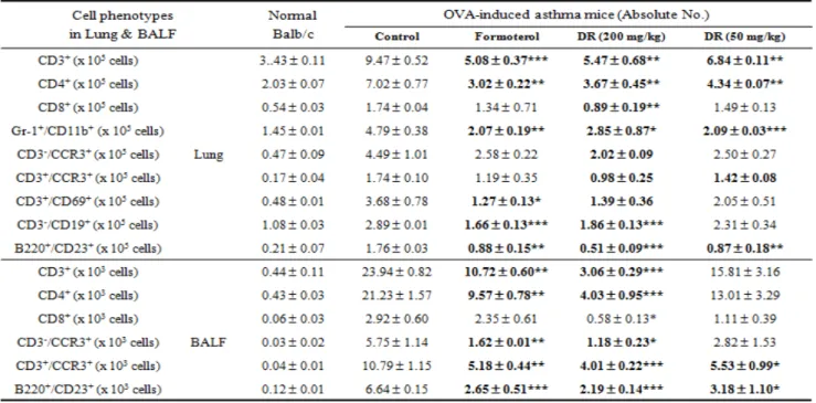
3.4. Inhibitory effect of DR on absolute number of immune cell subtypes in murine OVA-induced asthma lung and BALF
To evaluate effect of DR on immune cell subtypes flow cytometric analysis was accomplished. The numbers of CD3, CD4, CD8, CCR3, CD69, B220, CD23, CD11b, Gr-1 positive cells in the lungs of OVA-challenged mice were increased compared to the saline treated group, and generally, each values from DR-treated mice were significantly lower than those of OVA-challenged mice (Table 1). Formoterol administration resulted significant reduction in T cell subtypes similarly.
Effects of DR on leukocyte subsets in lungs and BALF were marked with change in numbers of CD3+
T cells, CD4+ helper T cells, Gr-1
+/CD11b
+granulocytes, CD3-/CCR3+ eosinophils, CD3+/CCR3+
Th2 cells, CD3 +/CD69+ early activated T cells, B220+/CD23+ B cells in a mouse model of asthma compared to control group, and the deficits in CD3-/CCR3+ eosinophils were accompanied by concurrent decreases eosinophils in BALF cytospin (Fig. 3d).
DR and formoterol groups treated with OVA resulted in significant reductions in CD3+ T cells (**P
< .01, ***P < .001), Gr-1
+/CD11b
+granulocytes(**P
< .01, ***P < .001), B220+/CD23+ B cells (*P < .05,
**P < .01, ***P < .001) in lung were decreased significantly and CD3-/CCR3+ (**P < .01), B220+/CD23+ B cells (***P < .001) in BALF were also decreased significantly (Table 1).
Table 1. Quantification by means of FACS analysis of various immune cell subtypes in lung and BALF.
Absolute numbers of various immune cell subtypes in lung were counted (described in Materials and Methods). Results are expressed as mean±S.E (N=5). Statistical significance between control and treatment groups was assessed by ANOVA (*p<0.05, **p<0.01, ***p<0.001).
N:Normal Balb/c mice, CT:Ovalbumin inhalation + vehicle, formoterol:OVA + formoterol (1 mg/kg), OVA+DR (200, 50 mg/kg).
3.5. Inhibition of Th2 cytokines (in vivo and in vitro) and OVA specific IgE production in BAL fluid and serum
To determine whether DR influenced cytokine and chemokine secretion in the BALF, the levels of IL-4, IL-5, and IL-13 in this fluid were measured using ELISA after the final challenge.
As shown in Figure 4A, 4B, IL-5, IL-13 and eotaxin levels were significantly reduced in DR-treated (200 mg/kg) mice. A important component of allergic asthma model is the production of OVA
specific IgE. Therefore, levels of anti-OVA IgE were measured in serum from the OVA-challenged mice, PBS, and DR treated groups. OVA specific IgE levels in serum from OVA-induced asthmatic mice were significantly increased compared with normal mice (PBS only), and DR-treated mice had significantly reduced OVA specific IgE (Fig. 4C). We also measured IL-4 and IFN-γ in the culture supernatants were measured by ELISA and found that DR (200, 50 mg/kg) inhibited Th2 cytokine (IL-4) production and increased Th1 cytokine (IFN-γ) in splenocytes (Fig.
4D).
Figure 4. Effect of DR on Th2 cytokines (IL-5, IL-13), eotaxin in BALF and OVA specific IgE in serum, and immunomodulatory effects of DR on OVA-specific Th1/Th2 cytokines production in spleen cells (described in Materials and Methods). Results are expressed the mean±S.E (N=5). Statistical significance between control and treatment groups was assessed by ANOVA or unpaired Student's t-test followed by Dunnett's multiple comparison test (*p<0.05, **p<0.01, ***p<0.001). N:Normal BALB/c mice, CT:Ovalbumin inhalation+vehicle, formoterol:OVA + formoterol (1 mg/kg), OVA + DR (50, 200 mg/kg).