Copyright ⓒ Korean Academy of Dental Technology
This is an Open Access article distributed under the terms of the Creative Commons Attribution Non-Commercial License (http://creativecommons.org/licenses/by-nc/4.0) which
INTRODUCTION
1990년대부터 가철성 보철물 제작을 위한 다양한 치과용 디지 털 장비 기술이 발전되어 임상에 도입되었다. 따라서 과거에 사용된 자가중합, 열중합 방식으로 제작하던 가철성 보철물을 CAD/CAM (computer-aided design/computer-aided manufacturing)을 이 용하여 제작하게 되었다[1]. 이러한 CAD/CAM의 제조 공정은 절삭 가 공(subtractive manufacturing)과 적층 가공(additive manufactur-
ing)으로 나눌 수 있다[2]. 절삭 가공 방식은 제조사가 고압에서 사전에 중합하여 공급한 PMMA 블록을 기계적으로 절삭(milling)하여 제작하 는 방식이다[3,4]. 적층 가공 방식은 흔히 3차원(three-dimensional, 3D) 프린팅 혹은 rapid prototyping으로 불리며 재료를 연속적으로 쌓아 올려 3D로 제작하는 방식으로 절삭 가공방식으로 제작하기 어려 운 복잡한 형태도 제작 가능하며 동시에 여러 개의 형상을 만들 수 있 다는 장점이 있다[5,6].
치과 분야에서는 다양한 방식의 3D 프린터(fused deposition
Purpose:
In this study, we added two kinds of photoinitiators (CQ and TPO) to prepare two kinds of denture base resins (Bis-GMA series and UDMA series) for three-dimensional (3D) printing to compare and analyze their mechanical and biological properties and to find the optimal composition.Methods:
Control specimens were made using the mold made of polyvinyl siloxane of the same size. Light curing was performed twice for 20 seconds on both the upper and lower sur- faces with LED (light emitting diode) light-curing unit (n=10). Experimental 3D printing dental resins were prepared, to which two photoinitiators were added. Digital light processing type 3D printer (EMBER, Autodesk, CA, USA) was used for 3D printing. The specimen size was 64 mm×10 mm×3.3 mm according to ISO 20795-1. The final specimens were tested for flexural strength and flexural modulus, and MTT test was performed. Furthermore, one-way analysis of variance was performed, and the post-test was analyzed by Duncan’s test at α=0.05.Results:
The flexural strength of both Bis-GMA+CQ (97.12±6.47 MPa) and UDMA+TPO (97.40±3.75 MPa) was significantly higher (p<0.05) in the experimental group. The flexural modulus in the experimental group of UDMA+TPO (2.56±0.06 GPa) was the highest (p<0.05). MTT test revealed that all the experimental groups showed more than 70% cell activity.
Conclusion:
The composition of UDMA+TPO showed excellent results in flexural strength, flexural modulus, and biocompatibility.Key Words:
Biocompatibility, Mechanical properties, Photoinitiator, Three-dimensional print- er, Three-dimensional printing dental resinArticle Info
Received October 30, 2020 Revised December 17, 2020 Accepted December 22, 2020
Corresponding Author Ju lee Son
Department of Dental Hygiene, VISION Col- lege of Jeonju, 235 Cheonjam-ro, Jeonju 55069, Korea
E-mail: [email protected] https://orcid.org/0000-0003-4611-8891
https://doi.org/10.14347/jtd.2020.42.4.355 pISSN: 1229-3954 eISSN: 2288-5218
J Tech Dent 2020;42(4):355-361 Original Article
광개시제에 따른 실험용 3D 프린팅 의치상 레진의 기계적 성질과 생체적합성 비교
박다령1, 손주리2
1
원광보건대학교 치기공과,
2전주비전대학교 치위생과
Comparison between mechanical properties and biocompatibility of experimental 3D printing denture resins according to photoinitiators
Da Ryeong Park
1, Ju lee Son
21Department of Dental Laboratory Technology, Wonkwang Health Science University, Iksan, Korea
2Department of Dental Hygiene, VISION College of Jeonju, Jeonju, Korea
JOURNAL OF TECHNOLOGIC DENTISTRY
J TD
JOURNAL OF TECHNOLOGIC DENTISTRY
J TD
modeling, FDM; polyjet photopolymer, PPP; stereolithography apparatus, SLA; digital light processing, DLP)가 사용되고 있는데 그중에서 DLP, SLA 등의 방식이 보철물 제작에 사용되고 있다. DLP 방식은 챔버에 광중합 액체 레진을 담아 아래에서 빛을 조사해 레진을 연속적으로 적층하여 제작한다. 따라서 환자에 직접적으로 적용하는 치과분야에서는 높은 정밀도를 가진 경화성 레진 소재를 이용한 SLA방 식과 높은 해상도와 빠른 조형 속도, 낮은 비용 등의 장점을 가진 DLP 방식이 주로 활용되고 있다. 복셀(voxel) 단위로 경화하기 때문에 제작 속도를 비교하면 적층 단면 단위로 동시 경화시키는 DLP방식이 치과 분야에서 더 선호되고 있다[7-9]. 현재 CAD/CAM 기술의 발전과 의치 상의 수요에 따라 3D 프린팅을 이용한 의치상과 의치상용 인공치의 제 작이 가능해지고 있다[10]. Methacrylate를 기반으로 하고 인공치까 지 출력이 가능한 Dentca denture, 색상과 우수한 강도를 가지고 있 는 가장 많이 사용되고 있는 NextDent, 국내 최초로 개발된 3D 프린 팅 의치상 레진인 DIOnavi-Denture 등 3D 프린팅 의치상용 레진 재 료에 대한 개발이 국내외에서 활발히 진행 중이다[11-13].
현재 치과 분야에서 3D 프린터를 이용한 형상 가공에 주로 이용되는 재료는 레진이며 레진을 제작하는 데 있어 광중합 방법을 사용되고 있 다. 일반적으로 3D 프린터로 출력된 레진의 경우 기존의 레진과 비교 시 기계적 물성이 낮은 것으로 나타났다. 기계적 물성을 개선하기 위한 방법으로는 광개시제나 광증감제를 이용하여 중합률을 높이거나 자외 선(ultraviolet, UV)나 LED를 사용하여 추가적인 중합을 통해 레진 내 부의 단량체(monomer) 중합을 유도하는 등 여러 가지 방법이 제시되 고 있다. 임상에서 사용하기 위해서는 최상의 기계적 성질을 가지며, 생체적합성이 우수하고, 수복물의 파절을 방지할 수 있는 중합과정을 가장 중요시 여겨야 한다[14-18].
SLA 또는 DLP 출력공정에 사용되는 광 중합체는 단량체, 올리고머 (oligomer) 및 광개시제의 화합물로서 고품질의 입체적인 3D 대상물 을 출력할 수 있다. Bisphenol A glycidyl methacrylate (Bis-GMA), urethane dimethacrylate (UDMA)와 triethylene glycol dimeth- acrylate (TEGDMA)는 치과용 복합재에서 가장 일반적으로 사용되는 단량체 중 일부이다[19,20]. 광중합 레진은 광원에 따라 반응하며 레진 의 원료로 사용되는 단량체, 올리고머와 다양한 첨가제들이 연속적으
로 반응하여 경화된다[21]. 치과용 수복용 레진의 가장 효과적이고 안 전한 광개시제로는 camphorquinone (CQ)이 있다[22]. 하지만 낮은 중합속도 및 전환 효율과 가속화하는 3차 아민의 존재가 필요하다. 이 에 대체될 수 있는 Diphenyl(2,4,6-trimethylbenzoyl) phosphine Oxide (TPO)는 여러 상업용 복합재에 포함되어 있으며 3D 프린팅 레 진에도 사용되고 있다[23-25]. 임상적용에 있어서 우수한 기계적 성 질, 생체적합성을 갖추어야 하며 수복물의 파절을 방지할 수 있는 중합 과정이 중요하다. 따라서 본 연구에서는 기반이 다른 실험용 3D 프린 팅 치과용 레진에 두 가지의 광개시제를 첨가하여 3D 프린터로 출력한 결과물의 기계적, 생물학적 성질을 비교 분석하고자 하였다.
MATERIALS AND METHODS
1. 레진 시편 제작
유동성 의치상용 레진(PRESS LT, Retec; Baesweiler, Germany) 을 대조군으로 선택하였다. 임상에서 사용되고 있는 3D 프린팅 용 의치상 레진인 NextDent (Base, Vertex Dental; Soesterberg, Netherlands)도 비교 분석하고자 하였다. 실험군은 단량체인 Bis- GMA (SHIN-NAKAMURA, Wakayama, Japan)와 UDMA (Sigma- Aldrich, St. Louis, MO, USA)를 기반으로 Bis-GMA계와 UDMA계 의 레진을 제작하였다. Bis-GMA를 제외한 다른 모든 재료는 Sigma- Aldrich 제품을 사용하였다. Table 1에 본 실험에 제작된 레진의 조성 과 함량을 나타내었다. 광개시제로 각각 CQ와 TPO을 첨가하여 각 그 룹당 10개씩(n=10) 시편을 제작하였다(Table 1).
대조군인 유동성 의치상용 레진(PRESS LT) 시편은 polyvinyl silox- ane으로 만든 몰드를 이용하여 제작하였다. 몰드에서 제작된 시편은 제거 후 SiC 연마지 200, 600, 800, 1,200번으로 순차적으로 시편의 양면을 연마하였다. LED light-curing unit (Dr’s light Clever, Good Doctors, Incheon, Korea)를 사용하여 양면에 20초씩 광중합을 시행 하였다(Fig. 1).
NextDent와 실험군으로 제작한 3D 프린팅 레진은 적층 가공 방식 인 DLP type 3D 프린터(EMBER, Autodesk, CA, USA)를 이용하여 100 μm씩 출력하였고 중합도를 높이기 위해 DLP-UV (LC-3D Print;
Table 1.
Table 1. Ratio of components for experimental 3D printing denture base resins
Contents Bis-GMA+CQ
(wt%)
Bis-GMA+TPO (wt%)
UDMA+CQ (wt%)
UDMA+TPO (wt%)
Bisphenol A glycol dimethacrylate (Bis-GMA) 38.7 38.7 - -
Urethane dimethacrylate (UDMA) 27.6 27.6 39.8 39.8
Di(trimethylolpropane) tetraacrylate - - 24 24
Triethylene glycol dimethacrylate (TEGDMA) 32.7 32.7 13.9 13.9
Pentaerythiritol tetraacrylate - - 21.75 21.75
4,4'-bis(diethylamino) benzophenone 0.5 0.5 0.5 0.5
Camphorquinone (CQ) 0.5 - 0.5 -
Diphenyl(2,4,6-trimethylbenzoyl) phosphine Oxide (TPO) - 0.5 - 0.5
Da Ryeong Park, Ju lee Son: 3D printing denture resins according to photoinitiators
NextDent)를 이용하여 20분 동안 후중합 처리를 시행하였다(Fig. 1).
실험에 사용된 모든 시편은 ISO 20795-1 규격에 따라 64 mm×10 mm×3.3 mm로 제작하였다.
2. 기계적 성질(굴곡강도와 굴곡계수)
최종시편은 굴곡강도를 측정하기 전까지 증류수가 담긴 밀폐 용기 에 넣어 37℃ 건조기(FO-600M, JEIO TECH, Gimpo, Korea)에서 24
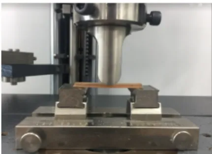
±2시간 동안 보관하였다. Support 간격이 50 mm로 고정되어 있는 만능시험기(Z020, Zwick, Ulm, Germany)를 이용하여 cross-head speed 5 mm/minute으로 굴곡강도와 굴곡계수를 측정하였다(Fig. 2).
3. 생물학적 성질(MTT test)
10% fetal bovine serum와 항생제(Penicillin Streptomycin; Life technologies, Carlsbad, CA, USA)를 RPMI (RPMI-1640 MEDIUM 1X+2.05 mML-Glutamine)에 첨가하여 배지를 제작하였다. 37℃를 유지하는 5% CO2 배양기(MCO175; SANYO Electric Biomedical, Osaka, Japan)에서 L-929 세포(NCTC clone 929: CCL 1, ATCC)를 배양하였다.
ISO 10993-5에 따라 모든 시편은 표면적이 3 cm2/mL가 되도록 계 산하여 배지의 양을 구한 후 시편과 함께 넣어 37℃ 건조기에서 24시 간 동안 추출하였다. 양성대조군(positive control)으로 1% phenol (OCI, Seoul, Korea)을 사용하였고, 음성대조군(negative control) 으로는 산화 에틸렌 가스(ethylene oxide gas)로 소독한 aluminium oxide ceramic rod (SAMHWA CERAMIC COMPANY, Seoul, Ko- rea)를 사용하였다. L-929 세포를 96 well plate에 1×10⁴ cells/well 가 되도록 100 μL씩 분주하고, 37℃의 5% CO2 배양기에서 24시간 동 안 세포를 부착시켰다.
그 후 37℃ 유지하는 CO2 배양기에서 90 μL 배지와 10 μL 추출액 을 첨가하여 24시간 동안 재배양하였다. PBS buffer에 1 mg/mL 녹인 MTT용액을 50 μL씩 넣고, 37℃ 유지하는 5% CO2 배양기에서 3시간 동안 보관한 후 배지를 제거하고 Isopropanol를 100 μL 넣고 혼합기 로 혼합하였다.
570 nm에서 흡광도를 분석하는 enzyme-linked immuno-assay
reader (Spectra MAX250; Molecular Devices, San Jose, CA, USA)를 이용하여 흡광도를 측정하였다. 음성대조군의 MTT 환원율 100%로 표준화하여 대조군과 비교하여 실험군의 세포활성도를 백분 율로 표시하였다. ISO 10993-5:2009(E) 규격에 따라 70% 이상일 경 우 독성이 없다고 판단하였다[17].
4. 통계분석
기계적 성질과 생물학적 성질의 결과는 IBM SPSS ver. 22.0 프로그 램(IBM, Armonk, NY, USA)을 사용하여 one-way ANOVA로 신뢰 수준 95%에서 분석하였고 사후검정으로 Duncan test를 시행하였다.
모든 검정에서 α=0.05를 기준으로 통계적 유의성을 판정하였다.
RESULTS
1. 기계적 성질 1) 굴곡강도
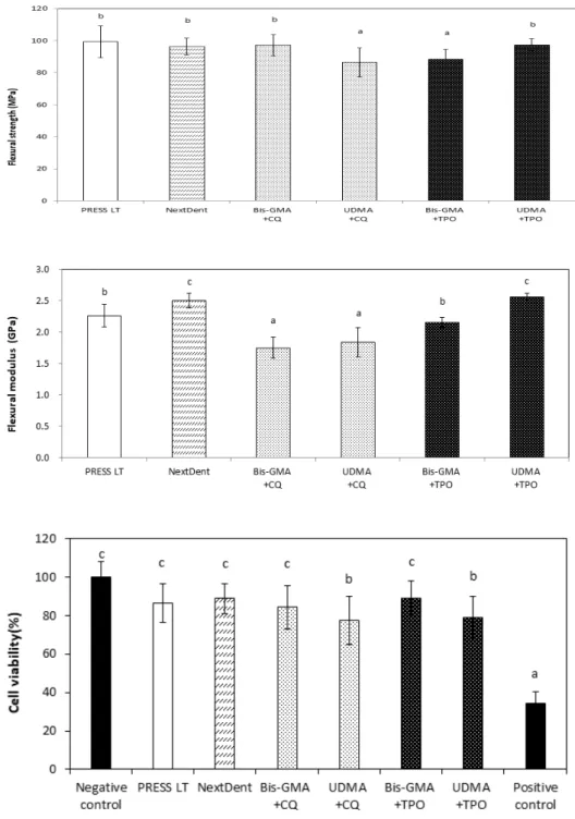
국제표준규격 제20795-1호(ISO 20795-1:2008)에서는 의치상용 열중합 레진의 굴곡강도 기준값은 65 MPa 이상, 자가중합 레진의 기 준값은 60 MPa 이상으로 규정되어 있다. 실험용 3D 프린팅용 의치상 레진은 모두 열중합 레진의 굴곡강도의 기준값인 65 MPa보다 높은 굴 곡강도값을 나타냈다.
굴곡강도를 측정한 결과는 Fig. 3과 같다. 실험군에서는 Bis- GMA+CQ (97.12±6.47 MPa), UDMA+TPO (97.40±3.75 MPa)의 굴곡강도가 유의성 있게 높았다(p<0.05). UDMA+CQ (86.48±9.03 MPa)와 Bis-GMA+TPO (88.31±6.17 MPa)의 조합이 낮은 굴곡강도 를 나타내었다(p<0.05).
2) 굴곡계수
굴곡계수를 측정한 결과, NextDent (2.50±0.12 GPa)와 UDMA+TPO (2.56±0.06 GPa)의 실험군이 가장 높은 굴곡계수를 나 타내었다(p<0.05). CQ를 첨가한 모든 실험군이 TPO을 첨가한 실험 군보다 굴곡계수가 유의하게 낮았다(p<0.05). Bis-GMA계 실험군보
Figure 2.
Figure 2. Testing of flexural strength and flexural modulus using a uni-
versal testing machine.
Figure 1.
Figure 1. (A) Light polymerization of control. (B) Specimen produced us-
ing three-dimensional printer.
B
A
JOURNAL OF TECHNOLOGIC DENTISTRY
J TD
다 UDMA계 실험군이 유의하게 높은 굴곡계수를 나타내었다(p<0.05;
Fig. 4).
국제표준규격 제20795-1호(ISO 20795-1:2008)에 따르면 의치상 용 열중합 레진의 굴곡계수 기준값은 2 GPa 이상, 자가중합 레진의 기 준값은 1.5 GPa 이상으로 규정되어있다. TPO을 첨가한 실험군은 의 치상용 열중합 레진의 기준을 만족시키는 결과값을 나타내었고 CQ를 첨가한 모든 실험군은 자가중합 레진의 기준값 이상의 굴곡계수를 나 타내었다.
2. 생물학적 성질(MTT test)
제작한 레진을 24시간 동안 용출시킨 후 세포의 활성도 측정값은 Fig. 1과 같다. 모든 실험군의 세포활성도가 70% 이상으로 나타났다 (p<0.05). Bis-GMA계 실험군이 UDMA계 실험군보다 세포활성도가 유의하게 높은 값을 나타내었다(p<0.05; Fig. 5).
Figure 4.
Figure 4. Flexural modulus of experi-
mental three-dimensional (3D) printing denture base resins in comparison of commercial 3D printing resins and con- ventional denture base resin (p<0.05).
Different lowercase letters (a, b, c) are significantly different among the resins by one-way ANOVA and Duncan test at α
α=0.05. Bis-GMA: bisphenol A glycidyl methacrylate, CQ: camphorquinone, UDMA: urethane dimethacrylate, TPO:
diphenyl(2,4,6-trimethylbenzoyl) phos- phine Oxide.
Figure 5.
Figure 5. Cell viability was expressed as
percentage means and standard devia- tions of MTT conversion, normalized to the 100% conversion of negative control (p<0.05). Different lowercase letters (a, b, c) are significantly different among the three-dimensional printing resins by Dun- can test at αα=0.05. Bis-GMA: bisphenol A glycidyl methacrylate, CQ: camphorqui- none, UDMA: urethane dimethacrylate, TPO: diphenyl(2,4,6-trimethylbenzoyl) phosphine Oxide.
Figure 3.
Figure 3. Flexural strengths of experi-
mental three-dimensional (3D) printing denture base resins in comparison of commercial 3D printing resins and con- ventional denture base resin (p<0.05). Dif- ferent lowercase letters (a, b) are signifi- cantly different among the resins by one- way ANOVA and Duncan test at αα=0.05.
Bis-GMA: bisphenol A glycidyl methacry-
late, CQ: camphorquinone, UDMA: ure-
thane dimethacrylate, TPO: diphenyl(2,4,6-
trimethylbenzoyl) phosphine Oxide.
Da Ryeong Park, Ju lee Son: 3D printing denture resins according to photoinitiators
DISCUSSION
본 연구의 목적은 CQ와 TPO 2종의 광개시제를 첨가하여 Bis-GMA 와 UDMA를 기반으로 한 3D 프린팅 의치상용 레진을 제작하여 기계 적 성질과 생물학적 성질이 비교 분석하여 현존하는 3D 프린팅용 치과 용 레진보다 우수한 레진을 제작하고자 하였다.
Bagheri와 Jin [26]은 디지털 기술의 발전과 3D 프린터의 설계로 인해 높은 인쇄 해상도를 얻을 수 있으며 이에 광경화성 재료는 중요 한 요인이 된다고 하였다. 3D 프린팅 기술을 더욱 발전시키기 위해 새 로운 감광성 조성물과 적절한 광개시 시스템을 만드는 것이 필요하다 [27]. 따라서 본 연구에서는 3D 프린터의 보다 높은 중합도를 위하여 치과용 복합체 및 접착제로 널리 사용되고 있는 CQ와 상업용 복합체 에 포함되고 있는 TPO을 첨가하여 새로운 3D 프린팅용 광경화 레진 을 제작하였다. 3D 프린팅용 레진은 점도가 중요한 요소이므로 의치상 용 레진 중 점도가 비슷한 유동성 의치상용 레진을 대조군으로 제작하 였다. 미중합을 우려하여 Janda 등[28]은 CQ가 390~480 nm의 넓은 광파장대를 지니고 있어 가장 일반적인 광개시제로 사용되지만 3차 아 민이 존재해야 하며 특유의 노란색을 나타내어 심미성이 낮다는 단점 이 있다고 하였다. 의치상을 제작하기 위한 레진은 심미성과 색안정성 이 중요한 요인이 되므로 CQ를 사용할 시 치주와 비슷한 색상을 나타 내기 위해 색의 조합을 이용하여 다른 색소의 첨가가 필요할 것으로 생 각된다.
반면 380~425 nm의 광파장대 가진 TPO는 CQ가 첨가된 레진 혼 합물과 유사하거나 더 나은 경화효율 및 색상안정성을 나타낸다[29].
본 실험에서는 광중합 후 무색을 나타내는 광개시제인 TPO을 선택함 으로써 레진을 제작할 시에 광개시제를 혼합하여도 색소의 종류에 따 라 색상을 쉽게 조절하여 술자가 원하는 색상의 의치상을 만들 수 있도 록 하였다. 또 2종의 광개시제 모두 DLP 3D 프린터의 통상적인 광파 장대인 395~410 nm를 포함하므로 광중합이 진행되어 경화되 3D 프 린터 레진으로 사용할 수 있을 것으로 생각된다. 후중합 공정을 시행 하여 보다 높은 중합도를 얻고자 하였다. 같은 조건을 위하여 대조군인 유동성 의치상용 레진 또한 미중합을 우려해 광중합기를 이용해서 후 중합 공정을 시행하였다.
Floyd와 Dickens [30]은 다양한 유형의 단량체는 서로 다른 반응성 이 나타난다. 다른 비율의 혼합물은 재료의 적절한 점도와 강도에 영향 을 미친다고 하였다. Bis-GMA는 시판되는 치과용 수복 재료에서 중 요한 역할을 한다. 유기상의 주된 염기성 모노머 중 하나인 UDMA는 Bis-GMA에 비해 낮은 점도를 나타내며 중합수축률이 낮다[31,32].
Lin 등[33]은 다양한 조성으로 3D 프린팅용 레진에 대한 연구를 시 행하였는데 UDMA와 Bis-GMA를 적절한 비율로 포함하는 실험군에 서 가장 높은 정확도, 만족스러운 기계적인 물성과 생체적합성을 얻었 다고 하였다. 본 연구에서도 Bis-GMA와 UDMA를 기반으로 하여 다 양한 단량체의 함량을 조절하며 3D 프린터로 출력 가능한 레진을 제작
하였다. Bis-GMA계 실험군은 굴곡강도가 높았으나 출력 시 챔버에 슬 러리가 남는 것을 볼 수 있었다. 이는 Bis-GMA계 실험군이 UDMA계 실험군보다 높은 점도로 인해 미반응된 슬러리들이 생성되는 것으로 생각된다.
Anseth 등[34]은 UDMA계 실험군에서는 entaerythritol tetraac- rylate와 Di(trimethylolpropane) tetraacrylate와 같은 comono- mer를 사용하였는데 이는 가교제, 반응성 희석제 및 화학 중간체와 같 은 다기능 단량체로 사용되며 경화 시 빠른 경화 반응과 높은 가교 밀 도를 가능하게 한다고 하였다. 본 연구에서도 이러한 단량체들을 첨가 하여 보다 높은 중합도와 강도를 얻고자 하였다. 그 결과, 시판되고 있 는 NextDent와 유의한 값의 강도를 나타내었다.
Alshali 등[35]은 레진의 굴곡강도는 경도와 파절에 대한 저항성 을 나타내고, 굴곡강도 시험은 저작력에 대한 의치상 레진의 강도를 결정하는 데 필수적이라고 하였다. 따라서 본 연구에서도 만능시험 기를 이용하여 굴곡강도와 굴곡계수를 알아보았다. 그 결과 UDMA 계 실험군이 Bis-GMA계 실험군보다 유의하게 높은 굴곡강도 값 을 나타냈다(p<0.05). 이는 단량체 사슬에 페놀고리가 없어 높은 유 연성과 인성을 가져 중합도에 영향을 미친 결과이다[31]. 본 연구에 서도 UDMA의 함유량과 첨가된 entaerythritol tetraacrylate와 Di(trimethylolpropane) tetraacrylate와 같은 comonomer의 작용 이 굴곡 강도를 향상시킨 것으로 보인다.
굴곡계수 또한 Bis-GMA계 실험군보다 UDMA계 실험군이 유의 하게 높은 굴곡계수를 나타내었다. 이는 Bis-GMA보다 인성이 높은 UDMA의 성질이 반영된 것으로 생각된다. TPO는 친수성 광개시제로 서 친수성 모노머에 혼합할 경우 일반적으로 이용되는 CQ보다 중합정 도를 높일 수 있는 효과를 나타낸다[24]. 본 연구의 결과도 TPO를 첨 가한 실험군이 CQ를 첨가한 모든 실험군이 보다 굴곡계수가 유의하게 높은 값을 나타냈다.
구강 내에서 사용 가능한 레진을 제작하기 위해 시행한 MTT test 결 과를 살펴보면 제작한 레진을 24시간 동안 용출시킨 후 세포의 활성도 측정값은 모든 실험군이 음성대조군보다 세포활성도가 유의하게 높았 다. ISO 10993-5:2009(E)에서 MTT test 결과 세포활성도가 70% 이 상이면 해당 재료는 독성이 없다고 규정하고 있다. 본 연구의 결과에서 도 사용된 모든 실험군이 70% 이상의 세포활성도를 보여 독성이 없음 이 판정되어 생체친화성이 있음을 나타낸다. 따라서 구강 내에 사용 가 능한 만족스런 생체적합성을 가지고 있으므로 임상적으로 사용할 수 있을 것으로 생각된다.
임상 적용을 위해서는 최적의 기계적 물성 가지고 우수한 생체적합 성과 수복물의 조기 파절을 방지하기 위해 중합단계가 가장 중요한 요 인이 되어야 한다. 광중합도를 높이기 위해 가장 효과적인 방법은 빛을 조사하였을 때 중합반응을 개시시키는 광중합 시스템을 개발하는 것이 다. 향후 더욱 다양한 광개시제와 기계적 물성과 생체적합성을 지닌 레 진의 최적의 조합을 찾아 새로운 3D 프린팅 레진 개발에 대한 연구가
JOURNAL OF TECHNOLOGIC DENTISTRY
J TD
필요할 것으로 보인다.
CONCLUSIONS
본 연구에서는 2종류의 광개시제(CQ, TPO)를 첨가하여 2종의 3D 프린팅용 의치상레진(Bis-GMA계와 UDMA계)을 제작하여 기계적 성 질과 생물학적 성질을 비교 분석하고 최적의 조성을 찾고자 하였다. 이 에 다음과 같은 결론을 얻었다.
1 . 제 작 한 3 D 프 린 팅 용 의 치 상 레 진 중 B i s - G M A + CQ , UDMA+TPO의 조합이 유의하게 높은 굴곡강도를 나타내었고 굴곡 계수 또한 UDMA+TPO의 실험군에서 가장 높은 값을 나타내었다 (p<0.05).
2. MTT test 결과 모든 실험군이 70% 이상의 세포활성도를 나타내 어 생체적합성이 우수한 것으로 판단된다.
따라서 굴곡강도와 굴곡계수, 생체합성까지 우수한 UDMA+TPO 조 성을 중심으로 다양한 성분을 조절하며 더욱 깊은 연구가 시행되어 진 다면 새로운 3D 프린팅이 가능한 레진을 개발할 수 있을 것으로 생각 된다.
CONFLICT OF INTEREST
No potential conflict of interest relevant to this article was reported.
ORCID
Da Ryeong Park, https://orcid.org/0000-0002-4801-0908 Ju lee Son, https://orcid.org/0000-0003-4611-8891