Comparison of or Changes in the Thicknesses and Ratios of the Deep and Superficial Multifidus Muscles According to the
Lumbar Stabilization Exercise Methods
Choi Mansoo, PT, MS
1․ Lee Sanyeol, PT, Ph.D
2ǂ1
Dept. of Physical Therapy, Daeho Pain Clinic, Team Leader
2ǂ
Dept. of Physical Therapy, Kyungsung University, Professor
Abstract
Purpose : Lower back pain is a common disorder experienced by approximately 90-% of the population at least once in a lifetime. This study examines changes in the thicknesses and ratios of the deep and superficial fibers of the multifidus according to the lumbar stabilization exercise used for spinal stabilization.
Methods : Ten different lumbar stabilization exercises were implemented by 20 healthy men in random order, and the thickness of multifidus muscle was measured ultrasound image during each exercise.
Results : The surface muscle fibers of the multifidus muscles significantly increased in the exercise method in which the arms and legs were lifted (p<.05), while the deep muscle fibers of the multifidus muscles increased significantly in the exercise in which the arms and legs were not lifted (p<.05). The ratio of the thickness of surface muscle fibers to the total thickness of muscle fibers was higher in the exercise method in which the arms and legs were lifted (p<.05), while the ratio of the thickness of deep muscle fibers to the total thickness of muscle fibers was higher in the hollowing and bracing exercise method in which the arms and legs were not lifted (p<.05).
Conclusion : When lumbar stabilization exercise should be performed at clinics to strengthen the deep muscle fibers of the multifidus muscles that have larger effects on the stability of spinal segments, taking the stability of the spine into consideration indicates that, hollowing and bracing exercise methods that do not that cause isotonic extension to the spine are appropriate.
Key-words : bracing, hollowing, lumbar stabilization exercise, multifidus deep muscle fiber, multifidus superficial muscle fiber
ǂ
Corresponding author : Lee Sangyeol, [email protected]
1)
Received : May 23, 2019 | Revised : June 12, 2019 | Accepted : June 21, 2019
※ This research supported by Kyungsung University Research Grants in 2018.
Ⅰ . Introduction
The lumbar multifidus plays a critical role in stabilizing the lumbar spine neutral zone, and atrophy of this muscle lowers its controlling power on the neutral zone (Freeman et al., 2010). Panjabi (1992) described the neutral zone as a part of the spinal motion range and a neutral position where minimal passive resistance is generated from the vertebrae. Wilke et al. (1995) argued that the multifidus muscle, which accounts for more than two-thirds of the lumbar stability function, is important for maintaining stability of the L4~L5 vertebral segments in the neutral zone. One of the functions of the lumbar multifidus muscle adjacent to the periphery of the vertebrae is spinal stability and segmental control. The lumbar multifidus plays various roles in maintaining the stability of lumbar vertebral segments (Bogduk et al., 1992; Macintosh et al., 1986;
Moseley et al., 2002; Panjabi, 1992; Sirca & Kostevc, 1985; Wilke et al., 1995).
The multifidus muscle can be divided into superficial and deep muscle fibers, the former of which has the optimal lever arm to provide enough torque to enable lumbar extension. The role of the superficial muscle fibers of the multifidus is to regulate the direction of the spine as well as to produce lumbar extension. Unlike the superficial muscle fibers, the deep muscle fibers are located near the center of rotation of the lumbar spine and control the shear and torsion of the vertebrae through intervertebral compression with minimal torque, while their major role is to provide the stability of the lumbar segments (Danneels, 2007; Jemmett et al., 2004; Kay, 2000; Macintosh et al., 1986). It is clinically important to educate patients with low back pain accompanied by dysfunction or atrophy of the lumbar multifidus muscle to perform muscle training to activate their lumbar multifidus muscle (Freeman et al., 2010). In the rehabilitation strategy for low back pain, exercise therapy is closely related to the restoration of the function of the multifidus muscle (Hides et al., 2001;
Hodges & Moseley, 2003). Richardson and Jull (1995) reported that for low back pain, exercise therapy improves the function of trunk muscles, and the increased muscle strength of the trunk muscles further stabilizes the spine.
High muscle activities were detected in the multifidus muscle after performing exercises that involve keeping the torso lifted in the prone position or lifting and maintaining the legs (Ng & Richardson, 1994). Abdominal hollowing exercise performed in the four-point kneeling position increased the thickness of the deep muscles fibers of the multifidus muscle and bilateral arm and leg lift exercise in the prone position increased the thickness of the superficial muscle fibers of the multifidus muscle (Kim et al., 2012).
Segmental stabilization exercises through abdominal hollowing relieved pain in patients with chronic low back pain and reduced the recurrence of low back pain (Arab &
Chehrehrazi, 2011). Arab and Chehrehrazi (2011) reported that abdominal bracing and abdominal hollowing were effective in lumbar stabilization. When these exercises are performed to activate the multifidus muscle, visual feedbacks provided through ultrasound imaging help patients enhance their ability (Kim et al., 2008). Kim et al.
(2008) reported that after 6-week lumbar stabilization exercise, the multifidus muscle area increased by about 6
%, while after segmental stabilization exercise, the multifidus muscle area increased by about 10~11 %. Akbari et al. (2008) reported that after controlled exercise through the abdominal draw-in maneuver, the cross-sectional area of the multifidus muscle increased in patients with chronic low back pain.
Although many studies on the roles of the multifidus
muscle in various exercises have been carried out, there
have been few studies that investigated the separate roles
for the superficial and deep muscle fibers of the multifidus
muscle. In addition, it is difficult to select an optimal
posture to activate the multifidus muscle in clinical practice
due to the lack of comparative analysis of the effects of
various postures on lumbar stabilization exercise in a same
subject. Therefore, this study aims to investigate the effects
of different lumbar stabilization exercise on the thickness of the deep and superficial muscle fibers of the multifidus, the thickness of the total muscle fibers of the multifidus, and the ratio of the thickness of the deep and superficial muscle fibers of the multifidus using an ultrasonographic measurement method.
Ⅱ . Methods
1. Participants
The participants in this study was conducted on 20 health males. The subjects had no experience of musculoskeletal disorders, neurological or lumbar pain during the past six months. In this study, multifidus muscles on the dominant side and non-dominant side were measured during a rest and the thicknesses of deep and superficial multifidus muscle fibers were measured through ultrasound imaging when the end motion of lumbar stabilization exercise was maintained in 20 healthy male adults. The multifidus muscles on the dominant side and non-dominant side were measured and compared before implementing lumbar stabilization exercise methods in random order. Each exercise method was maintained for 10 seconds and a rest time for 5 minutes. was given after completion of each exercise method to avoid the fatigue of the multifidus muscles due to continued exercise. Ultrasound images were measured three times per exercise method and the average values were compared and analyzed (Koppenhaver et al., 2009).
The participating study subjects were sufficiently informed about the purpose and method of this study so that they could understand the content of experiment and their agreement was obtained thereafter. The concrete selection criteria for the subjects participating in this study are as follows.
1) Those who had no musculoskeletal or nervous system
problems during the last six months.
2) Those who have no past history of surgery due to back pain of fracture.
3) Those who have no spinal deformities such as scoliosis, kyphosis, and lordosis.
4) Those who have not limit the range of motion of the joint in the lumbar region.
5) Those who have not performed ant exercise that affect regions around the waist for the last 3 months.
6) Those who have no significant difference in the thickness between the dominant and non-dominant sides.
7) Those whose right side is the dominant side.
This study received approval from the Ethics Committee of Kyungsung University for the protection of rights and safety of research subjects and obedience of law related to bioethics and safety. The approval number is KSU-17-06-001.
2. Measurement tool
1) Ultrasound image procedure
A 3.5-5 ㎒, 50 ㎜ curved probe of an ultrasonic device (Picus, ESAOTE Europe BV, Netherlands) was used to measure the deep and superficial muscle fibers of the multifidus on the non-dominant and dominant sides at rest, and the deep and superficial muscle fibers of the multifidus on the dominant side at the end of the exercise while the subject maintained the last posture (Koppenhaver et al., 2009). The inter-rater reliability of the ultrasound measurement method of measuring the thickness of the lumbar multifidus muscle fibers showed intra-class correlation coefficients in a range of .75~.98 thereby showing relatively high reliability (Kim et al., 2011).
According to the criteria of the previous studies, the side usually used for throwing and kicking the ball was defined as the dominant side (Jacobs et al., 2005; Van den Tillaar
& Ettema, 2009) and the subjects whose dominant side was
the right side were selected for the study. A pre-exercise ultrasonography was performed while the subject took the prone position with a rolled towel underneath his abdomen to minimize the lumbar lordosis (Van et al., 2006). The middle region of the curved probe was placed on the processus spinosus of the fourth lumbar vertebra (L4) in the longitudinal axis direction (Kisel et al., 2007).
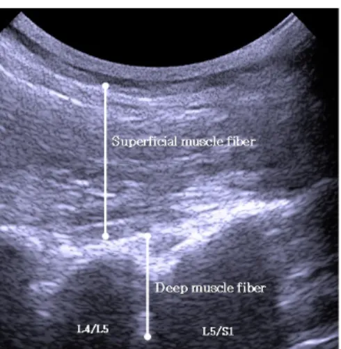
The probe was tilted slightly inward and moved outward until the zygapophysis between the L4/5 was able to be identified, then the probe was fixed and the image was stored. The stored image file was subjected to image J Software to measure the thickness of the deep and superficial muscle fibers of the multifidus. For the superficial muscle fibers of the multifidus, the vertical distance from the boundary between the subcutaneous tissue and the fascia to the zygapophyseal joint between the L4/5 of the lumbar vertebrae was measured, and for the deep muscle fibers of the multifidus, the vertical distance from the lowest point between the zygapophyseal joint between the L4/5 and L5/S1 of the sacral vertebrae to the bottom of the superficial muscle fibers was measured (Kisel et al., 2007)(Fig 1).
Fig 1. Ultrasound image of mulitifidus
Measurement of the thickness and proportion of deep and superficial muscle fibers of multifidus muscles based on the
previous study (Koppenhaver et al., 2009). This study the thickness of deep muscle fibers and superficial muscle fibers of multifidus muscles were measured from the ultrasound image and computed the thickness of entire muscle fibers of multifidus muscle and the ratio between the thickness of deep and superficial muscle fiber of multifidus muscle during exercise as follows.
(1) Measurement of the thickness of the superficial muscle fiber of multifidus muscle:
Vertical distance from the boundary between hypodermis and fascia to the zygapophysial joint of the fourth / fifth lumbar.
(2) Measurement of the thickness of the deep muscle fiber of multifidus muscle:
Vertical distance from the lowest part between the zygapophysial joint of the fourth / fifth lumbar and the zygapophysial joint of the fifth lumbar / first sacral vertebrae to the bottom of the superficial muscle fiber.
(3) Total thickness of the multifidus:
Sum of the thickness of the deep muscle fiber of multifidus and superficial muscle fiber of multifidus.
(4) Ratio between the deep muscle fiber and superficial muscle fiber of multifidus muscle during lumbar stabilization exercise:
High proportion of deep or superficial muscle fiber of multifidus muscle during motion implies the high share of the thickness of multifidus muscle during contraction occupied by the corresponding muscle fibers.
3. Lumbar stabilization exercise
1) Abdominal hollowing on prone position
The subject was instructed to flex both shoulders at 180
degrees on the floor in the prone position without moving
the pelvis and pull the navel toward the vertebrae, then to
maintain the position for about 10 seconds (Harringe et al.,
2007; O'Sullivan, 2000).
2) Abdominal bracing on prone position
The subject was instructed to flex both shoulders at 180 degrees on the floor in the prone position without moving the pelvis and keep the entire abdominal region at high tension to increase the intra-abdominal pressure, then to maintain the position for about 10 seconds (Harringe et al., 2007).
3) The lift dominant side leg and non-dominant side arm on prone position
The subject was instructed to flex both shoulders at 180 degrees on the floor in the prone position and lift ipsilateral leg and the opposite arm from the ground, then to hold the position for about 10 seconds (Sung, 2003).
4) The lift dominant side leg and non-dominant side arm on prone position during abdominal hollowing
The subject was instructed to flex both shoulders at 180 degrees on the floor in the prone position without moving the pelvis, pull the navel toward the vertebrae and maintain the position, and then to lift the ipsilateral leg and the opposite arm from the ground and hole the position for about 10 seconds (Harringe et al., 2007).
5) The lift dominant side leg and non-dominant side arm on prone position during abdominal bracing
The subject was instructed to flex both shoulders at 180 degrees on the floor in the prone position without moving the pelvis, keep the entire abdominal region at high tension to increase the intra-abdominal pressure and maintain the position for about 10 seconds, and then to lift the ipsilateral leg and the opposite arm from the ground and hold the position for about 10 seconds (Harringe et al., 2007).
6) Abdominal hollowing on four point kneeling position
The subject was instructed to place his hands on the floor just below the shoulder joints and take the four-point kneeling position with the knees positioned just below the hip joints, and then to pull the navel toward the vertebrae maintaining the chest and pelvis in place and hold the position for about 10 seconds (Harringe et al., 2007;
O'Sullivan, 2000).
7) Abdominal bracing on four point kneeling position The subject was instructed to place his hands on the floor just below the shoulder joints and take the four-point kneeling position with the knees positioned just below the hip joints, and then to keep the entire abdominal region at high tension to increase the intra-abdominal pressure maintaining the chest and pelvis in place and maintain the position for about 10 seconds (McGill, 2001).
8) The lift dominant side leg and non-dominant side arm on four point kneeling position
The subject was instructed to place his hands on the floor just below the shoulder joints and take the four-point kneeling position with the knees positioned just below the hip joints, and then to lift the ipsilateral leg and the opposite arm from the ground and hold the position for about 10 seconds (McGill, 1998).
9) The lift dominant side leg and non-dominant side arm on four point kneeling position during abdominal hollowing
The subject was instructed to place his hands on the floor just below the shoulder joints and take the four-point kneeling position with the knees positioned just below the hip joints, and then to pull the navel toward the vertebrae maintaining the chest and pelvis in place and hold the position for about 10 seconds lifting the ipsilateral leg and the opposite arm from the ground (Harringe et al., 2007;
O'Sullivan, 2000).
10) The lift dominant side leg and non-dominant side arm
on four point kneeling position during abdominal bracing The subject was instructed to place his hands on the floor just below the shoulder joints and take the four-point kneeling position with the knees positioned just below the hip joints, and then to keep the entire abdominal region at high tension to increase the intra-abdominal pressure maintaining the chest and pelvis in place and hold the position for about 10 seconds lifting the ipsilateral leg and the opposite arm from the ground (Harringe et al., 2007).
4. Statistical analysis
SPSS ver. 21.0 was used for data analysis and the paired t-test was used to determine the difference in the multifidus muscle between the non-dominant and dominant sides for the selection of subjects. Thereafter, the one-way ANOVA was used to compare the thickness of the deep and superficial muscle fibers of the multifidus, the thickness of the total muscle fibers of the multifidus, and the ratio of
the thickness of the deep and superficial muscle fibers of the multifidus after different exercises were applied, and the Scheffe analysis was used for post-hoc analysis. The statistical significance level 𝛂 was set to .05.
Ⅲ . Results
1. General characteristics of subjects
The age, height, body weight, the thickness of the deep and superficial muscle fibers of the multifidus, and the thickness of the total muscle fibers of the multifidus were as follows (Table 1).
There were no significant differences in the thicknesses of the superficial and deep muscle fibers of the multifidus between the non-dominant and dominant sides of the subjects before exercise (p>.05) (Table 2).
Age (years) height (㎝) weight (㎏) SM (㎝) DM (㎝) Multifidus (㎝)
26.00±4.66 172.70±6.40 68.05±5.84 3.11±0.26 2.16±0.24 5.27±0.42
SM; Multifidus superficial muscle fiber thickness, DM; Multifidus deep muscle fiber thickness
Table 1. General characteristics of study subjects (n=20)
Non-dominant Dominant p
SM 3.06±0.31 3.11±0.26 0.09
DM 2.14±0.26 2.16±0.24 0.52
Values are presented as mean (SD).
Table 2. Non-dominant and dominant multifidus thickness of subjects before exercise (unit : ㎝)
2. Total thickness of multifidus muscle
There was no statistically significant difference in the
thickness of the total muscle fibers of the multifidus
between the exercise methods performed by the subjects
(p>.05) (Table 3).
HP BP DLNAL -P
DLNAL -HP
DLNAL
-BP HF BF DLNAL
-F
DLNAL -HF
DLNAL
-BF F p
Multifidus Thickness
6.06±0.
40
6.04±
0.44
5.98±
0.43
6.08±
0.30
6.06±
0.33
6.14±
0.40
6.12±
0.36
5.96±
0.40
6.08±
0.29
6.04±
0.39 0.44 0.90 note. P; prone position, F; four point kneeling position, H; hollowing exercise, B; bracing exercise, DLNAL; dominant side leg and
non-dominant arm lift
Table 3. Total thickness of multifidus muscle fiber on each lumbar stabilization exercise (unit : ㎝)
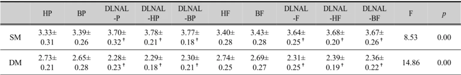
3. Thickness of deep and superficial multifidus muscle fiber on each lumbar stabilization exercises
Each lumbar stabilization exercise was compared. There was a difference in the in the thicknesses of the superficial and deep muscle fibers of the multifidus. According to the exercise methods of the subjects.
The thickness of the superficial muscle fibers significantly increased in the dominant side leg and non-dominant arm lift at prone position (DLNAL-P), dominant side leg and non-dominant arm lift during hollowing exercise at prone position (DLNAL-HP), dominant side leg and non-dominant arm lift during bracing exercise at prone position (DLNAL-BP), dominant side leg and non-dominant arm lift at four point kneeling position (DLNAL-F), dominant side leg and non-dominant arm lift
during hollowing exercise at four point kneeling position (DLNAL-HF), dominant side leg and non-dominant arm lift during bracing exercise at four point kneeling position (DLNAL-BF) (p<.05) (Table 4), while that of the deep muscle fibers significantly increased in the hollowing exercise at prone position (HP), bracing exercise at prone position (BP), the hollowing exercise at four point kneeling position (HF), the hollowing exercise at four point kneeling position (BF) (p<.05) (Table 4).
Thickness of the deep muscle fiber of multifidus muscle was high in case of the lumbar stabilization exercise without lifting arms and legs. In case of the lumbar stabilization exercise with lifted arms and legs, the thickness of the superficial muscle fiber of multifidus muscle was high.
HP BP DLNAL
-P
DLNAL -HP
DLNAL
-BP HF BF DLNAL
-F
DLNAL -HF
DLNAL
-BF F p
SM 3.33±
0.31
3.39±
0.26
3.70±
0.32
✝3.78±
0.21
✝3.77±
0.18
✝3.40±
0.28
3.43±
0.28
3.64±
0.25
✝3.68±
0.20
✝3.67±
0.26
✝8.53 0.00 DM 2.73±
0.21
2.65±
0.28
2.28±
0.23
✝2.29±
0.18
✝2.30±
0.21
✝2.74±
0.25
2.69±
0.27
2.31±
0.25
✝2.39±
0.19
✝2.36±
0.22
✝14.86 0.00
✝
The value with different superscripts(
✝) in the same column are significantly different (p<.05) by scheffe’s method.
note. P; prone position, F; four point kneeling position, H; hollowing exercise, B; bracing exercise, DLNAL; dominant side leg and non-dominant arm lift
Table 4. Compared thickness of deep and superficial multifidus muscle fiber on each lumbar stabilization
exercise (unit : ㎝)
4. Ratio of deep and superficial multifidus muscle fiber about total multifidus total thickness
There was a difference in the ratio of the superficial
muscle fibers of the multifidus to the deep multifidus
muscle fibers between the exercise methods performed by the subjects (Table 5).
The ratio of the superficial muscle fibers significantly increased in the DLNAL-P, DLNAL-HP, DLNAL-BP, DLNAL-F, DLNAL-HF, DLNAL-BF exercise methods (p<.05) and the deep muscle fibers significantly increased in the HP, BP, HF, BF exercise methods (p<.05) (Table 5).
Share of the thickness of the deep muscle fiber out of the total muscle fiber thickness of multifidus muscle was high in case of the lumbar stabilization exercise without lifting arms and legs. In case of the lumbar stabilization exercise with lifted arms and legs, the proportion of the thickness of superficial muscle fiber out of the total muscle fiber thickness of multifidus muscle was high.
HP BP DLNAL
-P
DLNAL -HP
DLNAL
-BP HF BF DLNAL
-F
DLNAL -HF
DLNAL
-BF F p
SM ratio (%)
54.98±
2.75
56.23±
2.75
61.79±2.
89
✝62.27±2.
12
✝62.19±2.
01
✝55.33±
2.86
56.11±
3.50
61.21±2.
68
✝60.64±2.
20
✝60.86±2.
50
✝26.76 0.00 DM ratio
(%)
44.99±
2.70
43.77±
2.75
38.21±2.
89
✝37.72±2.
12
✝37.81±2.
01
✝44.67±
2.86
43.89±
3.50
38.79±2.
67
✝39.36±2.
19
✝39.14±2.
50
✝26.78 0.00
✝