Introduction
Better range of motion (ROM) following knee arthroplasty is an important factor with regard to better clinical outcomes13). In Asian cultures, highly flexed knee positions are required more frequently than in other cultures because of cultural or religious reasons1,35). Theoretically, unicompartmental knee arthroplasty (UKA) has the benefit of increasing ROM compared to total knee
arthroplasty (TKA)68); however, high flexion can also increase the rate of complications such as dislocation of the polyethylene bearing in mobile bearing systems4,5). Currently, improved opera
tive techniques and new implants have been developed to permit higher knee flexion and to minimize complications911).
In general, increased flexion angle of femoral components may permit higher flexion3). However, as the angle increases, the risk of dislocation can also increase due to subsequently increased flexion gap with abnormal gap balancing in mobile bearing sys
tems4,12). Given these facts, the Oxford group suggested a flexion angle of the femoral component between 5° extension and 10°
flexion for mobile bearing UKA13). Many articles regarding mo
bile bearing UKA have reported the mean flexion angle of the femoral component ranged between 0.8° extension and 2.1° flex
ion in their series1316). Those angles are close to neutral 0° flexion and far from the 10° flexion of acceptable limit suggested by them. Nonetheless, we could not find any clinical report present
ing beyond 2.1° of average femoral component flexion angle in mobile bearing UKAs.
Intentionally Increased Flexion Angle of the Femoral Component in Mobile Bearing Unicompartmental Knee Arthroplasty
KyeYoul Cho, MD
1,2, KangIl Kim, MD
2,3, SangJun Song, MD
3, and KyuJin Kim, MD
21Department of Medicine, Graduate School, Kyung Hee University, Seoul; 2Department of Orthopaedic Surgery, Center for Joint Diseases and Rheumatism, Kyung Hee University Hospital at Gangdong, Seoul; 3Department of Orthopaedic Surgery, Kyung Hee University College of Medicine, Seoul, Korea
Purpose: The purpose of this study was to determine the results of mobile bearing unicompartmental knee arthroplasty (UKA) with an intentionally increased flexion angle of the femoral component in patients requiring high flexion.
Materials and Methods: We investigated 45 knees treated by UKA. Clinically, we measured the range of motion (ROM) and the American Knee Society (AKS) score preoperatively and at final followup and investigated complications. Radiologically, we measured the flexion angle of the femoral component, the posterior slope angle of the tibial component, the femorotibial angle and mechanical axis of the limb postoperatively.
Results: The ROM was increased from 123° preoperatively to 139° at the final followup. The AKS knee and function scores increased from 59 and 68, respectively, preoperatively to 94 and 96, respectively, at the final followup. The flexion angle of the femoral component was 9.1°, and the posterior slope angle of the tibial component was 8.6°. There was one case of bearing dislocation in the largest femoral flexion angle case.
Conclusions: The results might reflect the positive effect of an increased flexion angle of the femoral component up to 10° on ROM in mobile bearing UKA, which would contribute to better quality of life after UKA especially in populations requiring deep knee flexion.
Keywords: Knee, Arthroplasty, Unicompartmental, Flexion angle, Femoral component, Range of motion pISSN 2234-0726 · eISSN 2234-2451
Knee Surgery & Related Research
Received June 23, 2017; Revised August 21, 2017;
Accepted September 18, 2017 Correspondence to: KangIl Kim, MD
Department of Orthopaedic Surgery, Center for Joint Diseases and Rheumatism, Kyung Hee University Hospital at Gangdong, 892 Dongnamro, Gangdonggu, Seoul 05278, Korea
Tel: +8224406151, Fax: +8224406296 Email: [email protected]
23
This is an Open Access article distributed under the terms of the Creative Commons Attribution NonCommercial License (http://creativecommons.org/licenses/bync/4.0/) which permits unrestricted noncommercial use, distribution, and reproduction in any medium, provided the original work is properly cited.
Copyright © 2018 KOREAN KNEE SOCIETY www.jksrr.org
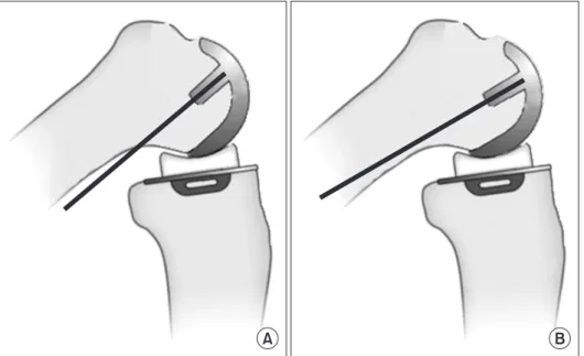
The purpose of this study was to determine the effects of mobile bearing UKA in patients requiring high flexion knees, receiving about 10° of flexion angle of the femoral component which is the nearest numerical value as acceptable limit in flexion angle of the femoral component suggested by the Oxford group13). We hypothesized that the intentionally increased angle of the femoral component close to 10° of flexion would increase the postopera
tive ROM (Fig. 1) without increasing the rate of bearing disloca
tion or additional complications. To support the hypothesis, we compared our findings with those presented in the literatures on flexion angles of the femoral component in mobile bearing UKA.
Materials and Methods
We retrospectively investigated 43 patients (45 knees) treated by UKA using Oxford phase 3 (Biomet, Bridgend, UK) components.
The mean followup period was 51 months (range, 23 to 75 months), except one patient due to death from lung cancer. There were 7 males and 35 females with a mean age of 61 years (range, 48 to 78 years). Written informed consent was obtained from all patients before this institutionally approved study was initiated.
The preoperative diagnosis was medial unicompartmental osteo
arthritis of the knee in all cases. The operation was performed by a senior author in all cases.
We assessed the preoperative magnetic resonance imaging scans in all patients to verify the status of cruciate ligaments, me
nisci and degenerative changes in the cartilaginous lesions. All patients had varus deformities and flexion contractures less than 15° with ROM greater than 100° preoperatively6). Patients with
asymptomatic degenerative changes of the patellofemoral joint were included6,8). We excluded patients with anterior or posterior instability and those with grade 2 degenerative lesions in the lat
eral compartment according to the Kellgren and Lawrence clas
sification6,8).
For the operation, about 8 cm longitudinal incision was made at about 1 cm medial to the proximal part of the patella. After opening the joint, without everting the patella, we removed all os
teophytes and verified the status of intraarticular structures such as cartilage, anterior cruciate ligament and menisci. Next, we per
formed a medial tibial cut perpendicular to the tibial mechanical axis using a tibial saw guide aimed at about 7° of the posterior tibial slope angle. We then performed an excision of the ante
rior part of the medial meniscus. Then we drilled a hole at 1 cm anterior to the anteromedial corner of the intercondylar notch, inserted an intramedullary (IM) rod and positioned a femoral drill guide based on the IM rod. From a sagittal view, the upper
most surface of the drill guide had been recommended parallel to the IM rod, however, we positioned the drill guide at about 10°
flexed to the IM rod using a goniometer in the lateral view. Then we cut the posterior side of the medial femoral condyle using a cutting guide based on the drill guide with about 10° flexion. We measured the flexion and extension gap using a filler gauge and matched the gap by gradual milling of the distal femoral condyle.
After checking the balanced flexion and extension gap with a trial implant inserted, we fixed the real tibial and femoral components with bone cement and inserted the mobile bearing polyethylene.
All patients performed anklepumping exercises and active mo
tion exercises on the day of operation as well as passive motion
A B
Fig. 1. Illustrations showing different sagit
tal positions of the femoral component.
Flexed position of the femoral component can increase posterior contact surface and range of motion. (A) Neutral position. (B) Flexed position.
exercises from postoperative day one. We educated patients on crutch ambulation and allowed painfree distance ambulation.
Outpatient followup was performed at 6 weeks, 3 months, 6 months, and 1 year postoperatively and then once every year. Ra
diologically, we measured the femorotibial angle pre and post
operatively as well as the mechanical axis. Furthermore, we mea
sured the flexion angle of the femoral component and posterior slope angle of the tibial component on postoperative radiographs.
Clinically, we measured the American Knee Society (AKS) score and ROM preoperatively and at the latest followup and investi
gated complications including polyethylene bearing dislocation at the last followup.
Results
The mean preoperative femorotibial angle was 2.5° valgus (range, 4.8° varus to 8.1° valgus), which was corrected to 6.0°
valgus (range, 0.2° valgus to 12.8° valgus) postoperatively, with the mean preoperative mechanical axis of 4.8° varus (range, 12.0°
varus to 3.1° valgus) corrected to 0.7° varus (range, 6.7° varus to 6.9° valgus). The average flexion angle of the femoral component was 9.1° (range, 5.0° to 15.3°), and the average posterior slope angle of the tibial component was 8.6° (range, 4.6° to 10.0°).
The average AKS knee score increased from 59 (range, 52 to 70) preoperatively to 94 (range, 70 to 100) and the average AKS func
tion score increased from 68 (range, 40 to 70) to 96 (range, 80 to 100) at the last followup. The average ROM was increased from 123° to 139° and the flexion contracture decreased from 4.7° to 0°
at the last followup (Table 1).
There was one case of bearing dislocation. It developed at post
operative 6 weeks in a patient with 15.3° of flexion of the femoral component which was the largest flexion angle among our series.
Consequently, it was converted to TKA. There was no other ad
ditional postoperative complication such as infection or early implant loosening till the last followup.
Discussion
The principal finding of this study was that a better ROM was achieved with an intentionally increased flexion angle of the fem
oral component in mobile bearing UKA. To achieve satisfactory clinical outcomes with UKA, it is crucial to determine the proper position of components1719). Radiologically, the femoral compo
nent’s varus/valgus angle or mediolateral distance in the coronal plane is important because of impingement or edge loading on the polyethylene bearing especially in fixed bearing UKA14,15). However, there has been little knowledge about the sagittal posi
tioning of the femoral components, and much contention exists surrounding the normal ranges of femoral component flexion and extension angles13).
In the current study, we hypothesized that the postoperative ROM would increase after UKA performed with the target femo
ral component angle of 10° as suggested by the Oxford group as the acceptable high flexion angle of the femoral component13). We conjectured that increasing the flexion angle of femoral com
ponents would facilitate better flexion of the knee joints through gradual but stable widening and lengthening of the articular surface in contact with the posterior surface of the femoral com
ponent and bearing during deep knee flexion (Fig. 1). Up to now, existing literatures have reported average flexion angle of the
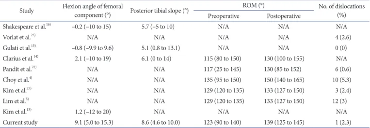
Table 1. Comparison of Previous Studies about Component Angles, Range of Motion (ROM) and Dislocation Study Flexion angle of femoral
component (°) Posterior tibial slope (°) ROM (°) No. of dislocations
Preoperative Postoperative (%)
Shakespeare et al.16) –0.2 (–10 to 15) 5.7 (–5 to 10) N/A N/A N/A
Vorlat et al.23) N/A N/A N/A N/A 4 (2.6)
Gulati et al.15) –0.8 (–9.9 to 9.6) 5.1 (0.8 to 13.1) N/A N/A 0 (0)
Clarius et al.14) 2.1 (–10 to 19) 6.1 (0 to 14) 115 (80 to 150) 130 (100 to 155) N/A
Pandit et al.22) N/A N/A 117 (25 to 145) 130 (85 to 152) 6 (0.6)
Choy et al.4) N/A N/A 135 (95 to 150) 150 (140 to 165) 10 (5.3)
Kim et al.25) N/A N/A 129 (120 to 135) 133 (127 to 150) 3 (2.4)
Lim et al.5) N/A N/A 129 (120 to 135) 133 (127 to 150) 12 (3)
Kim et al.13) 1.2 (–12 to 20) N/A N/A N/A N/A
Current study 9.1 (5.0 to 15.3) 8.6 (4.6 to 10.0) 123 (90 to 140) 139 (125 to 145) 1 (2.3) Values are presented as mean (range).
N/A: not available.
femoral components between 0.8° extension and 2.1° flexion1316). Therefore, we intended to evaluate the postoperative ROM of the knee with an intentionally increased flexion angle of the femoral component of about 10° and compare with previous reports.
The postoperative ROM documented in previous Western articles using the Oxford phase 3 ranges from 130° to 133°8,14,20). Among Asian studies, Lim et al.5) reported 133° of postopera
tive ROM compared to 129° of preoperative ROM, and Kim et al.21) reported 133.5° as a mean postoperative ROM. The average postoperative ROM in the current study (139°) was in agreement with the results of these previous articles. However, there was no information on the flexion angle of the femoral component in those studies5,8,20,21). Therefore, we could not compare with those studies in term of radiographic flexion angle of the femoral com
ponent.
There were few articles reporting the femoral component angle in the sagittal plane. To the best of our knowledge, only one ar
ticle by Clarius et al.14) reported a relationship between flexion
extension angles of the femoral components and clinical scores in UKA using Oxford phase 3 implants. They inserted femoral components with an average 2.1° of flexion and there was no dif
ference in clinical scores between the properly implanted group and the outlier group according to the guidelines proposed by the Oxford group14). In all the other reports with Oxford phase 3, the radiographic mean flexion angle of the femoral compo
nents was considerably lower than that in the current study1416) (Table 1). Among these, comparison on the postoperative ROM was possible only with the study of Clarius et al.14): postoperative knee flexion was greater in our study compared to the study with a different flexion angle of the femoral component. This might indicate the positive effect of the increased flexion angle of the femoral component on postoperative ROM.
Although an increased flexion angle of the femoral component may allow a better ROM of the knee joint3), bearing dislocation can occur due to the increased flexion gap resulting from gradual widening of the flexion gap in deep flexion4). In the current study, the flexion angle of the components was aimed at about 10°, which is the maximum permissible angle according to the Oxford group recommendation13), and there was no dislocation observed up to this angle. We experienced one case of bearing dislocation, but it was an exceptional case with 15.3° of flexion of the femoral component; this was far beyond our target angle and was the largest flexion angle in our series. So, we carefully suggest that the risk of bearing dislocation can increase in case of overly greater flexion angle of the femoral component.
The incidence of bearing dislocation in mobile bearing UKA
was 0.6% to 2.6% in recent studies2225). However, considering these results were all from the Western countries, it has limited applicability to Asian countries where kneeling and crosslegged positions are required much more frequently. Indeed, the studies in East Asia reported dislocation rates of 3% to 5.3%4,5), which might suggest the influence of different lifestyles on the rate of dislocation in different populations. On the other hand, Lim et al.5) reported the nonanatomical bearing resulted in a higher rate of dislocation in the early period than the anatomical bearing (3.2% vs. 2.8%). Similarly, Choy et al.4) also suggested the impact of nonanatomical bearing on dislocation in their series. There
fore, we think the nonanatomic type of bearing could be consid
ered as one of the causative factors of bearing dislocation.
The newly designed Microplasty (Zimmer Biomet, Bridgend, UK), the twin peg Oxford partial knee, adopted the extra peg, lengthened the posterior flange and the arc by 15° and conse
quently increased the contact with a bearing at high knee flex
ion11). Although White et al.11) reporting the new twin peg design with a 5.4° of flexion angle of the femoral component did not demonstrate an increase in the actual ROM compared to the conventional one peg design, we anticipate an increased postop
erative ROM in the twin peg design model with an intentionally increased flexion angle of about 10° based on our results.
The limitations of our study include a relatively small number of cases (45 knees) without a comparison group; therefore, we could not compare with knees with a neutral flexion angle of the femoral components in a single surgeon series. On the risk of dislocation, other related factors such as the angle of posterior tibial slope or the tension of medial ligamentous structures were not investigated thoroughly. However, the degree and range of posterior tibial slope were similar among patients included in the current study and the operation technique was the same in all cases since it was a single surgeon’s series. Last, the followup period was relatively short for arthroplasty, and therefore further investigation with a longer term followup would be required.
Conclusions
We think that the technique of intentionally increasing the femoral component flexion angle to about 10° in mobile bear
ing UKA may produce a better ROM without increasing the incidence of bearing dislocation. This would contribute to better quality of life after UKA especially in the population demanding deep knee flexion.
Conflict of Interest
No potential conflict of interest relevant to this article was re
ported.
References
1. Ueo T, Kihara Y, Ikeda N, Kawai J, Nakamura K, Hirokawa S.
Deep flexionoriented bisurfacetype knee joint and its tibial rotation that attributes its high performance of flexion. J Ar
throplasty. 2011;26:47682.
2. Miner AL, Lingard EA, Wright EA, Sledge CB, Katz JN;
Kinemax Outcomes Group. Knee range of motion after total knee arthroplasty: how important is this as an outcome mea
sure? J Arthroplasty. 2003;18:28694.
3. Kurosaka M, Yoshiya S, Mizuno K, Yamamoto T. Maximiz
ing flexion after total knee arthroplasty: the need and the pitfalls. J Arthroplasty. 2002;17(4 Suppl 1):5962.
4. Choy WS, Kim KJ, Lee SK, Yang DS, Lee NK. Midterm re
sults of oxford medial unicompartmental knee arthroplasty.
Clin Orthop Surg. 2011;3:17883.
5. Lim HC, Bae JH, Song SH, Kim SJ. Oxford phase 3 unicom
partmental knee replacement in Korean patients. J Bone Joint Surg Br. 2012;94:10716.
6. Murray DW. Mobile bearing unicompartmental knee re
placement. Orthopedics. 2005;28:9857.
7. Newman J, Pydisetty RV, Ackroyd C. Unicompartmental or total knee replacement: the 15year results of a prospective randomised controlled trial. J Bone Joint Surg Br. 2009;91:
527.
8. Pandit H, Jenkins C, Barker K, Dodd CA, Murray DW. The Oxford medial unicompartmental knee replacement using a minimallyinvasive approach. J Bone Joint Surg Br. 2006;88:
5460.
9. Murray DW, Goodfellow JW, O’Connor JJ. The Oxford medial unicompartmental arthroplasty: a tenyear survival study. J Bone Joint Surg Br. 1998;80:9839.
10. Price AJ, Webb J, Topf H, Dodd CA, Goodfellow JW, Mur
ray DW; Oxford Hip and Knee Group. Rapid recovery after oxford unicompartmental arthroplasty through a short inci
sion. J Arthroplasty. 2001;16:9706.
11. White SH, Roberts S, Jones PW. The Twin Peg Oxford partial knee replacement: the first 100 cases. Knee. 2012;19:3640.
12. Lewold S, Goodman S, Knutson K, Robertsson O, Lidgren L. Oxford meniscal bearing knee versus the Marmor knee in unicompartmental arthroplasty for arthrosis: a Swedish
multicenter survival study. J Arthroplasty. 1995;10:72231.
13. Kim JG, Kasat NS, Bae JH, Kim SJ, Oh SM, Lim HC. The radiological parameters correlated with the alignment of the femoral component after Oxford phase 3 unicompartmental knee replacement. J Bone Joint Surg Br. 2012;94:1499505.
14. Clarius M, Hauck C, Seeger JB, Pritsch M, Merle C, Aldinger PR. Correlation of positioning and clinical results in Oxford UKA. Int Orthop. 2010;34:114551.
15. Gulati A, Chau R, Simpson DJ, Dodd CA, Gill HS, Murray DW. Influence of component alignment on outcome for uni
compartmental knee replacement. Knee. 2009;16:1969.
16. Shakespeare D, Ledger M, Kinzel V. Accuracy of implanta
tion of components in the Oxford knee using the minimally invasive approach. Knee. 2005;12:4059.
17. Kaya Bicer E, Servien E, Lustig S, Demey G, Ait Si Selmi T, Neyret P. Sagittal flexion angle of the femoral component in unicompartmental knee arthroplasty: is it same for both medial and lateral UKAs? Knee Surg Sports Traumatol Ar
throsc. 2010;18:92833.
18. Bert JM. 10year survivorship of metalbacked, unicompart
mental arthroplasty. J Arthroplasty. 1998;13:9015.
19. Cartier P, Sanouiller JL, Grelsamer RP. Unicompartmental knee arthroplasty surgery. 10year minimum followup pe
riod. J Arthroplasty. 1996;11:7828.
20. Rees JL, Price AJ, Beard DJ, Dodd CA, Murray DW. Mini
mally invasive Oxford unicompartmental knee arthroplasty:
functional results at 1 year and the effect of surgical inexpe
rience. Knee. 2004;11:3637.
21. Kim KT, Lee S, Park HS, Cho KH, Kim KS. A prospective analysis of Oxford phase 3 unicompartmental knee arthro
plasty. Orthopedics. 2007;30(5 Suppl):158.
22. Pandit H, Jenkins C, Gill HS, Barker K, Dodd CA, Murray DW. Minimally invasive Oxford phase 3 unicompartmental knee replacement: results of 1000 cases. J Bone Joint Surg Br.
2011;93:198204.
23. Vorlat P, Putzeys G, Cottenie D, Van Isacker T, Pouliart N, Handelberg F, Casteleyn PP, Gheysen F, Verdonk R. The Ox
ford unicompartmental knee prosthesis: an independent 10
year survival analysis. Knee Surg Sports Traumatol Arthrosc.
2006;14:405.
24. Price AJ, Waite JC, Svard U. Longterm clinical results of the medial Oxford unicompartmental knee arthroplasty. Clin Orthop Relat Res. 2005;(435):17180.
25. Kim SJ, Bae JH, Lim HC. Factors affecting the postoperative limb alignment and clinical outcome after Oxford unicom
partmental knee arthroplasty. J Arthroplasty. 2012;27:12105.