• 검색 결과가 없습니다.

Botryoid Odontogenic Cyst on Mandibular Anterior and Both Body Area: a Case Report

N/A
N/A
Protected

Academic year: 2021

Share "Botryoid Odontogenic Cyst on Mandibular Anterior and Both Body Area: a Case Report"

Copied!
5
0
0

로드 중.... (전체 텍스트 보기)

전체 글

(1)

Introduction

Botryoid odontogenic cyst (BOC) has been a rare multilocular variant of the lateral periodontal cyst. It is a developmental cyst of odontogenic epithelial ori- gin. It has been known to originate from rests of the dental lamina and represented the counterpart of the gingival cyst of the adult. The most common site of this lesion is in the periodontal space of vital tooth.

1)

Botryoid odontogenic cyst was the term first employed by Weathers & Waldron for a multilocular variant of the lateral periodontal cyst.

2)

The BOC is apparently uncommon, and it was reported to have a tendency to recur. The BOC is more likely found in middle-aged and older adults, and the teeth more likely affected are mandibular canines and premolar area. On radiography, the cyst appears grape-like appearance.

1)

This report describes a case of botryoid odontogenic cyst, which occurred from right mandibular 2nd pre- molar to left mandibular 1st premolar of a 67-year- old male.

Case report

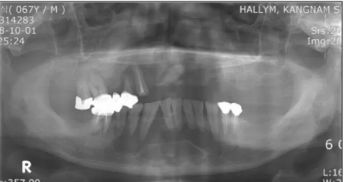
A 67-year-old male visited with chief complaint of a painless, bluish, fluctuant, swelling mass in his mandibular anterior area, and severe mobility of mandibular incisors. Radiological examination and CT scan revealed a large, multilocular radiolucent, cystic lesion with distinct radiopaque borders (Figs. 1, 2).

The lesion extended from the right mandibular 2nd premolar to left mandibular 1st premolar area. The width of the periodontal ligament space seemed nor- mal. There was a slightly compressible swelling of the mandibular anterior area, but there were no paresthesia, tenderness, or other changes in sensa- tion. All mandibular anterior teeth were asympto- matic, and they had neither caries nor restorations.

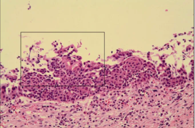
All mandibular anterior teeth except the left 1st incisor proved to be vital on electric pulp test. At the operation, the lesion consisted of multiple and sepa- rated cystic cavities with a very thin wall. The loca- tion and radiographic appearance of the abnormality and the age of the patient suggested a differential diagnosis of botryoid odontogenic cyst, odontogenic Jeong-Hun Nam, Da-Young Kim, Young-Ju Park, Jang-Hoon Ahn, Tae-In Gang,

Mi-Hee Park, Woo-Geun Yu, Bo-Gyun Kim, Jung-Won Lee, Jung-Hee Kim

1

Department of Oral and Maxillofacial Surgery, Kangnam Sacred Heart Hospital, Seoul, Korea

1

Dental Hospital, Graduate School of Clinical Dentistry, College of Medicine, Hallym University, Seoul, Korea

Botryoid Odontogenic Cyst on Mandibular Anterior and Both Body Area:

a Case Report

Botryoid odontogenic cyst (BOC) is considered a rare multilocular variant of the lateral periodontal cyst. In this report, a 67-year-old male visited with chief complaint of severe mobility on mandibular incisors.

Multilocular radiolucent lesion was seen from the right premolar to the left premolar area, involving almost the whole mandible. Histologically, the botryoid odontogenic cyst showed focal nodular thickening of the lining epithelium. These thickening often showed swirling appearance of the cells. Cyst enucleation and bone graft on mandible anterior and both body area were performed under general anesthesia, and postop- erative healing was favorable without recurrence.

Key words: Botryoid odontogenic cyst, Lateral periodontal cyst

Abstract

(2)

keratocyst and ameloblastoma. Finally, cyst enucle- ation and bone graft with xenograft (Biocera

, Osscotec, Korea) and allograft (Oragraft

, Megazen, Korea) materials.

During postoperative follow-up period, bony expan- sion disappeared and normal bone structure was formed on labial surface at 6 months after surgery (Fig. 3). Additionally, new bone formation occurred at operative site and there was no recurrence at 11 months after operation (Fig. 4).

The surgical specimen of the initial lesion consisted of several separate cyst cavities. Histologically, these were generally lined by one to three cell layers with thin and cuboidal epithelium. There was prominent separation of the epithelium from surrounding fibrous connective tissue wall. Botryoid odontogenic cyst showed focal nodular thickening of the lining epithe- lium. Focal thickening often showed swirling appear- ance of the cells (Figs. 5-7).

Fig. 1. Pre-operative panoramic view. Multilocular radiolu- cent lesion was seen from right mandibular 2nd premolar to left mandibular 1st premolar. There was an slight dis- placement of root area between right mandibular 2nd incisor and right mandibular canine. There was no root resorption and displacement of other involved teeth.

Fig. 2. Pre-operative axial and coronal CT scan.

Multilocular radiolucent lesion was seen mandibu- lar anterior and both body area. Bone expansion and thinning change of bucco- labial cortical bone were seen on mandibular anteri- or area.

Fig. 3. 3-D CT scan at post-

operative 6 months. New

bone formation noted

from right mandibular 2nd

premolar to left mandibu-

lar 1st premolar. Bone

expansion disappeared on

labial surface and there

was seen normal bone

healing structure.

(3)

Discussion and Conclusion

The first reported case of multilocular cystic lesion in the jaws resembled a cluster of grapes and was called botryoid odontogenic cyst.

3)

Since this original description, the BOC has been widely regarded as a variant of the lateral periodontal cyst and publica- tions on the subject have not distinguished these from the smaller multicystic lateral periodontal cysts.

4)

Microscopically, the lesion is quite similar to the lateral periodontal cyst. Radiographically, most are monocystic; but occasional polycystic or multiloc- ular cases have been encountered.

5)

The most fre- quent location of the BOC is the mandibular premo- lar area.

6)

In this case, the BOC extended from the roots of the right premolar to the left premolar of the mandible. However, this multilocular radiolucency is not characteristic of the BOC. Radiographically and histologically, there are similarities between the BOC and some odontogenic cysts and odontogenic tumors such as ameloblastoma, odontogenic myxoma, glan- dular odontogenic cyst

7)

and various others in terms of appearance.

Odontogenic keratocyst (OKC) is more likely found from 10 to 40 years of age. The mandible is involved in 60 to 80% of cases, with a marked tendency to involve the posterior body and ascending ramus. The OKC tends to grow in an antero-posterior direction within the medullary cavity of the bone without

Fig. 4. Panoramic view at post-operative 11 months.

Healing was favorable without recurrence.

Fig. 5. Botryoid odontogenic cyst has a thin, fibrous wall with an epithelial lining that is one to three cells' thickness in most area. This epithelium usually consists of flattened squamous cells, but sometimes the cells are cuboidal in shape (H&E stain, × 100 magnification).

Fig. 6. Botryoid odontogenic cyst shows focal nodular thick- ening of the lining epithelium, which are composed chiefly of clear cells (H&E stain, × 200 magnification).

Fig. 7. There was a thin epithelial lining with focal nodular

thickening. These thickening often shows swirling appear-

ance of the cells (H&E stain, × 200 magnification).

(4)

causing obvious bone expansion. This feature may be useful in differential clinical and radiographic diagno- sis. The diagnosis of odontogenic keratocyst is based on the histopathologic features. The epithelial lining is 6 to 8 cells thick, with a hyperchromatic and pal- isaded basal cell layer. There was corrugated parak- eratotic surface. The reported frequency of recurrence in various studies ranges from 5% to 62%.

8)

Ameloblastoma is encountered in patients over a wide age range. A painless swelling of expansion of the jaw is the usual clinical presentation. The most typical radiographic feature is that of a multilocular radiolucent lesion. The lesion is often described as having a “soap-bubble”or “honey-combed”appear- ance. The follicular and plexiform patterns are the most common. Recurrence rates of 50 to 90% have been reported in various studies after curettage.

9)

Odontogenic myxoma is more likely found in 25 to 30 years. Radiographically, the myxoma appears a unilocular or multilocular radiolucency that may dis- place or cause resorption of teeth in the area of the tumor. Histopathologically, A loose, myxomatous tumor can be seen filling the marrow spaces between the bony trabeculae. The inset shows stellate-shaped cells and fine collagen fibrils.

10)

Preoperative differen- tial diagnosis can be carried out by means of incision- al biopsy.

The BOC is known to be a recurrent odontogenic cyst. The recurrence rate may range from 15 to 20%.

11,12)

The proliferation rate of the epithelial lining has to be determined to estimate the possibility of recurrence. The risk of recurrence of the BOC is sim- ilar to that of odontogenic keratocyst.

13)

Histologically, the BOC has a distinctive thin, nonkeratinized epithelium which is one to five cell layers thick, and it has cystic spaces lined by squa- mous epithelium with thickening and clear cells.

14)

The BOC shows focal nodular thickening of the lining epithelium and swirling appearance. Clinicopathologic analysis of ten cases with three recurrences of BOC was reported. Eight of ten lesions were located in the mandible; the anterior mandible being the dominant site. Five of the ten lesions were unilocular, the largest measuring 4.5 × 1.2 cm. Only two of the ten botryoid odontogenic cysts were radiographically mul- tilocular. Three lesions represented recurrences; 8,

10 and 10 years after previous surgical intervention, respectively. Perhaps the most significant informa- tion gained from this investigation of botryoid odon- togenic cysts is the fact three of ten lesions recurred after initial surgical removal. It is important that practitioners take note of the fact that: 1) there may be a heightened recurrence potential for the botryoid odontogenic cyst that has gone unrecognized 2) recurrence may not occur until a decade after initial surgery; and, 3) lesions may occasionally become destructive.

15)

Histologically, the surgical specimen of the 1st recurrence was covered by a hyperplastic oral epithe- lium, and the connective tissue between the surface epithelium and cyst epithelium contained several microcysts.

16)

These small cysts were lined by thin squamous epithelium and many of them were filled with keratin. It can be assumed that remnants of the thin cyst wall had been behind, resulting in multiple recurrences. In this report, histologically, there was little hyperplastic oral epithelium. Furthermore, cys- tic lesion was not filled with keratin. There has been no recurrence during postoperative follow-up, but further long-term check-up will be necessary in order to diagnose possible recurrences in time.

In conclusion, the BOC was commonly removed with cyst enucleation. But, size and multilocular pat- terns of lesion could be the main factors associated to recurrence in botryoid odontogenic cyst. The botryoid odontogenic cyst is rare but often found in cystic lesion when it is found multilocular pattern on dental X-ray screening, the clinician should include botryoid odontogenic cyst to the differential diagnosis and to optimal operation.

References

1. Neville BW, Damm DD, Allen CM et al : Gingival cyst of adult, Oral & Maxillofacial pathology, 2nd ed.

Philadelphia, WB Saunders, 2002, p.602.

2. Redman RS, Whitestone BW, Winne CE : Botryoid odon- togenic cyst : report of a case with histologic evidence of multicentric origin. Int J Oral Maxillofac Surg 19 : 144, 1990.

3. Ucok O, Aman ZY, Aykul TB : Botryoid odontogenic cyst : report of a case with extensive epithelial proliferation. Int J Oral Maxillofac Surg 34 : 693, 2005.

4. Gurol M. Burkes EJ, Jacoway J : Botryoid odontogenic

cyst : analysis of 33 cases. J Periodontol 66 : 1069, 1995.

(5)

5. Weathers DR, Waldron CA : Unusual multilocular cysts of the jaws (botryoid odontogenic cysts). Oral Surg 36 : 235, 1973.

6. Altini M, Shear M : The lateral periodontal cyst. J Oral Pathol Med 21 : 245, 1992.

7. Oh JS, Kim SG, Kim HK et al : Clinical and histopatho- logic analysis of glandular odontogenic cysts of the jaws. J Kor Acad Maxillofac Plast Reconstr Surg 29 : 451, 2007.

8. Neville BW, Damm DD, Allen CM et al : Odontogenic ker- atocyst, Oral & Maxillofacial pathology, 2nd ed.

Philadelphia, WB Saunders, 2002, p.594.

9. Neville BW, Damm DD, Allen CM et al : Ameloblastoma, Oral & Maxillofacial pathology, 2nd ed. Philadelphia, WB Saunders, 2002, p.611.

10. Neville BW, Damm DD, Allen CM et al : Odontogenic myxoma, Oral & Maxillofacial pathology, 2nd ed.

Philadelphia, WB Saunders, 2002, p.635.

11. Heikinheimo K, Happonen RP, Forssell K : A botryoid odontogenic cyst with multiple recurrences. Int J Oral Maxillofac Surg 18 : 10, 1989.

12. Mendes PD : Botryoid odontogenic cyst; clinical and pathological analysis in relation to recurrence. Med Oral Pathol Oral Cir Bucal 12 : 594, 2007.

13. Standish SM, Shafer WG : The lateral periodontal cyst. J Periodontol 29 : 27, 1958.

14. Lynch DP, Madden CR : The botryoid odontogenic cyst.

Report of the a case and review of the literature. J Periodontol 56 : 163, 1985.

15. Robert O, Green JR : Botryoid odontogenic cyst; clinico- pathologic analysis of ten cases with three recurrences. J Oral Maxillofac Surg 46 : 574, 1988.

16. Shear M, Pindborg JJ : Microscopic features of the lateral periodontal cyst. Scand J Dent Res 83 : 103, 1975.

저자 연락처

우편번호 150-950

서울시 영등포구 대림1동 948-1

한림대학교 의과대학 강남성심병원 구강악안면외과 남 정 훈

원고 접수일 2010년 05월 11일 게재 확정일 2010년 07월 07일

Reprint Requests Jeong-Hun Nam

Department of Oral and Maxillofacial Surgery, Kangnam Sacred Heart Hospital, College of Medicine, Hallym University 948-1, Daerim-1 Dong, Yeongdeungpo-Gu, Seoul, 150-950, Korea Tel: +82-2-829-5233

E-mail: [email protected]

Paper received 11 May 2010

Paper accepted 7 June 2010

수치

Fig. 1. Pre-operative  panoramic  view.  Multilocular  radiolu- radiolu-cent lesion was seen from right mandibular 2nd premolar to  left  mandibular  1st  premolar
Fig. 7. There was a thin epithelial lining with focal nodular thickening. These thickening often shows swirling  appear-ance of the cells (H&E stain, × 200 magnification).

참조

관련 문서