Two Cases of Caseous Calcification of the Mitral Annulus
4
0
0
전체 글
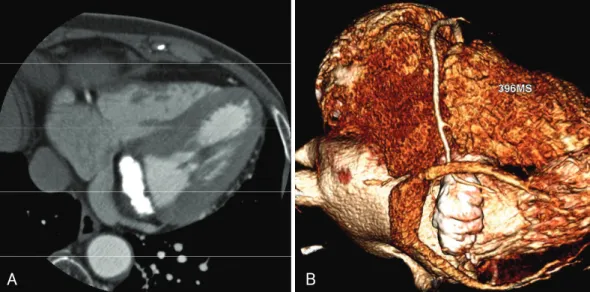
(2) Kyeong Min Byeon, et al.·83. (CT) was performed in order to characterize this masslike lesion. Cardiac CT revealed a dense calcified mass in the mitral valvular annulus, measuring 3.6×16×18 mm. Attenuation in the center of the mass was lower than that seen in the border. The coronary arteries were normal,. with no stenosis (Fig. 3). We thought there was a slight possibility that the caseous MAC was associated with the patient’s symptoms, and we decided to follow her condition. She returned three months later and had no symptoms.. Fig. 1. Plain chest radiograph (posteroanterior projection) shows a 23 mm-sized nodule in the left lower lung zone, retrocardiac area (arrow).. Case 2 A 65-year-old woman was brought to a local hospital in an unconscious state. A brain CT scan had revealed no significant findings. As there was no improvement in her clinical status, she was referred to our hospital. She had no history of relevant clinical illness. Initial physical examination revealed a blood pressure of 241/ 134 mmHg, a pulse rate of 106 beats/min, a respiratory rate of 30 breaths/min, and a body temperature of 37.4℃. The patient was semi-comatose. Her heart rhythm was regular, and no abnormal heart sounds were detected. Abnormal laboratory parameters were as follows: white blood cell count, 15,350/μL (segmented neutrophils, 92.1%); hemoglobin, 16.5 g/dL; platelet count, 143× 103/μL; prothrombin time, 15.5 seconds (International Normalized Ratio, 1.26); activated prothrombin time, 39.5 second; total cholesterol, 240 mg/dL; low-density lipoprotein cholesterol, 165 mg/dL; high-density lipoprotein cholesterol, 63 mg/dL; triglyceride 93 mg/dL; ionized calcium, 1.39 mmol/L; creatinine kinase MB fraction, 26.14 ng/mL; cardiac troponin I, 5.151 ng/mL;. A. B. LA. LA. C. LA. D. LV. LA. LV. LA. Fig. 2. Echocardiograph of case 1. A: parasternal long axis view shows a mitral annular calcification. B: zoomed image of the mitral valve shows a large, spherical echogenic mass with a smooth border. C: apical 2-chamber view of this calcified mass has a central echolucent area. D: color Doppler image shows no vascularity within the mass..
(3) 84·Mitral Annular Calcification. and N-terminal prohormone brain natriuretic peptide, 2,509 pg/mL. No significant ST-T segment changes were noted on the initial electrocardiogram, and the LV ejection fraction and wall motion were normal on echocardiography performed in the emergency room. The next day, the patient’s creatinine kinase MB fraction level had decreased to normal. Brain magnetic resonance imaging showed multiple embolic infarctions, so she was started on anticoagulation therapy. On the third day after presentation, the. A. patient underwent transthoracic echocardiography to assess cardiac abnormalities relevant to the multiple embolic events. A round, echogenic mass, measuring 25.5 ×12.7 mm, was visualized in the posterior-to-posterolateral mitral leaflet, extending to the endocardium of the LV base. The mass had a central echolucent area, measuring 12.8×9.6 mm. The left and right atria were enlarged, and there was mild mitral stenosis and regurgitation (Fig. 4A and B). On the fourth day, her orientation and consciousness were restored to normal, but. B. Fig. 3. Cardiac computed tomography. In the contrast-enhanced axial (A) and reconstructed volume rendering images (B), a dense calcified mass in the mitral annulus measuring 3.6×16×18 mm is shown. The coronary arteries are normal.. A. B. C. D. Fig. 4. Echocardiograph of case 2. Transthoracic echocardiography (A and B) and transesophageal echocardiography (C and D). A: parasternal long axis view of a spherical bright echogenic mass in the mitral annular area. B: parasternal short axis view of echogenic material around the mitral annular area. C: egg-shell like mass. D: color Doppler image shows mild mitral regurgitation and no significant flow communication between the central echolucent area and the cardiac chamber..
(4) Kyeong Min Byeon, et al.·85. there was residual dysarthria and difficulty swallowing. Transesophageal echocardiography was performed after two weeks of anticoagulation to better characterize the mass and to evaluate for the presence of an intracardiac thrombus. The mass, measuring 17.3×14.0 mm, had a smooth border with central echolucency (11.0× 9.5 mm). Mild mitral stenosis was noted, with a mean pressure gradient of 3.4 mmHg, but there was no intracardiac thrombus (Fig. 4C and D). The patient was treated with anticoagulant and antihypertensive medications. She was followed up in the outpatient clinic, and no additional embolic events occurred over the next six months. At that time, anticoagulation therapy was discontinued, and the patient was switched to an antiplatelet agent.. Discussion The prevalence of caseous MAC has been reported to be 0.0557)-0.067%2) in general populations referred for echocardiographic evaluation. The hallmark feature of this variant form of MAC is central liquefaction. Histology shows a calcified envelope and an amorphous acellular eosinophilic material surrounded by macrophages and lymphocytes. The mechanism of central liquefaction is unknown. Both of the current cases showed the typical echocardiographic appearance of caseous MAC: large, round, echo-dense masses with smooth borders, containing central echolucent areas along the posterior mitral annulus. The first patient had mild dyspnea, but her symptoms had little correlation with the echocardiographic findings. Most of the symptoms associated with caseous MAC that lead to referral for echocardiography are non-specific. The echocardiographic findings of caseous calcification are incidental and are usually unrelated to the symptoms.2) Because caseous MAC is known to be indolent and benign, most patients are treated conservatively.2) However, in some cases, surgery4-8) is performed based on suspicion of an intracardiac tumor, for prevention of embolism, or for treatment of severe mitral valvular dysfunction. Although MAC is regarded as a risk factor for systemic embolism and stroke,1) the risk of these entities has not been established in the setting of caseous MAC. Harpaz et al.2) reported that during a mean follow-up period of 3.8 years, 7 (37%) patients had unchanged repeat echocardiographic studies, which suggest the stability of this condition. Most case reports have shown good prognosis. with no cerebrovascular events. Interestingly, one study reported spontaneous resolution at the one-year followup visit.10) However, at least one case has been reported in association with embolic stroke;6) therefore, patients presenting with a decrease in mass size and flattening in the echolucent center warrant careful follow-up. The second patient in the current study had cerebral embolic infarction. Neither the transthoracic echocardiography performed on the third day after presentation nor the transesophageal echocardiography performed two weeks after the start of anticoagulation revealed any potential source of embolism. However, it is possible that a thrombus undetected on transthoracic echocardiography resolved after anticoagulation. Therefore, the relationship between caseous mitral calcification and embolism in our cases is still uncertain. In addition, the size of the mass decreased during the short period of 10 days, but the echolucent center did not flatten. Unfortunately, interpretation is limited because earlier measurements were done through the transthoracic window, whereas later measurement were done through the transesophageal window. REFERENCES 1) Zipes DP, Libby P, Bonow RO, Braunwald E. Braunwald’s He-. 2). 3). 4). 5). 6). 7) 8). 9) 10). art Disease: A Textbook of Cardiovascular Medicine. 7 ed. Philadelphia: W.B. Saunders; 2005. Harpaz D, Auerbach I, Vered Z, Motro M, Tobar A, Rosenblatt S. Caseous calcification of the mitral annulus: a neglected, unrecognized diagnosis. J Am Soc Echocardiogr 2001;14:825-31. Teja K, Gibson RS, Nolan SP. Atrial extension of mitral annular calcification mimicking intracardiac tumor. Clin Cardiol 1987; 10:546-8. Kato M, Nakatani S, Okazaki H, Tagusari O, Kitakaze M. Unusual appearance of mitral annular calcification mimicking intracardiac tumor prompting early surgery. Cardiology 2006;106: 164-6. de Very EA, Scholte AJ, Krauss XH, et al. Intracardiac pseudotumor caused by mitral annular calcification. Eur J Echocardiogr 2006;7:62-6. Poh KK, Wood MJ, Cury RC. Prominent posterior mitral annular calcification causing embolic stroke and mimicking left atrial fibroma. Eur Heart J 2007;28:2216. Kronzon I, Winer HE, Cohen ML. Sterile, caseous mitral anular abscess. J Am Coll Cardiol 1983;2:186-90. Gilbert HM, Grodman R, Chung MH, Hartman G, Krieger KH, Hartman BJ. Sterile, caseous mitral valve “abscess” mimicking infective endocarditis. Clin Infect Dis 1997;24:1015-6. Stone E, Cohn D, Deal C, Pollock C. Calcific atrial mass in endstage renal failure. Nephrol Dial Transplant 1997;12:807-10. Gramenzi S, Mazzola AA, Tagliaferri B, et al. Caseous calcification of the mitral annulus: unusual case of spontaneous resolution. Echocardiography 2005;22:510-3..
(5)
수치

관련 문서
• 이명의 치료에 대한 매커니즘과 디지털 음향 기술에 대한 상업적으로의 급속한 발전으로 인해 치료 옵션은 증가했 지만, 선택 가이드 라인은 거의 없음.. •
q March 28, 1906: County Magistrate Shim Heung-taek of Uldo-gun[Uldo County] heard this news from the Japanese survey team, he reported to the Korean central government
In a statement to Kuwait News Agency (KUNA) on the sidelines of a meeting of the Arab Parliament's Foreign Affairs Political and National Security
The meeting was attended by Assistant Foreign Minister for GCC Affairs, Ambassador, Nasser Al-Muzayyen, and Deputy Assistant Foreign Minister for the Office of the
“ Sheikh Nasser has a written message from HH the Amir, Sheikh Sabah Al-Ahmad Al-Jaber Al-Sabah to the Chinese President, Chi Gen Beng related to enhancing mutual
On his part, CEO of Express Roads Authority, Saud Al-Naqqi said that the heavy rains of the previous day led to clogging parts of the express
Kuwait will celebrate on Sunday the fourth anniversary of the UN honoring and proclamation of His Highness the Amir, Sheikh Sabah Al-Ahmad Al-Jaber Al-Sabah as
The Joseon government designed and promulgated the Taegeukgi as a national flag for diplomatic and political purposes, but it was the independence movement that made it
