224
Open Access
The Value of Brachial-Ankle Pulse Wave Velocity as a Predictor of Coronary Artery Disease in High-Risk Patients
Won-Woo Seo, MD
1,2, Hyuk-Jae Chang, MD
1,2, IkSung Cho, MD
1,2, Yeon-Yee Yoon, MD
1,2, Jung-Won Suh, MD
1,2, Kwang-Il Kim, MD
1,2, Yong-Seok Cho, MD
1,2, Tae-Jin Youn, MD
1,2, In-Ho Chae, MD
1,2,
Dong-Ju Choi, MD
1,2, Cheol-Ho Kim, MD
1,2, Eun-Ju Chun, MD
3and Sang-Il Choi, MD
31Department of Internal Medicine, Seoul National University College of Medicine, Seoul,
2Division of Cardiology and 3Radiology, The Cardiovascular Center, Seoul National University Bundang Hospital, Seongnam, Korea
ABSTRACT
Background and Objectives
: Arterial stiffness has been known as an independent contributory factor for coronary artery disease (CAD). Brachial-ankle pulse wave velocity (baPWV) is widely used as a simple noninvasive measure of arterial stiffness. The aim of our study was to test whether baPWV had predictive value for CAD in the subset of patients with high pretest probability.
Subjects and Methods: We enrolled 174 consecutive patients who were referred for evaluation of suspected CAD, and who underwent both baPWV measurement and computed tomo- graphy (CT) for coronary artery calcium scoring (CACS) as part of a diagnostic work-up. Subsequently, 160 of those patients underwent invasive coronary angiography. The CAD indices consisted of 1) CACS, 2) modified Gensini scoring system, and 3) presence of obstructive CAD and 4) multi-vessel obstructive CAD.
Results: baPWV correlat- ed with CACS (r=0.25, p=0.001), but not with modified Gensini scoring (r=0.10, p=0.19). However, after adjust- ment for factors influencing PWV, baPWV no longer correlated with CACS (r=0.14, p=0.14). By receiver operat- ing characteristic (ROC) curve analysis, baPWV was neither a sensitive nor specific index for predicting the pre- sence of obstructive CAD or multi-vessel obstructive CAD (sensitivity: 53% and 59%; specificity: 50% and 55%, respectively).
Conclusion: Our findings demonstrated that baPWV is associated with CACS, however, this may be primarily attributed to common risk factors, such as age. Furthermore, baPWV may be of limited value in identify- ing patients at risk for CAD.
(Korean Circ J 2010;40:224-229)KEY WORDS:
Coronary artery disease; Compliance.
Introduction
It is well known that arterial stiffness is increased by cardiovascular risk factors such as age, hypertension, dia- betes, and chronic kidney disease. As a consequence of elevated arterial stiffness and decreased vascular dis- tensibility, there is an increase in systolic blood pressure,
which causes an increase in left ventricular workload and subsequent left ventricular hypertrophy, and a de- crease in diastolic blood pressure, which leads to im- paired coronary perfusion.
1)2)There are various nonin- vasive methods for measuring arterial stiffness, such as ultrasound and MRI. These technologies detect changes in the diameter (or area) of an artery in response to dis- tending pressure, and pulse wave velocity (PWV). Among these measurements, PWV is a simple index derived from pressure-sensitive transducers, doppler ultrasound, or ap- planation tonometry. Thus, it is widely used in clinical settings.
3)4)PWV reflects the degree of arterial sclerosis, and aortic stiffness assessed by PWV is an independent risk factor for cardiovascular diseases.
1)4)5)However, it is not known whether an increase in peripheral artery PWV is related to severity of coronary artery disease (CAD), or whether PWV can be used to predict CAD.
Invasive coronary angiography is the gold standard
Received: March 27, 2009
Revision Received: September 24, 2009 Accepted: November 22, 2009
Correspondence: Hyuk-Jae Chang, MD,Division of Cardiology, The Cardio- vascular Center, Seoul National University Bundang Hospital, 166 Gumi- ro, Bundang-gu, Seongnam 463-707, Korea
Tel: 82-31-787-7009, Fax: 82-31-787-4051 E-mail: [email protected]
This is an Open Access article distributed under the terms of the Creative Commons Attribution Non-Commercial License (http://creativecommons.
org/licenses/by-nc/3.0) which permits unrestricted non-commercial use, distribution, and reproduction in any medium, provided the original work is properly cited.
cc
for detecting CAD. It is useful not only for the diagno- sis of obstructive CAD, but also for determining the se- verity of CAD by evaluating the degree of luminal nar- rowing and identifying the anatomical location of the obstruction.
6)Coronary artery calcification develops as part of the atherosclerotic process, and therefore occurs almost ex- clusively in atherosclerotic arteries and is absent in nor- mal vessel walls.
7)Cardiac computed tomography (CT) is highly sensitive for detecting coronary artery calcium and is being used with increasing frequency to screen for CAD in both asymptomatic and symptomatic pa- tients. Previous studies with cardiac CT reported that the severity of coronary atherosclerosis could be evaluated noninvasively, and that the prognosis of CAD could be predicted by coronary artery calcium scoring (CACS).
7)8)Brachial-ankle pulse wave velocity (baPWV) is the most widely used PWV index because of its convenience.
The aim of this study was to evaluate the usefulness of baPWV as a predictor of the severity of coronary athero- sclerosis. Furthermore, we assessed its value for predict- ing the severity and presence of obstructive CAD in high- risk patients suspected of having CAD.
Subjects and Methods
Study population
Between March 2005 and February 2008, 174 conse- cutive patients were enrolled, who had been referred for evaluation of suspected CAD and who had undergone both baPWV measurement and cardiac CT for CACS at the Seoul National University Bundang Hospital. Pa- tients with a history of obstructive CAD were excluded.
Based on clinical judgment, 92% of patients (160 of 174) underwent invasive coronary angiography.
Brachial-ankle pulse wave velocity
After a subject was in the supine position for 5 or more minutes, blood pressure and baPWV were measured us- ing an automated waveform analyzer (Colin VP-2000, Colin Medical Instruments Corp., Komaki, Japan). Blo- od pressure was measured in both arms using the blood pressure cuffs of the device, and the higher value was used for further analysis. The mean of baPWV measur- ed bilaterally was used for analysis.
Cardiac computed tomography
Scanning was performed using a 64-slice multi-de- tector CT scanner (Brillance 64; Philips Medical systems, Best, The Netherlands) with a tube voltage of 120 kV, tube current of 55 mAs, 200 mm, collimation of 40×
0.625 mm, and gantry rotation speed of 0.40 second.
The score for each calcified focus of coronary artery was determined by the method of Agatston et al.
9)and the total value for CACS was obtained by summing indivi-
dual focus scores from each of the 4 epicardial arteries (left main, left anterior descending, circumflex, and right coronary arteries).
Coronary angiography
Coronary angiography was performed at the discre- tion of the examining physician. The procedure was per- formed through the right radial artery, or, if that app- roach proved difficult, through the right femoral artery.
Obstructive CAD was defined as coronary artery lumi- nal narrowing greater than 50% by gross measurement.
Moreover, if luminal narrowing greater than 50% in- volved the left main coronary artery or more than 3 co- ronary arteries, the patient was diagnosed with multi-ves- sel obstructive CAD.
10)The severity of CAD was classifi- ed using the modified Gensini scoring system.
6)Statistical analysis
Values are expressed as means±standard deviation.
Continuous variables were compared using the unpaired t-test, and categorical variables were compared using the χ
2test. Pearson product-moment correlation was used to assess the correlations between baPWV and CACS and modified Gensini scoring values. Multivariate logistic regression analysis was carried out to assess the odds ra- tios of baPWV for obstructive CAD. Receiver operating characteristic (ROC) curve analyses were used to deter- mine the optimal baPWV cut-off values for predicting obstructive CAD. Statistical analysis was performed us- ing the Statistical Package for the Social Sciences (SP- SS) version 12 (SPSS Inc., Chicago, IL, USA), and p<0.05 was considered significant.
Results
Baseline clinical characteristics
The mean age was 63±9 years (n=174; range 33-92 years); 68% were male (118 patients), 74% were hyper- tensive (129 patients), 27% had diabetes (47 patients), and 49% had smoked within 3 months (86 patients).
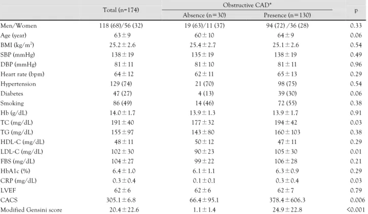
Among 160 patients who underwent coronary angio- graphy, obstructive CAD was detected in 130 patients (81%). Among them, 36 patients were diagnosed with multi-vessel obstructive CAD. The mean modified Ge- nsini score of the 160 patients was 20.4±22.6. Patients with obstructive CAD had significantly higher values of total cholesterol (TC), low density lipoprotein cho- lesterol, C-reactive protein levels, CACSs, and modified Gensini scores compared with the patients without ob- structive CAD, and there were no statistical differences observed in the other indicators (Table 1).
Correlation analysis of brachial-ankle pulse wave velocity and coronary artery calcium scoring
Among the entire study population, the baPWV sig-
nificantly correlated with CACS by bivariate correla- tion analysis (r=0.25; p=0.001). However, after adjust- ing for factors influencing PWV, including sex, age, body mass index, blood pressure, heart rate, TC, and fasting blood sugar, baPWV no longer correlated with CACS (r=0.14; p=0.14) (Table 2).
Correlation between brachial-ankle pulse wave velocity and coronary artery disease severity
To evaluate the correlation between baPWV and CAD severity, simple correlation analysis was carried out be- tween the baPWV and modified Gensini score. There was no significant correlation (r=0.10, p=0.19) seen, and also no significant correlation after adjustment for fac- tors that influence baPWV (r=0.07, p=0.40) (Table 2).
The usefulness of brachial-ankle pulse wave velocity as a predictor of obstructive coronary artery disease
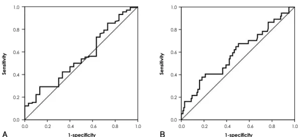
By multivariate logistic regression analysis, significant predictors of obstructive CAD were age, TC level, and CACS; whereas baPWV was not revealed to be predic- tive of obstructive CAD (Table 3). In order to assess the usefulness of baPWV as a predictor of obstructive CAD, we attempted to determine the optimal cut-off baPWV value by ROC curve analysis. The cut-off point 1,546 cm/s showed 53% sensitivity and 50% specificity in pre- dicting obstructive CAD, and the cut-off point 1,593 cm/s
Table 1. Baseline characteristics of patients
Obstructive CAD*
Total (n=174)
Absence (n=30) Presence (n=130) p
Men/Women 118 (68)/56 (32) 19 (63)/11 (37) 94 (72) /36 (28) 0.33
Age (year) 63±9 60±10 64±9 0.06
BMI (kg/m2) 25.2±2.6 25.4±2.7 25.1±2.6 0.54
SBP (mmHg) 138±19 135±19 138±19 0.49
DBP (mmHg) 81±11 81±10 81±11 0.96
Heart rate (bpm) 64±12 62±11 65±13 0.29
Hypertension 129 (74) 21 (70) 98 (75) 0.54
Diabetes 47 (27) 4 (13) 39 (30) 0.06
Smoking 86 (49) 14 (46) 72 (55) 0.38
Hb (g/dL) 14.0±1.7 13.9±1.3 13.9±1.7 0.91
TC (mg/dL) 191±40 177±32 194±42 0.03
TG (mg/dL) 155±97 143±80 160±103 0.38
HDL-C (mg/dL) 48±11 50±12 47±11 0.29
LDL-C (mg/dL) 102±30 90±23 105±30 0.01
FBS (mg/dL) 104±27 99±22 106±28 0.21
HbA1c (%) 6.4±1.0 6.1±1.1 6.3±0.9 0.29
CRP (mg/dL) 0.3±0.4 0.1±0.1 0.3±0.4 0.03
LVEF 62±6 62±6 62±7 0.79
CACS 305.1±6.8 66.4±95.1 378.4±606.3 0.006
Modified Gensini score 20.4±22.6 1.1±1.4 24.9±22.8 <0.001
Data were presented as n (%) or mean±standard deviation. *Obstructive CAD was defined as >50% luminal narrowing of coronary artery.
CAD: coronary artery disease, BMI: body mass index, SBP: systolic blood pressure, DBP: diastolic blood pressure, Hb: hemoglobin, TC: total cholesterol, TG: triglyceride, HDL-C: high density lipoprotein-cholesterol, LDL-C: low density lipoprotein-cholesterol, FBS: fasting blood sugar, HbA1c: Hemoglobin A1c, CRP: C-reactive protein, LVEF: left ventricular ejection fraction, CACS: coronary artery calcium score Table 2. Correlation analyses of baPWV with CACS and seve-
rity of CAD
BaPWV
Bivariate analysis Partial analysis*
r p r p
CACS 0.25 0.001 0.14 0.14
Modified Gensini score 0.10 0.190 0.07 0.40
*After adjusting for factors influencing PWV (sex, age, body mass in- dex, blood pressure, heart rate, total cholesterol, fasting blood sugar).
baPWV: brachial-ankle pulse wave velocity, CACS: coronary artery calcium score, CAD: coronary artery disease, r: correlation coeffi- cient
Table 3. Logistic regression analyses of various factors in the pre- sence of obstructive coronary artery disease*
Factors p Odds ratio 95%
confidence interval
Sex 0.12 0.465 0.176-1.227
Age 0.03 1.049 1.003-1.097
BMI 0.39 0.926 0.777-1.105
TC 0.004 1.018 1.006-1.031
FBS 0.15 1.014 0.995-1.034
CACS 0.003 1.006 1.002-1.010
baPWV 0.10 1.001 1.000-1.003
*Obstructive CAD was defined as >50% luminal narrowing of co- ronary artery. BMI: body mass index, TC: total cholesterol, FBS:
fasting blood sugar, CACS: coronary artery calcium score, baPWV:
brachial-ankle pulse wave velocity
showed 59% sensitivity and 55% specificity in predict- ing multi-vessel obstructive CAD (Fig. 1).
Discussion
Aging induces structural and functional changes in vessel walls. These changes include arterial wall hyper- trophy, increased calcium deposition, and quantitative and qualitative changes in the collagen of the medial layer, which leads to disorganized microarchitecture. As a result, vessel wall compliance and elasticity decrease, and the PWV increases because of increased arterial stiff- ness.
11)In addition, changes in blood vessel tension and elasticity caused by increased blood pressure also have an impact on measured PWV, along with gender, diabetes, renal failure, and salt intake.
12)13)Increase in arterial stiffness measured as increased PWV, has been reported to be associated with atherosc- lerosis and the CAD.
5)14)Previous studies using aortic pulse wave velocity (aPWV) reported increases in odds ratio by 1.34 for cerebrovascular accident,
15)1.38 for CAD,
5)and 1.23 for cardiovascular mortality,
16)for each increase of aPWV by 340 cm/s. There are several me- chanisms that might explain the association between increased PWV and CAD. Arterial stiffness causes pre- mature return of the reflected pulse wave in late systole, leading to an increase in central pulse pressure and, con- sequently, to increased load on the ventricle. This results in decreased ejection fraction and increased myocardial oxygen demand.
5)In addition, the decreased absorption capacity of the arterial wall leads to wall injury, which ac- celerates the progression of atherosclerosis.
17)This study also evaluated the usefulness of baPWV, the most widely-used PWV index, for prediction of obst- ructive CAD, and whether baPWV correlated with the severity of coronary artery calcification or CAD in high-
risk patients. Although the CACS is a good reflection of the severity of coronary atherosclerosis and the prog- nosis of CAD, its value is limited for predicting the pre- sence and severity of obstructive CAD.
18)To compen- sate for the limitations of CACS, we evaluated the pre- sence of obstructive CAD with modified Gensini scoring and invasive coronary angiography to identify CAD.
baPWV correlated neither with CACS nor with mo- dified Gensini scoring after adjusting for factors influ- encing PWV. By multivariate logistic regression ana- lysis, baPWV failed to predict obstructive CAD. In ad- dition, baPWV showed low sensitivity and specificity for predicting the presence of obstructive CAD and mul- ti-vessel obstructive CAD by ROC curve analyses.
Several studies have investigated the relationship be- tween arterial stiffness and atherosclerotic burden. Meg- nien et al.
19)reported that aPWV did not correlate with the degree of CACS, and that aPWV was mainly affected by aging and blood pressure, etc.; therefore aPWV could not predict coronary atherosclerosis in asymptomatic patients with risk factors for cardiovascular disease. But study results were inconsistent. Haydar et al.
20)reported that aPWV was closely associated with CACS in patients with chronic kidney disease, and Kullo et al.
14)showed similar results in 401 asymptomatic patients.
While most of the previous studies were performed in asymptomatic patients or in those at low risk for CAD, most of our study patients either had obstructive CAD or they were at high risk for CAD. Accordingly, our mean CACS (305.1±6.8) and baPWV (1,651±354 cm/s) were higher than those of other study populations. Tsu- chiya et al.
21)showed that a cut-off value of 1,800 cm/s for baPWV predicted CACS values greater than 100 with 70% sensitivity and 79% specificity in asymptoma- tic patients with diabetes. In our study, however, the same cut-off value predicted a CACS greater than 100
1.0
0.8
0.6
0.4
0.2
0.0
Sensitivity
0.0 0.2 0.4 0.6 0.8 1.0 1-specificity
1.0
0.8
0.6
0.4
0.2
0.0
Sensitivity
0.0 0.2 0.4 0.6 0.8 1.0 1-specificity
A B
Fig. 1. ROC curve analysis of baPWV for obstructive CAD. A: for prediction of presence of obstructive coronary artery disease, baPWV cut-off value at 1,550 cm/s showed sensitivity 53%, specificity 50%, AUC 0.557. B: for prediction of multi-vessel obstructive CAD, 1,593 cm/s showed sensitivity 59%, specificity 55%, AUC 0.590. ROC: receiver operating characteristic, baPWV: brachial-ankle pulse wave velocity, CAD: coronary artery disease, AUC: area under curve.
with only 29% sensitivity and 87% specificity. This re- sult may demonstrate the wide variation among study populations.
Xu et al.
22)reported on a study of 272 patients with documented CAD showing that baPWV and ankle-bra- chial index were useful in predicting CAD and periph- eral artery disease, and that a baPWV greater than 1,800 cm/s was suggestive of severe atherosclerosis as well as obstructive CAD. In the current study, we applied the method of Xu, and divided the patients into 4 groups according to their modified Gensini score as follows: Ge- nsini score=0, 0.01-3.00, 3.01-32.67, >32.68. There was a significant difference in baPWV between the highest and lowest modified Gensini score group (p=0.049) without adjustment for age. It is well known that incre- asing age is the most decisive factor for increasing PWV regardless of the presence of atherosclerosis.
23)In our study, there was also a highly significant correlation be- tween age and baPWV (r=0.57, p<0.001), and after ad- justing for age, there was no significant correlation be- tween baPWV either with CACS or with modified Gen- sini scoring. By multivariate logistic regression analysis that included age, baPWV was not revealed to be a pre- dictor of obstructive CAD.
Marchand discovered that fatty degeneration was in- volved with arterial stiffening in 1904, and introduced the term “atherosclerosis”. Although the increase in ar- terial stiffness and the progression of atherosis share the same pathophysiological factors, their association and causality are not well understood. Besides the progres- sion of atherosclerosis, increased arterial stiffness is as- sociated with vessel wall changes such as increase in col- lagen fibers, and decrease in elastin and vascular smooth muscle tone, in addition to “extrinsic factors” such as hormones, salt, and glucose.
24)After excluding age, ch- olesterol plays an important role in the progression of atherosis, while blood pressure seems to play a more important role in the progression of arterial stiffness.
25)Therefore the increase in PWV, which represents arte- rial wall stiffness, does not simultaneously increase to the same degree as progression of atherosis.
While previous studies have used aPWV, which repre- sents central arterial wall stiffness, in this study we used baPWV, which has been increasingly used as an index of arterial stiffness because of its convenience. However, baPWV has some limitations. First, the pulse wave does not propagate directly from the brachial arteries to the posterior tibial arteries in the same arterial tree; thus, baPWV is indirectly estimated using the PWV from the heart to the brachial and posterior tibial arteries. Second, compared to the central arteries, peripheral arteries do not represent aging and disease-related changes as well.
26)Although Sugawara et al.
27)reported that baPWV pro- vided similar qualitative information to information de- rived from central arterial stiffness, the best indicator re-
flecting central artery stiffness is considered to be aPWV measured as the carotid-femoral PWV.
28)Because of its convenience, the examination of peri- pheral artery stiffness is widely used in various patient groups with risk factors for CAD. Although some stu- dies reported that patients at high risk for CAD can be screened using baPWV, the current study demonstrates that baPWV does not directly correlate with coronary artery calcification and severity of CAD in high-risk patients. Furthermore, baPWV also has low sensitivity and specificity for predicting obstructive CAD. Conse- quently, it may be of limited value as a screening tool for CAD in high-risk patient groups.
As mentioned previously, the current study popul- ation included 130 patients with obstructive CAD out of 160 patients who underwent coronary angiography.
Our study population differed significantly from those in other studies. Therefore extrapolating the findings of our study to the general population is difficult, and further studies of various study populations are needed.
Furthermore, since there is a significant difference be- tween aPWV and baPWV, future studies evaluating arte- rial wall stiffness should include these different indices.
REFERENCES
1) Mattace-Raso FU, van der Cammen TJ, Hofman A, et al. Arterial stiffness and risk of coronary heart disease and stroke: the Rot- terdam study. Circulation 2006;113:657-63.
2) Kim SH, Choi SI, Shin JH, et al. Arterial stiffness and abdomi- nal obesity are associated with the relaxation abnormality of left ventricle in human hypertension. Korean Circ J 2006;36:221-8.
3) Rhee MY, Lee HY, Park JB. Measurements of arterial stiffness:
methodological aspects. Korean Circ J 2008;38:343-50.
4) Oliver JJ, Webb DJ. Noninvasive assessment of arterial stiffness and risk of atherosclerotic events. Arterioscler Thromb Vasc Biol 2003;23:554-66.
5) Boutouyrie P, Tropeano AI, Asmar R, et al. Aortic stiffness is an independent predictor of primary coronary events in hypertensive patients: a longitudinal study. Hypertension 2002;39:10-5.
6) Gensini GG. A more meaningful scoring system for determining the severity of coronary heart disease. Am J Cardiol 1983;51:606.
7) O’Rourke RA, Brundage BH, Froelicher VF, et al. American Col- lege of Cardiology/American Heart Association Expert Consen- sus Document on electron-beam computed tomography for the diagnosis and prognosis of coronary artery disease. J Am Coll Cardiol 2000;36:326-40.
8) Budoff MJ, Gul KM. Expert review on coronary calcium. Vasc Health Risk Manag 2008;4:315-24.
9) Agatston AS, Janowitz WR, Hildner FJ, Zusmer NR, Viamonte M Jr, Detrano R. Quantification of coronary artery calcium using ul- trafast computed tomography. J Am Coll Cardiol 1990;15:827-32.
10) Kim HJ, Nam JS, Park JS, et al. Usefulness of brachial-ankle pulse wave velocity as a predictive marker of multiple coronary artery occlusive disease in Korean type 2 diabetes patients. Diabetes Res Clin Pract 2009;85:30-4.
11) Tomiyama H, Yamashina A, Arai T, et al. Influences of age and gender on results of noninvasive brachial-ankle pulse wave velo- city measurement: a survey of 12517 subjects. Atherosclerosis 2003;166:303-9.
12) Hopkins KD, Lehmann ED, Jones RL, Turay RC, Gosling RG. A family history of NIDDM is associated with decreased aortic dis- tensibility in normal healthy young adult subjects. Diabetes Care 1996;19:501-3.
13) Benetos A, Waeber B, Izzo J, et al. Influence of age, risk factors, and cardiovascular and renal disease on arterial stiffness: clinical applications. Am J Hypertens 2002;15:1101-8.
14) Kullo IJ, Bielak LF, Turner ST, Sheedy PF 2nd, Peyser PA. Aor- tic pulse wave velocity is associated with the presence and quan- tity of coronary artery calcium: a community-based study. Hyper- tension 2006;47:174-9.
15) Laurent S, Katsahian S, Fassot C, et al. Aortic stiffness is an in- dependent predictor of fatal stroke in essential hypertension.
Stroke 2003;34:1203-6.
16) Laurent S, Boutouyrie P, Asmar R, et al. Aortic stiffness is an in- dependent predictor of all-cause and cardiovascular mortality in hypertensive patients. Hypertension 2001;37:1236-41.
17) Demer LL. Effect of calcification on in vivo mechanical response of rabbit arteries to balloon dilation. Circulation 1991;83:2083-93.
18) Greenland PG, Bonow RO, Brundage BH, et al. ACCF/AHA 2007 clinical expert consensus document on coronary artery calcium scoring by computed tomography in global cardiovascular risk assessment and in evaluation of patients with chest pain. J Am Coll Cardiol 2007;49:378-402.
19) Megnien JL, Simon A, Denarie N, et al. Aortic stiffening does not predict coronary and extracoronary atherosclerosis in asympto- matic men at risk for cardiovascular disease. Am J Hypertens 1998;11:293-301.
20) Haydar AA, Covic A, Colhoun H, Rubens M, Goldsmith DJ. Co- ronary artery calcification and aortic pulse wave velocity in chro-
nic kidney disease patients. Kidney Int 2004;65:1790-4.
21) Tsuchiya M, Suzuki E, Egawa K, et al. Stiffness and impaired blood flow in lower leg arteries are associated with severity of co- ronary artery calcification among asymptomatic type 2 diabetic patients. Diabetes Care 2004;27:2409-15.
22) Xu Y, Wu Y, Li J, et al. The predictive value of brachial-ankle pulse wave velocity in coronary atherosclerosis and peripheral artery diseases in urban Chinese patients. Hypertens Res 2008;
31:1079-85.
23) Avolio AP, Chen SG, Wang RP, Zhang CL, Li MF, O’Rourke MF.
Effects of aging on changing arterial compliance and left ventri- cular load in a northern Chinese urban community. Circulation 1983;68:50-8.
24) Zieman SJ, Melenovsky V, Kass DA. Mechanisms, pathophysio- logy, and therapy of arterial stiffness. Arterioscler Thromb Vasc Biol 2005;25:932-43.
25) Blankenhorn DH, Kramsch DM. Reversal of atherosis and scle- rosis: the two components of atherosclerosis. Circulation 1989;
79:1-7.
26) Benetos A, Laurent S, Hoeks AP, Boutouyrie PH, Safar ME. Ar- terial alterations with aging and high blood pressure: a nonin- vasive study of carotid and femoral arteries. Arterioscler Thromb 1993;13:90-7.
27) Sugawara J, Hayashi K, Yokoi T, et al. Brachial ankle pulse wave velocity: an index of central arterial stiffness? J Hum Hypertens 2005;19:401-6.
28) Laurent S, Cockcroft J, van Bortel L, et al. Expert consensus do- cument on arterial stiffness: methodological issues and clinical applications. Eur Heart J 2006;27:2588-605.