The Effectiveness of Carvedilol, a New Antioxidant and Antiproliferative Beta-Blocker, on Prevention of Restenosis after Coronary Stent Implantation:a Prospective,
Randomized, Multicenter Study
Kwang-Soo Cha, MD
1, Moo-Hyun Kim, MD
1, Jin-Woo Kim, MD
2, Doo-Il Kim, MD
3, Hje-Jin Kim, RN
1, Young-Dae Kim, MD
1, Dong-Soo Kim, MD
3and Jong-Seong Kim, MD
11Department of Cardiology, Dong-A University Hospital, Busan, 2Department of Cardiology, Ulsan University Hospital, Ulsan, 3Department of Cardiology, Pusan Paik Hospital, Busan, Korea ABSTRACT
Background
: Carvedilol is a direct inhibitor of vascular smooth muscle cell migration and proliferation through inhibition of mitogen-activated protein kinase activity and regulation of cell cycle progression. It produced an 84% suppression of neointimal hyperplasia in rat carotid angioplasty model, but no data are available regarding its effect on stent restenosis in patients. We tested whether a sustained oral administration of carvedilol reduces restenosis after coronary stenting in patients.
Methods: One hundred fifty nine patients were randomized to receive either carvedilol (50 mg/day, n=80) or atenolol (50 mg/day, n=79) at least 1 day before stenting and continued on the same medication over 3 months. The primary end point was angiographic restenosis (>50% diameter stenosis) at follow-up angiography.
Results: Baseline clinical and angiographic variables were similar between the carvedilol and atenolol group. The carvedilol dose was tolerable in most patients after adjustment of other medications, but reduced in 3 patients due to hypotension and dizziness. Angiographic follow-up was done in 137 patients (86%) and the restenosis rate was not different significantly between both groups (17.1% versus 19.4%, p=0.732).
Conclusions: A sustained oral administration of carvedilol is not effective to reduce stent restenosis. With carvedilol targeting regulators of cell cycle progression and having a profound neointimal inhibition with a high blood concentration in an experiment, further investigations using a stent-based delivery to achieve a high local concentration may be warranted.
(Korean Circulation J 2004;34 (1):35-40)KEY WORDS
: Carvedilol; Stents; Coronary restenosis; Drug therapy.
Introduction
The long-term clinical efficacy of intracoronary sten- ting is limited by restenosis, which occurs in 15% to 30% of patients.
1)2)Stent restenosis is due solely to neointimal hyperplasia.
3-5)Stent-induced mechanical
arterial injury and a foreign body response to the pro- sthesis incite acute and chronic inflammation in the vessel wall, with elaboration of cytokines and growth factors that induce multiple signaling pathways to activate smooth muscle cell (SMC) migration and prolifer- ation.
3-5)Therefore, the delivery of the agents inhibiting cell cycle progression via stent platforms
6-8)or systemi- cally
9)produces effective inhibition of in-stent neoin- timal hyperplasia.
Carvedilol is a neurohumoral antagonist with multiple actions.
10)11)It was originally discovered to be a beta- adrenoreceptor antagonist.
11)However, subsequent re- search revealed that this agent possessed potent antioxi-
Received:June 30, 2003Revision received:October 6, 2003 Accepted:October 17, 2003
Correspondence:Kwang-Soo Cha, MD, Department of Cardi- ology, Dong-A University Hospital, 3 Ga 1, Dongdaesin-dong, Seo-gu, Busan 602-715, Korea
Tel:82-51-240-2880, Fax:82-51-247-5875 E-mail:[email protected]
Korean Circulation J 2004;34(1):35-40 36
dant and free radical scavenger properties.
12)In addition, carvedilol inhibits vascular SMC proliferation induced by a broad group of mitogens, such as platelet derived growth factor, fibroblast growth factor, endothelin-1, serum and thrombin.
13)14)Recently, the mechanisms res- ponsible for these actions of carvedilol have been proven due to the inhibition of mitogen-activated protein kinase activity and regulation of cell cycle progression.
15)16)Carvedilol produced an 84% suppression of neointimal hyperplasia in rat carotid injury model,
13)but no data is available regarding its effect on restenosis after stenting in patients. Therefore, the purpose of this study was to test whether a sustained oral administration of high-dose carvedilol reduces restenosis after coronary stenting in patients.
Methods
Study population
One hundred fifty nine patients who underwent elective coronary stenting for single or multiple lesions were eligible for this study. Inclusion criteria were sympto- matic coronary artery disease, documented myocardial ischemia with exercise stress electrocardiogram or my- ocardial perfusion scan, and angiographic evidence of
≥75% diameter stenosis of de novo lesion in native coronary arteries. We attempted to perform single stenting to cover a whole lesion in all patients, and patients with a long lesion over 30 mm requiring multiple overlapping stents were excluded.
Severely calcified and diffuse lesions requiring rota- tional atherectomy were also excluded. The other ex- clusion criteria included stenting for left main coronary artery stenosis, primary or rescue stenting for acute myocardial infarction, contraindication of anti-platelet agents, and inability to take high-dose β-blocker;
severe left ventricular dysfunction (<30% of ejection fraction), hypotension (<90 mmHg of systolic blood pressure), sinus bradycardia (<50 bpm), second- or third-degree atrioventricular block, or obstructive lung disease. The study was carried out with approval of the
institutional review board at each center, and written informed consent was obtained from every patient.
Stent implantation procedure
All eligible patients were randomly assigned to receive either carvedilol (50 mg/day) or atenolol (50 mg/day) according to a randomization list. The medications were administered at least 1 day before stenting and thereafter continued for 3 months. Dosage was reduced among other concomitant drugs causing hypotension or brady- cardia, such as calcium channel antagonists, or angio- tensin converting enzyme inhibitors.
Coronary angioplasty procedure was performed with the standard technique via femoral or radial approach.
Aspirin (200 mg/day) and ticlopidine (500 mg/day) were administered for at least 3 days before stenting. All the procedures were performed with intravenous admi- nistration of heparin (10,000 U), and 200 μg of intraco- ronary nitroglycerin was repeatedly injected during the procedure, if necessary. The selection of appropriate stents for lesions was done at the discretion of the operators. The stents were deployed using an inflation pressure of 12 to 16 atmosphere for 15 to 30 seconds.
After intracoronary administration of 200 μg nitrogly- cerin, angiography was repeated to confirm adequate stent expansion and vessel patency. Procedural success was defined as successful stenting at the desired position with <25% residual stenosis (visual estimation) and normal antegrade flow.
Follow-up and end points
All patients underwent clinical examination as outpa- tients every 4 weeks after discharge. Follow-up coronary angiography was performed approximately 6 months after stenting, or earlier if clinically indicated. The pri- mary end point was angiographic restenosis (>50%
diameter stenosis) at the follow-up angiography. The
secondary end points were minimal luminal diameter
( MLD), late luminal loss, and loss index at the angio-
graphy and major adverse cardiac events (MACE) at 6
months, such as death of cardiac origin, myocardial
infarction, and percutaneous or surgical target lesion revascularization (TLR).
Restenosis was defined as >50% diameter stenosis of the treated lesion. Myocardial infarction was diagnosed when creatine kinase-MB level was elevated 2-fold or more with chest pain lasting ≥20 minutes or when new electrocardiographic changes were seen. TLR was per- formed in patients with stenosis of ≥90% or of ≥75%
with symptoms.
Quantitative coronary angiography
One experienced angiographer blinded to the patients’
assigned groups analyzed the angiographic results with the off-line method using an electronic caliper. The mea- surements were performed on the end-diastolic frames.
Reference diameter, lesion length, and MLD were mea- sured before and immediately after stenting and at the time of follow-up angiography. Acute gain (MLD after stenting-MLD baseline), late loss (MLD after stenting- MLD at follow-up), and late loss index (the ratio of late loss to acute gain) for each lesion were determined.
Statistical analysis
Sample size was determined based on the assumption
that the restenosis rate would be less than 10% in the carvedilol group and around 25% in the atenolol group, with a 2-tailed α of 0.05 and a β of 0.20, and required 89 cases in each group. The data are expressed as mean±
standard deviation or frequency (%) based on the ch- aracteristics of variables. Comparison of clinical and an- giographic variables between the 2 groups was performed with independent samples using the t-test for the con- tinuous variables and the χ
2test or Fisher’s exact test for the categorical variables. Differences were considered statistically significant at a p<0.05.
Table 1. Baseline clinical characteristics Carvedilol
(n=80)
Atenolol (n=79) p Age (year) 58.9±09.8 56.7±08.8 0.190 Male gender 50 (62.5%) 46 (58.2%) 0.582
Clinical diagnosis 0.506
Stable angina 31 (38.8%) 37 (46.8%) Unstable angina 24 (30.0%) 23 (29.1%) Acute or recent
myocardial infarction
25 (31.3%) 19 (24.1%) Previous myocardial
infarction
08 (10.0%) 09 (11.4%) 0.776
Risk factors
Hypertension 45 (56.3%) 40 (50.6%) 0.478 Diabetes mellitus 24 (30.0%) 26 (32.9%) 0.693 Current smoking 18 (22.5%) 21 (26.6%) 0.550 Hypercholesterolemia 27 (33.8%) 24 (30.4%) 0.649 Left ventricular ejection
fraction (%)
60.8±10.7 58.5±13.0 0.367
Table 2. Angiographic and lesion characteristics and stent types
Carvedilol (n=80)
Atenolol (n=79) p Extent of coronary artery disease 0.631
1 vessel disease 34 (42.5%) 34 (43.0%) 2 vessel disease 34 (42.5%) 37 (46.8%) 3 vessel disease 12 (15.0%) 08 (10.1%)
Lesion location 0.048
Left anterior descending
24 (30.0%) 38 (48.1%)
Left circumflex 15 (18.8%) 14 (17.7%) Right coronary 41 (51.3%) 27 (34.2%)
ACC/AHA lesion type 0.486
A or B1 49 (61.3%) 45 (57.0%) B2 21 (26.3%) 27 (34.2%) C 10 (12.5%) 07 (08.9%) Lesion morphology
Bifurcation lesions 15 (18.8%) 17 (21.5%) 0.406 Ostial lesions 06 (07.5%) 07 (08.9%) 0.490 Calcification,
mild to moderate
10 (12.5%) 07 (08.9%) 0.314
Diffuse disease 08 (10.0%) 06 (07.6%) 0.400 Thrombus-containing
lesions
05 (06.3%) 10 (12.7%) 0.133 Total occlusion,
non-thrombotic
05 (06.3%) 07 (08.9%) 0.374
Stent diameter (mm) 03.24±0.44 03.17±0.34 0.329 Stent length (mm) 16.10±4.50 17.40±5.12 0.126
Stent types 0.693
Tube-typed 56 (70.0%) 53 (67.1%) Coil-typed 24 (30.0%) 26 (32.9%) ACC/AHA: American college of cardiology/American heart association
Korean Circulation J 2004;34(1):35-40 38
Results
Clinical, angiographic, and procedural characteristics A total of 159 patients were recruited in the carvedilol or atenolol groups. Both patient groups were not different with respect to age, gender, diagnosis, previous myocar- dial infarction, risk factors, and heart function (Table 1).
Except for lesion location, the vessels affected, lesion type and morphology, and stent size and types were not significantly different between the two groups (Table 2).
Angiographic results
Successful stent implantation was achieved in all cases.
At the time of stenting, the target vessel lesion length, reference diameter and MLD as well as percent stenosis were similar between the carvedilol and atenolol groups.
There were no statistically significant differences in angiographic variables immediately after the stenting or at follow-up (Table 3). Acute gain and late loss were not different between the two groups. These translated into a
non-significant difference of angiographic restenosis rates between the two groups (17.1% vs. 19.4%, p=0.732).
Clinical follow-up
After dose reduction of other concomitant medications, the initial carvedilol dose (50 mg/day) was tolerable and continued for 3 months after stenting in most patients. In 3 patients, however, the dose was reduced to 25 mg/day after 1 week due to persistent hypotension and dizziness.
The rate of MACE at 6 months was not different in both groups (Table 4).
Table 4. Major adverse cardiac events at 6 months after stenting
Carvedilol (n=80)
Atenolol (n=79) p Death of cardiac origin 0 0 - Myocardial infarction 0 0 - Surgical revascularization 0 0 - Percutaneous
revascularization
7 (8.8%) 8 (10.1%) 0.767
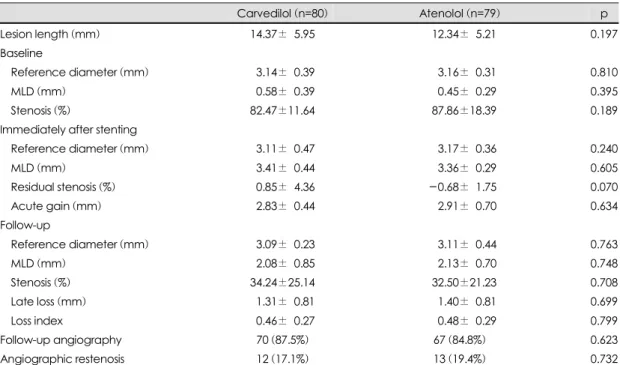
Table 3. Results of quantitative angiographic analysis
Carvedilol (n=80) Atenolol (n=79) p
Lesion length (mm) 14.37±05.95 -12.34±05.21 0.197
Baseline
Reference diameter (mm) 03.14±00.39 0-3.16±00.31 0.810
MLD (mm) 00.58±00.39 0-0.45±00.29 0.395
Stenosis (%) 82.47±11.64 -87.86±18.39 0.189
Immediately after stenting
Reference diameter (mm) 03.11±00.47 0-3.17±00.36 0.240
MLD (mm) 03.41±00.44 0-3.36±00.29 0.605
Residual stenosis (%) 00.85±04.36 0-0.68±01.75 0.070
Acute gain (mm) 02.83±00.44 -02.91±00.70 0.634
Follow-up
Reference diameter (mm) 03.09±00.23 0-3.11±00.44 0.763
MLD (mm) 02.08±00.85 0-2.13±00.70 0.748
Stenosis (%) 34.24±25.14 -32.50±21.23 0.708
Late loss (mm) 01.31±00.81 -01.40±00.81 0.699
Loss index 00.46±00.27 -00.48±00.29 0.799
Follow-up angiography 70 (87.5%) 67 (84.8%) 0.623
Angiographic restenosis 12 (17.1%) 13 (19.4%) 0.732
MLD: minimal luminal diameter, Acute gain: post-stenting MLD-baseline MLD, Late loss: post-stenting MLD-follow-up MLD, Loss index: late loss/acute gain
Discussion
This study shows that a sustained oral administration of carvedilol is not effective to reduce stent restenosis in patients. The results imply that the inhibition of neoin- timal hyperplasia in experiments, usually based on higher concentrations of carvedilol, cannot be achieved by oral administration of tolerable high-dose carvedilol in patients.
The ineffectiveness of carvedilol to reduce stent re- stenosis might result primarily from the inability of achieving a sufficient local or tissue concentration by oral administration, although carvedilol blood levels were not measured concomitantly in this study. Any significant antiproliferative effect in ex vivo studies
14)17)was ob- served with a high concentration of carvedilol (>1 μmol), and the profound inhibition of neointimal growth in rat was achieved at the concentration of approximately 5 ( mol/kg/day by intraperitoneal delivery of 1 mg/kg carvedilol twice daily for 17 days.
13)Oral administration of carvedilol (50 mg/day) in human provided the plasma concentration of 66 μg/mL (0.157 μmol) in a study,
18)although it was suggested that the concentration of carvedilol needed to produce antiproliferative effects might be achievable in in vivo settings.
14)Oral administration of carvedilol to prevent restenosis in patients was attempted in a trial (the EUROCARE) of directional coronary atherectomy (DCA), but failed to reduce restenosis after atherectomy.
19)At the time the EUROCARE trial was planned in 1994, stenting was in the early phase of clinical evaluation; the BENESTENT and STRESS trials had not yet been completed. It was assumed that restenosis after DCA was due to neointimal hyperplasia. For this reason, the investigation of an agent with antioxidant, antichemotactic, and direct antipro- liferative effects seemed ideally suited to an athere- ctomy-treated patient population. Intravascular ultrasound studies have since demonstrated that vessel remodeling accounts primarily for the restenotic response after DCA,
20)21)which reduces the target for antiproliferative drugs. This study represents the first clinical trial re- garding the effect of oral administration of carvedilol on
neointimal hyperplasia in stented patients, the population with an appropriate target for carvedilol.
In spite of the negative result from oral administration, carvedilol may deserve further investigations using a stent-based delivery
22)to achieve a high local concen- tration, because carvedilol targets regulators of cell cycle progression and the extent of neointimal inhibition with a high blood concentration in rat model far exceeds those reported for other compounds in animal models.
Limitations
The type and length of the stents used were not ho- mogenous and might be biased upon each operator’s experience although he wanted to choose an appropriate stent for a specific lesion. The late loss obtained from quantitative coronary angiography was used as an index of neointimal hyperplasia in this study. However, intra- vascular ultrasound study would allow closer observation of in-stent neointimal formation. Follow-up angiography was not performed in all the study patients, although patients who did not receive follow-up study did not show any recurrence of symptoms. The randomization was performed in each center and single-blinded.
Conclusions
This first clinical study shows that a sustained oral administration of carvedilol is not effective to reduce stent restenosis in patients. With carvedilol targeting regulators of cell cycle progression and having a profound neointimal inhibition with a high blood concentration in an experiment, further investigations using a stent-based delivery to achieve a high local concentration may be warranted.
■
Acknoewledgments
This work was supported by the Industry-Academy Coll- aboration Grant (No. 2000-9) from the Korean Circulation Society and 2001 research fund of University of Ulsan.
REFERENCES
1) Williams DO, Holubkov R, Yeh W, Bourassa MG, al-Bassam M, Block PC, et al. Percutaneous coronary interventions in
Korean Circulation J 2004;34(1):35-40 40
the current era compared with 1985-1986. Circulation 2000;
102:2945-51.
2) Mehran R, Dangas G, Abizaid AS, Mintz GS, Lansky AJ, Satler LF, et al. Angiographic patterns of in-stent restenosis:
angiographic classification and implications for long-term clinical outcome. Circulation 1999;100:1872-8.
3) Farb A, Sangiorgi G, Carter AJ, Walley VM, Edwards WD, Schwartz RS, et al. Pathology of acute and chronic coronary stenting in humans. Circulation 1999;99:44-52.
4) Grewe P, Deneke T, Machraoui A, Barmeyer J, Muller KM.
Acute and chronic tissue response to coronary stent im- plantation: pathologic findings in human specimen. J Am Coll Cardiol 2000;35:157-63.
5) Kornowski R, Hong MK, Tio FO, Bramwell O, Wu H, Leon MB. In-stent restenosis: contributions of inflammatory res- ponses and arterial injury to neointimal hyperplasia. J Am Coll Cardiol 1998;31:224-30.
6) Braum-Dullaeus RC, Mann MJ, Dzau VJ. Cell cycle pro- gression: new therapeutic target for vascular proliferative disease. Circulation 1998;98:82-9.
7) Sousa JE, Costa MA, Sousa AG, Abizaid AC, Seixas AC, Abizaid AS, et al. Two-year angiographic and intravascular ultrasound follow-up after implantation of sirolimus-eluting stents in human coronary arteries. Circulation 2003;107:
381-3.
8) Hong MK, Mintz GS, Lee CW, Song JM, Han KH, Kang DH, et al. Paclitaxel coating reduces in-stent intimal hyperplasia in human coronary arteries: a serial volumetric intrava- scular ultrasound analysis from the Asian Paclitaxel-Eluting Stent Clinical Trial (ASPECT). Circulation 2003;107:517-20.
9) Farb A, John M, Acampado E, Kolodgie FD, Prescott MF, Virmani R. Oral everolimus inhibits in-stent neointimal growth.
Circulation 2002;106:2379-84.
10) Feuerstein GZ, Ruffolo RR Jr. Carvedilol, a novel multiple action antihypertensive agent with antioxidant activity and the potential for myocardial and vascular protection. Eur Heart J 1995;16(Suppl F):38-42.
11) Ruffolo RR Jr, Boyle DA, Brooks DP, Feuerstein GZ, Venuti RP, Lukas MA, et al. Carvedilol: a novel cardiovascular drug with multiple actions. Cardiovasc Drug Rev 1992;10:127-57.
12) Yue TL, Cheng HY, Lysko PG, McKenna PJ, Feuerstein R,
Gu JL, et al. Carvedilol, a new vasodilator and beta adreno- ceptor antagonist, is an antioxidant and free radical sca- venger. J Pharmacol Exp Ther 1992;263:92-8.
13) Ohlstein EH, Douglas SA, Sung CP, Yue TL, Louden C, Arleth A, et al. Carvedilol, a cardiovascular drug, prevents vascular smooth muscle cell proliferation, migration and neointimal formation following vascular injury. Proc Natl Acad Sci U S A 1993;90:6189-93.
14) Sung CP, Arleth AJ, Ohlstein EH. Carvedilol inhibits va- scular smooth muscle cell proliferation. J Cardiovasc Phar- macol 1993;21:221-7.
15) Sung CP, Arleth AJ, Eichman C, Truneh A, Ohlstein EH.
Carvedilol, a multiple-action neurohumoral antagonist, in- hibits mitogen-activated protein kinase and cell cycle pro- gression in vascular smooth muscle cells. J Pharmacol Exp Ther 1997;283:910-7.
16) Fattori R, Piva T. Drug-eluting stents in vascular intervention.
Lancet 2003;361:247-9.
17) Patel MK, Chan P, Betteridge LJ, Schachter M, Sever PS.
Inhibition of human vascular smooth muscle cell proliferation by the novel multiple-action antihypertensive agent carvedilol.
J Cardiovasc Pharmacol 1995;25:652-7.
18) Neugebauer G, Akpan W, von Mollendorff E, Neubert P, Reiff K. Pharmacokinetics and disposition of carvedilol in humans. J Cardiovasc Pharmacol 1987;10(Suppl 11):S85-8.
19) Serruys PW, Foley DP, Hofling B, Puel J, Glogar HD, Seabra- Gomes R, et al. Carvedilol for prevention of restenosis after directional coronary atherectomy. Circulation 2000;101:
1512-8.
20) Mintz GS, Popma JJ, Picard AD, Kent KM, Satler LF, Wong C, et al. Arterial remodeling after coronary angioplasty: a serial intravascular ultrasound study. Circulation 1996;94:
35-43.
21) Kimura T, Kaburagi S, Tamura T, Yokoi H, Nakagawa Y, Yokoi H, et al. Remodeling of human coronary arteries undergoing coronary angioplasty or atherectomy. Circulation 1997;96:475-83.
22) Kim W, Jeong MH, Cha KS, Hong YJ, Cho JG, Park JC, et al. The effect of carvedilol-loaded BiodyvYsio stent on neoin- timal hyperplasia in a porcine coronary stent restenosis mo- del. J Am Coll Cardiol 2003;41(Suppl A):31A. Abstract