Lumbar spinal stenosis (LSS) is a clinical condition presenting with back and leg pain due to the compression of intraspinal vascular and neuronal structures by narrowed spinal canal. Pain, paresthesia, and cramping in one or both legs, described as neurogenic claudication, is present in 62% of patients, due to ischemia the nerve roots.1-3Lumbar spinal anteroposterior diameter under 11 mm and a dural area below 100 mm2in imaging procedures depict stenosis.4-6However, as narrowing of the lumbar spinal canal is present in 20% of asymptomatic individuals, clinical findings are highly important in diagnosis and leg pain described as neurogenic claudication emerges as a valuable parameter in the observation of recovery.7
Conservative treatment is effective in LSS patients with mild or occasionally moderate pain.6Daily life style adjustments, back training, exercise programs to stretch, strengthening the lumbar region, and general conditioning exercises both prescribed alone or together with physical therapy yield good clinical results.8-10
The Efficacy of Physical Therapy and Physical Therapy Plus Calcitonin in the Treatment
of Lumbar Spinal Stenosis
Fusun Sahin, Figen Yilmaz, Nurdan Kotevoglu, and Banu Kuran
Department of Physical and Rehabilitation Medicine, Sisli Etfal Training and Research Hospital, Istanbul, Turkey.
Purpose:The aim of our study was to compare the efficacy of physical therapy alone and in combination with calcitonin in patients with neurogenic claudication (NC). Materials and Methods: In this single blind, and randomized study, patients with lumbar spinal canal stenosis who were diagnosed by clinical findings and MRI and having NC were included. Patients were observed for 8 weeks and evaluated before and after treatment. Patients were randomized between the salmon calcitonin 200 U/day + physical therapy (n = 23) (Group 1) and paracetamol 1,500 mg/day + physical therapy (n = 22) (Group 2) treatment groups. Both groups received the same physical therapy (interferential current + hot pack + short wave diathermy) and exercise protocol. The association of various clinical and functional parameters was assessed statistically by using paired and unpaired t test, chi square test and McNemar’s test. p < 0.05 indicated statistical significant. Results:Mean age of the patients in Group 1 was 57.6 ± 11.2 and in Group 2 54.5 ± 10.6 years. Before treatment, there were no significant differences between groups with respect to age, body mass index, spinal axial diameter, Visual Analogue Scale (VAS), spinal mobility, functional status and walking distance (p > 0.05). After 8 weeks of treatment, both groups benefited significantly with respect to VAS, functional status and walking distance (p < 0.001). There was no statistically significant difference between groups (p > 0.05). Conclusion:In 45 patients with lumbar spinal stenosis who received 8 weeks of treatment, concomitant use of calcitonin with physical therapy and exercise did not have any benefical effect on the patient’s pain, functional status, lumbar mobility and walking distance.
Key Words : Lumbar spinal stenosis, calcitonin, physical therapy
Received: June 4, 2007 Revised: October 5, 2007 Accepted: October 5, 2007
Corresponding author: Dr. Fusun Sahin, Department of Physical and Rehabilitation Medicine, Sisli Etfal Training and Research Hospital, Serakent Sitesi Menekse Blok No 14/40 Kagithane, Istanbul, Turkey.
Tel: 90-212-2949102, Fax: 90-258-3741375 E-mail: [email protected]
∙The authors have no financial conflicts of interest.
© Copyright:
Yonsei University College of Medicine 2009
INTRODUCTION
Calcitonin is a hormone secreted by the para-follicular cells of the thyroid, and used in the treatment of osteopo- rosis and Paget’s disease. Due to its analgesic effect, it has also been used in LSS. Analgesic efficacy is considered to be through an increase of beta endorphin levels.11,12Since the narrowing of the spinal canal is the result of soft tissue hypertrophy and edema, its success in treatment is due to anti-inflammatory and anti-edematous efficacy of calcito- nin.13Baker, et al.14reported that, the deterioration of micro- circulation in older patients causes neurogenic claudication in degenerative spinal stenosis, and calcitonin produces a decline in symptoms through its arterial dilator effect.
The aim of our study was to evaluate in a short term the effect of physical therapy alone and in combination with calcitonin on pain, physical examination results and the functional status of patients with neurogenic claudication, and diagnosis of lumbar spinal stenosis.
Patients referred to our outpatient clinic with lower back and leg pain, which described neurogenic claudication, and was diagnosed as LSS by physical examination and imag- ing studies were included in the study.
Clinical diagnosis criteria15-17
1) Pain in one or both legs after walking, diminishing of pain upon sitting or bending forward, and increase of pain with activities that increased extension of the spine such as descending stairs, 2) Restriction of spinal extension and pain in the low back/leg determined physical examination.
In order to exclude the presence of arterial claudication during physical examination, care was taken to ascertain palpable peripheral pulse and the absence of trophic disor- ders due to arterial insufficiency. Neurogenic claudication was also confirmed by the bicycle test. In the bicycle test, patients mounted on a static bicycle (Tunturi Static Bicycle) and instructed to push pedals for 5 minutes at a tolerable resistance.18The absence of leg pain was evaluated in favor of neurogenic claudication.
Radiological diagnosis criteria
After clinical assessment, patients with the narrowest level or levels with an axial diameter below 10 mm, measured by lumbar MRI, were included in the study by a definition of absolute stenosis, while those with an axial diameter of 10-12 mm were included by the definition of relative ste- nosis in the presence of clinical findings.19
Exclusion criteria
Worsening of motor weakness, patients with bladder/gut
dysfunction, previous spinal surgery, presence of inflamma- tory, infectious, or metastatic disease, neurological diseases affecting ambulation ability (such as stroke, Parkinsonism, peripheral entrapment neuropathy, etc), knee pain affecting ambulation ability, hip osteoarthritis, lower extremity peri- pheral arterial insufficiency, and current calcitonin use for osteoporosis.
In this single blind and randomized study, patients were consecutively randomized into 2 groups. Group 1 received 200 U/day intranasal calcitonin, while Group 2 received a maximum of 1,500 mg/day paracetamol. All patients took part in a physical therapy and exercise program. Physical therapy consisted of vacuum interference (20 min) + Hot Pack (20 min) + short wave diathermia (10 min) to the lum- bar region, 5 days a week for 15 sessions. Exercises con- sisted of pelvic tilt, abdominal strengthening, hip flexion and hamstring stretching, and lumbar mobilization exercises.10 Exercises were taught and performed in the hospital by a physiotherapist during physical therapy session, and patients were thereafter recommended to repeat the same exercises twice a day at home.
Randomization (FY) and physical examinations (FS) were carried out by the same physiatrist, thus maintaining a single-blind design. No financial support was accepted for the study. The same nasal calcitonin product which could be obtained easily in the market was prescribed, and the other group was permitted to take paracetamol as analgesic. In order to evaluate the analgesic effect of calci- tonin in a short term, a dosage of 200 U/day was prescribed, which is the dosage available in our country. All patients who would use calcitonin were informed about storage condition of the bottle (before opening, bottles were stored between 36-46 F in the fridge, and open bottles were stored at room temperature) and also application style. Patients were informed not to pump the calcitonin spray after 14 sprays, because each bottle contained 14 sprays.
Patients were evaluated at baseline and 8 weeks after treatment. Age, occupation, and Body mass index (BMI) of patients were recorded. Assessment parameters:
- Pain (at rest and with movement); patients were req- uested to evaluate the severity of pain on a 10 mm scale using the Visual Analogue Scale (VAS).
- Range of motion: measured by lumbar Schober (cm), finger-to-floor distance (cm), extension (degree-by lumbar goniometry).
- Functional status: evaluated by the Roland-Morris Scale.20
- Walking distance: expressed in meters.
The 24-item Roland-Morris Scale adapted to Turkish was used for functional assessment.20 In this scale, each item was answered by “yes” or “no”, and “yes” responses took 1 and “no” responses 0 points, and the total score was
MATERIALS AND METHODS
calculated. A total score of 0 points meant “no disability”, while a total score of 24 points meant “extremely severe disability”.20
Walking distance was measured by a treadmill (Ferrox- Fulmine Treadmill) at 0Oinclination, with the body at a vertical position, at a speed of 2 km/h.21The test was termi- nated when patients suffered of leg pain, and walking distance was determined.
Statistical analyses were managed by the GraphPad Prisma V.3 program. Besides descriptive statistical methods (mean, standard deviation), independent t test in the com- parison of paired groups, paired t test in the comparison of repeated measures in paired groups, the χ2-test in the com- parison of qualitative data, and the McNemar’s test in the comparison of repeated measures of qualitative data were
used. Results were accepted significant at p < 0.05.
A total of 45 patients were included in the study. Group 1 consisted of 23, and Group 2 of 22 patients. The most commonly affected lumbar level was L4-L5, followed by narrowing at the L3-L4 level (Fig. 1).
Concomitant diseases were hypertension in 8, diabetes mellitus in 4, hypercholesterolemia in 4, ischemic heart disease in 2, and asthma in 1 patient.
In the group using calcitonin, only one patient had nasal irritation that did not necessitate cessation of therapy, and all patients were comfortable with the drug. In the group using paracetamol, patients used average 1.8 ± 0.8 tablets (each tablet of 500 mg). All patients who were enrolled in the study and completed the follow-up protocoland were included in the analysis.
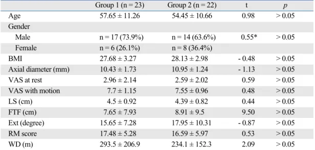
With respect to baseline values, there was no significant difference between groups in patients’ age, gender, BMI values, axial diameter averages, VAS, lumbar ROM, func- tional status, and walking distance parameters (Table 1).
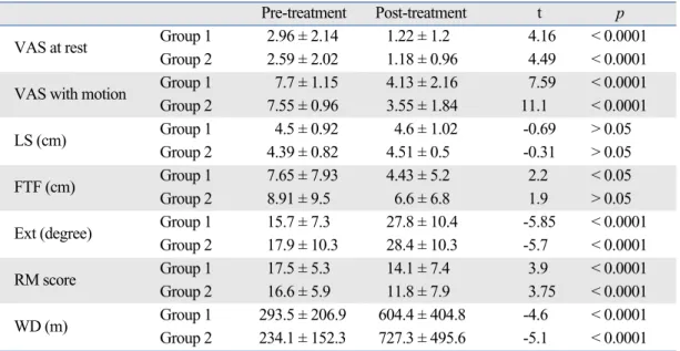
In the inter-group evaluation performed at week 8, lumbar Schober was similar in both groups, while the finger-to-floor distance significantly improved in the calcitonin group. All other parameters showed significant improvement in both groups (Table 2).
There was no significant difference between the groups with respect to improved parameters during the follow up period as well as their percent changes (Table 3).
RESULTS
Percent
L1-L5
Levels 0
60
10 20 30 40 50
L2-L3 L2-L4 L3-L4 L3-L5 L4-L5 L4-S1 L1-S1
Fig. 1. Level of disease in patients.
Table 1. The Comparison of Patients’ Age, Gender, BMI Values, Axial Diameter Averages and Pre-Treatment Follow-up Parameters
Group 1 (n = 23) Group 2 (n = 22) t p
Age 57.65 ± 11.26 54.45 ± 10.66 0.98 > 0.05
Gender
Male n = 17 (73.9%) n = 14 (63.6%) 0.55* > 0.05
Female n = 6 (26.1%) n = 8 (36.4%)
BMI 27.68 ± 3.27 28.13 ± 2.98 - 0.48 > 0.05
Axial diameter (mm) 10.43 ± 1.73 10.95 ± 1.24 - 1.13 > 0.05
VAS at rest 2.96 ± 2.14 2.59 ± 2.02 0.59 > 0.05
VAS with motion 7.7 ± 1.15 7.55 ± 0.96 0.48 > 0.05
LS (cm) 4.5 ± 0.92 4.39 ± 0.82 0.44 > 0.05
FTF (cm) 7.65 ± 7.93 8.91 ± 9.5 9.50 > 0.05
Ext (degree) 15.65 ± 7.28 17.95 ± 10.31 - 0.87 > 0.05
RM score 17.48 ± 5.28 16.59 ± 5.97 0.53 > 0.05
WD (m) 293.5 ± 206.9 234.1 ± 152.3 2.09 > 0.05
BMI, body mass index; VAS, visual analogue scale; LS, lumbar schober; FTF, finger-to-floor distance; Ext, extension; RM, Roland-Morris score; WD, walking distance.
*χ2.
The generally accepted practice for the treatment of LSS is the use of conservative therapy in mild and moderately symptomatic patients, and surgery in patients with severe symptoms.6,22,23Another approach in patients in whom con- servative treatment fails is to perform surgery.24-26 Naturally, conservative therapy is the treatment of choice in patients without motor disturbances and/or bladder or intestinal dysfunction.27 Trials examined the natural course of the disease actually confirm the use of conservative therapy as the first-line treatment. Johnsson, et al.,22in the 49-month follow-up of 32 patients, determined improvement accord- ing to VAS, and found improvement in 15% of patients, no change in 70%, and worsening in 15%. Physical examination established improvement in 41% of patients, no change in 41%, and worsening in 18%. Sengupta and Herkowitz28 also states in his review that 15% of patients improved, 30% worsened within 2-3 years and needed
surgery, while 45% were stable for a long time.
Conservative treatment options include physical therapy and exercise, epidural steroid injections, calcitonin and other analgesic/anti-inflammatory medications, and these are usually prescribed in combination.29Studies on exercise treatment particularly emphasize the importance of flexion exercises, and recommend the addition of general condi- tioning exercises.8,10,30Among trials using conservative treatment options, Onel, et al.31combined physical therapy with superficial and deep heat with calcitonin, and imple- mented a one-month inpatient rehabilitation with an aggres- sive exercise program. Following the one-month observation period, 70% of patients achieved good outcomes. Simotas, et al.32 administered physical therapy to 96%, epidural steroid injections to 78%, acupuncture to 58%, and orthesis, bed rest, transcutanous electrical nerve stimulation (TENS) and manipulation to decreasing percentages of 40 patients with LSS, and observed decrease in especially pain para- meters but no changes in functional assessments (walking distance, walking frequency) after a follow-up of average Table 2. Comparison of Pre-Treatment and Post-Treatment Assessments in Group 1 and Group 2
Pre-treatment Post-treatment t p
VAS at rest Group 1 2.96 ± 2.14 1.22 ± 1.2 4.16 < 0.0001
Group 2 2.59 ± 2.02 1.18 ± 0.96 4.49 < 0.0001
VAS with motion Group 1 7.7 ± 1.15 4.13 ± 2.16 7.59 < 0.0001
Group 2 7.55 ± 0.96 3.55 ± 1.84 11.1 < 0.0001
LS (cm) Group 1 4.5 ± 0.92 4.6 ± 1.02 -0.69 > 0.05
Group 2 4.39 ± 0.82 4.51 ± 0.5 -0.31 > 0.05
FTF (cm) Group 1 7.65 ± 7.93 4.43 ± 5.2 2.2 < 0.05
Group 2 8.91 ± 9.5 6.6 ± 6.8 1.9 > 0.05
Ext (degree) Group 1 15.7 ± 7.3 27.8 ± 10.4 -5.85 < 0.0001
Group 2 17.9 ± 10.3 28.4 ± 10.3 -5.7 < 0.0001
RM score Group 1 17.5 ± 5.3 14.1 ± 7.4 3.9 < 0.0001
Group 2 16.6 ± 5.9 11.8 ± 7.9 3.75 < 0.0001
WD (m) Group 1 293.5 ± 206.9 604.4 ± 404.8 -4.6 < 0.0001
Group 2 234.1 ± 152.3 727.3 ± 495.6 -5.1 < 0.0001 VAS, visual analogue scale; LS, lumbar schober; FTF, finger-to-floor distance; Ext, extension; RM, Roland Morris score; WD, walking distance.
Table 3. The Comparison of Percent Changes in Both Groups
Group 1 Group 2 t p
VAS at rest 54.6 ± 40.4 49.89 ± 33.5 0.39 > 0.05
VAS with motion 45.7 ± 28.2 53.6 ± 21.9 -1.04 > 0.05
LS (cm) 2.9 ± 19.2 10.3 ± 20.3 1.26 > 0.05
FTF (cm) 44.6 ± 41.1 16.9 ± 59.6 1.43 > 0.05
Ext (degree) 39.5 ± 29.03 37.5 ± 27.9 0.24 > 0.05
RM score 24.7 ± 33.1 32.9 ± 34.6 -0.81 > 0.05
WD (m) 40.8 ± 32.9 6 ± 56.2 ± 31.8 -1.61 > 0.05
VAS, visual analogue scale; LS, lumbar schober; FTF, Finger-to-floor distance; Ext, Extension; RM, Roland Morris score; WD, walking distance.
DISCUSSION
33 months. The authors concluded that aggressive conser- vative treatment was a good option. In a study by Tadokoro, et al.,27inpatient conservative therapy was administered to a group of patients with LSS over 70 years of age, and it was concluded after a follow-up of 57 weeks, that particul- arly those with radicular pain were good candidates for conservative treatment, and had a relatively good prognosis.
Our study also combined the conservative treatment options of physical therapy and exercise, and investigated the additive effect of short-term calcitonin as an analgesic.
We administered low frequency currents for analgesia, Hot Pack for superficial heat, and short-wave diathermia for deep heat as physical therapy modalities. Exercises consis- ting particularly of lumbar flexion, mobilization, and stretching exercises were taught to patients during physical therapy sessions, and adviced to be repeated at home. Cal- citonin was given at a dose of 200 U/day as a nasal spray, and evaluations were made at the end of 8 weeks.
The use of calcitonin in LSS was first published by Porter.33In this study, 10 patients with neurogenic claudi- cation symptoms were found to have improved with calci- tonin, and calcitonin was considered to be affective by increasing the blood flow. A follwing study by Porter com- pared 100 U calcitonin with plasebo in 42 patients, and found no statistical significance although calcitonin inc- reased walking distance.12A study by Eskola, et al.13with 15 patients followed for 3 months, also using 100 U calci- tonin, and reported a decrease in pain and an increase in performance. Another double-blind, randomized placebo- controlled study by Eskola, et al.34followed 39 patients for one year, and found that calcitonin was effective on the parameters like pain and walking distance, but less effec- tive in patients with a walking distance below 200-300 meters. In another randomized, double-blind, controlled trial by Podichetty et al.35with 36 patients with a VAS score over 6 which was observed for 6 weeks, no signifi- cant difference was found between 2 groups in terms of walking period, walking distance, or functional assess- ment by SF-36. Walking distance was limited to 130 m in this trial, and the authors suggested that an inadequate efficacy of calcitonin might be due to the severity of symp- toms in this patient group. In our study, although the average pain score was 7.5 with VAS which was similar to other studies, walking distance was an average of 300 m which was longer than the others, i.e. symptom severity was also lower than others. In spite of these differences, calcitonin did not add any benefit to physical therapy and exercise program. In the present study, the Roland-Morris score used for functional assessment showed significant increase in all patients, but there was no significant difference between the 2 groups in terms of the percent change.
A comparison between conservative treatment and sur-
gical therapy appears to be in favor of surgical therapy. In a long-term study with a large patient number, Atlas, et al.36followed, 119 of 148 patients for 4 years. The operated group with more severe symptoms at baseline, compared with the non-operated group at the end of 4 years, showed significantly fewer symptoms, more treatment satisfaction, and better functional status. In the non-surgical group, improvement was modest and stable for 4 years. Amun- dsen, et al.25followed 100 patients for 10 years, and reported that operated patients showed better outcomes and patients with severe symptoms especially benefited from surgery, and that conservative therapy should be administered to patients with moderate symptoms, as a delay in surgical treatment did not have negative impact on the outcome.
In conclusion, in our 45 patients with an average age of 55 years, neurogenic claudication, and moderate LSS symptoms in terms of walking distance, short-term physical therapy and exercise therapy for 3 months signi- ficantly improved pain, physical examination findings, and walking distance and functional parameters. The addition of 200 U/day calcitonin did not lead to a significant impro- vement in follow-up parameters. We, therefore, conclude that the addition of calcitonin as an analgesic in the short- term treatment of LSS along with physical therapy and exercise administration is not necessary.
1. Amundsen T, Weber H, Nordal HJ, Magnaes B, Abdelnoor M, Lilleas F. Lumbar spinal stenosis: conservative or surgical mana- gement?: A prospective 10-year study. Spine (Phila Pa 1976) 2000;25:1424-35; discussion 1435-6.
2. Porter RW. Spinal stenosis and neurogenic claudication. Spine (Phila Pa 1976) 1996;21:2046-52.
3. Turner JA, Ersek M, Herron L, Deyo R. Surgery for lumbar spinal stenosis. Attempted meta-analysis of the literature. Spine (Phila Pa 1976) 1992;17:1-8.
4. Shönström N, Lindahl S, Willén J, Hansson T. Dynamic changes in the dimensions of the lumbar spinal canal: an experimental study in vitro. J Orthop Res 1989;7:115-21.
5. Schonstrom NS, Bolender N, Spengler DM. The pathomorphology of spinal stenosis as seen on CT scans of the lumbar spine. Spine (Phila Pa 1976) 1985;10:806-11.
6. Fritz JM, Delitto A, Welch WC, Erhard RE. Lumbar spinal ste- nosis: a review of current concepts in evaluation, management, and outcome measurements. Arch Phys Med Rehabil 1998;79:
700-8.
7. Boden SD, Davis DO, Dina TS, Patronas NJ, Wiesel SW. Abnor- mal magnetic-resonance scans of the lumbar spine in asymptoma- tic subjects. A prospective investigation. J Bone Joint Surg Am 1990;72:403-8.
8. Fritz JM, Erhard RE, Vignovic M. A nonsurgical treatment approach for patients with lumbar spinal stenosis. Phys Ther 1997;
77:962-73.
REFERENCES
^
9. Simotas AC, Dorey FJ, Hansraj KK, Cammisa F Jr. Nonopera- tive treatment for lumbar spinal stenosis: clinical outcome results and 3-year survivorship analysis. Spine (Phila Pa 1976) 2000;25:
197-203.
10. Bodack MP, Monteiro M. Therapeutic exercise in the treatment of patients with lumbar spinal stenosis. Clin Orthop Relat Res 2001;384:144-52.
11. Porter RW, Hibbert C. Calcitonin treatment for neurogenic clau- dication. Spine (Phila Pa 1976) 1983;8:589-92.
12. Porter RW, Miller CG. Neurogenic claudication and root claudi- cation treated with calcitonin. A double-blind trial. Spine 1988;
13:1061-4.
13. Eskola A, Alaranta H, Pohjolainen T, Soini J, Tallroth K, Slätis P.
Calcitonin treatment in lumbar spinal stenosis: clinical observa- tions. Calcif Tissue Int 1989;45:372-4.
14. Baker AR, Collins TA, Porter RW, Kidd C. Laser Doppler study of porcine caudate equina blood flow. The effect of electrical sti- mulation of the rootlets during single and double site, low pres- sure compression of the cauda equina. Spine 1995;20:660-4.
15. Penning L, Wilmink JT. Posture-dependent bilateral compression of L4 or L5 nerve roots in facet hypertrophy. A dynamic CT- myelographic study. Spine (Phila Pa 1976) 1987;12:488-500.
16. Takahashi K, Miyazaki T, Takino T, Matsui T, Tomita K. Epidural pressure measurements. Relationship between epidural pressure and posture in patients with lumbar spinal stenosis. Spine (Phila Pa 1976) 1995;20:650-3.
17. Katz JN, Dalgas M, Stucki G, Lipson SJ. Diagnosis of lumbar spinal stenosis. Rheum Dis Clin North Am 1994;20:471-83.
18. Dyck P, Doyle JB Jr. “Bicycle test” of van Gelderen in diagnosis of intermittent cauda equina compression syndrome. Case report.
J Neurosurg 1977;46:667-70.
19. Tuite GF, Stern JD, Doran SE, Papadopoulos SM, McGillicuddy JE, Oyedijo DI, et al. Outcome after laminectomy for lumbar spinal stenosis. Part I: clinical correlations. J Neurosurg 1994;81:699-706.
20. Küçükdeveci AA, Tennant A, Elhan AH, Niyazoglu H. Valida- tion of the Turkish version of the Roland-Morris Disability Ques- tionnaire for use in low back pain. Spine (Phila Pa 1976) 2001;
26:2738-43.
21. Fritz JM, Erhard RE, Delitto A, Welch WC, Nowakowski PE.
Preliminary results of the use of a two-stage treadmill test as a clinical diagnostic tool in the differential diagnosis of lumbar spinal stenosis. J Spinal Disord 1997;10:410-6.
22. Johnsson KE, Rosén I, Udén A. The natural course of lumbar spinal stenosis. Clin Orthop Relat Res 1992;82-6.
23. Johnsson KE, Udén A, Rosén I. The effect of decompression on the natural course of spinal stenosis. A comparison of surgically
treated and untreated patients. Spine (Phila Pa 1976) 1991;16:
615-9.
24. Gunzburg R, Szpalski M. The conservative surgical treatment of lumbar spinal stenosis in the elderly. Eur Spine J 2003;12 Suppl 2:S176-80.
25. Amundsen T, Weber H, Nordal HJ, Magnaes B, Abdelnoor M, Lilleas F. Lumbar spinal stenosis: conservative or surgical mana- gement?: A prospective 10-year study. Spine (Phila Pa 1976) 2000;
25:1424-35.
26. Jolles BM, Porchet F, Theumann N. Surgical treatment of lumbar spinal stenosis. Five-year follow-up. J Bone Joint Surg Br 2001;83:
949-53.
27. Tadokoro T, Miyamoto H, Sumi M, Shimornura T. The prognosis of conservative treatmants for lumbar spinal stenosis; analysis of patients over 70 years of age. Spine (Phila Pa 1976) 2005;30:
2458-63.
28. Sengupta DK, Herkowitz HN. Lumbar spinal stenosis. Treatment strategies and indications for surgery. Orthop Clin North Am 2003;34:281-95.
29. Vo AN, Kamen LB, Shih VC, Bitar AA, Stitik TP, Kaplan RJ.
Rehabilitation of orthopedic and rheumatologic disorders. 5.
Lumbar spinal stenosis. Arch Phys Med Rehabil 2005;86:S69-76.
30. Bridwell KH. Lumbar spinal stenosis. Diagnosis, management, and treatmant. Clin Geriatr Med 1994;10:677-701.
31. Onel D, Sari H, Dönmez C. Lumbar spinal stenosis: clinical/
radiologic therapeutic evaluation in 145 patients. Conservative treatment or surgical intervention? Spine (Phila Pa 1976) 1993;18:
291-8.
32. Simotas AC, Dorey FJ, Hansraj KK, Cammisa F Jr. Nonopera- tive treatment for lumbar spinal stenosis. Clinical and outcome results and a 3-year survivorship analysis. Spine (Phila Pa 1976) 2000;25:197-203.
33. Porter RW, Hibbert C. Calcitonin treatment for neurogenic clau- dication. Spine (Phila Pa 1976) 1983;8:585-3.
34. Eskola A, Pohjolainen T, Alaranta H, Soini J, Tallroth K, Slätis P.
Calcitonin treatment in lumbar spinal stenosis: a randomized, placebo-controlled, double-blind, cross-over study with one-year follow-up. Calcif Tissue Int 1992;50:400-3.
35. Podichetty VK, Segal AM, Lieber M, Mazanec DJ. Effectiveness of salmon calcitonin nasal spray in the treatment of lumbar canal stenosis: a double-blind, randomized, placebo-controlled, parallel group trial. Spine (Phila Pa 1976) 2004;29:2343-9.
36. Atlas SJ, Keller RB, Robson D, Deya RA, Singer DE. Surgical and nonsurgical management of lumbar spinal stenosis: four-year outcomes from the maine lumbar spine study. Spine (Phila Pa 1976) 2000;25:556-62.
^