Prognostic Value of 18 F-Fluorodeoxyglucose Positron Emission Tomography in Patients with Resectable Pancreatic Cancer
Hye Jin Choi,
1,2Chang Moo Kang,
1,3Woo Jung Lee,
1,3Si Young Song,
1,4Arthur Cho,
5Mijin Yun,
1,5Jong Doo Lee,
5Joo Hang Kim,
2and Jae-Hoon Lee
51Yonsei Pancreatico-Biliary Cancer Clinic, Severance Hospital, Seoul;
2Division of Oncology, Department of Internal Medicine, 3Division of Hepatobiliary and Pancreas, Department of Surgery, 4Division of Gastroenterology, Department of Internal Medicine,
5Department of Nuclear Medicine, Yonsei University College of Medicine, Seoul, Korea.
Received: October 31, 2012 Revised: January 13, 2013 Accepted: January 23, 2013
Corresponding author: Dr. Jae-Hoon Lee, Department of Nuclear Medicine, Yonsei University College of Medicine, 50 Yonsei-ro, Seodaemun-gu, Seoul 120-752, Korea.
Tel: 82-2-2228-2350, Fax: 82-2-312-0578 E-mail: [email protected]
∙ The authors have no financial conflicts of interest.
© Copyright:
Yonsei University College of Medicine 2013 This is an Open Access article distributed under the terms of the Creative Commons Attribution Non- Commercial License (http://creativecommons.org/
licenses/by-nc/3.0) which permits unrestricted non- commercial use, distribution, and reproduction in any medium, provided the original work is properly cited.
Purpose: We evaluated the prognostic value of
18F-2-fluoro-2-deoxyglucose posi- tron emission tomography (FDG PET) in patients with resectable pancreatic cancer.
Materials and Methods: We retrospectively reviewed the medical records of pan- creatic cancer patients who underwent curative resection, which included 64 con- secutive patients who had preoperative FDG PET scans. For statistical analysis, the maximal standardized uptake value (SUVmax) of primary pancreatic cancer was measured. Survival time was estimated by the Kaplan-Meier method, and Cox’s proportional hazard model was used to determine whether SUVmax added new predictive information concerning survival together with known prognostic factors.
p<0.05 indicated statistical significance. Results: Overall survival (OS) and dis- ease-free survival (DFS) were respectively 42.9 months (27.6-58.2; 95% CI) and 14.9 months (10.1-19.7; 95% CI). When subjects were divided into two groups ac- cording to SUVmax with a cutoff value of 3.5, the high SUVmax group (n=32; SU- Vmax >3.5) showed significantly shorter OS and DFS than the low SUVmax group. Multivariate analysis of OS and DFS showed that both high SUVmax and poor tumor differentiation were independent poor prognostic factors. Conclusion:
Our study showed that degree of FDG uptake was an independent prognostic factor in pancreatic cancer patients who underwent curative resection.
Key Words: FDG PET, pancreatic cancer, prognosis, curative resection
INTRODUCTION
Pancreatic cancer is a deadly disease and carries a poor prognosis; for all stages
combined, the 5-year survival rate is less than 5%. Only 20% of patients with pan-
creatic cancer have resectable disease at the time of presentation, and in the event
of resectable disease, the 5-year survival rate is about 20%.
1,2Prognostic factors
for pancreatic cancer have been well studied, and include gender, age, size and lo-
cation of the tumor, stage, lymph node metastasis, tumor grade, and serum carbo-
hydrate antigen 19-9 (CA19-9) level.
3-8FDG PET imaging
All patients fasted for at least 4 hours before the FDG PET scan. Blood glucose levels were measured before each PET study. Patients were scanned when their plasma glucose levels were below 130 mg/dL. Scanning was initiated 60 min after the administration of FDG. Images from the neck to the proximal thigh were obtained either on an Advance PET scanner (GE Healthcare, Milwaukee, WI, USA) with a spatial resolution of 5 mm in the center of the field of view or on an Allegro PET scanner (Philips-ADAC medi- cal systems, Cleveland, OH, USA) with a spatial resolution of 5.3 mm in the center of the field of view. When using the Advance scanner, approximately 370 MBq of FDG were injected intravenously, and an emission scan was acquired for 5 min per bed position in the two-dimensional mode.
When the Allegro scanner was used, data were acquired in the three-dimensional mode after the administration of 5.18 MBq (0.14 mCi)/kg of FDG. Transmission scans (3 min per bed position) were obtained to correct for nonuniform atten- uation using
68Ge and
137Cs point sources for the Advance and Allegro scanners, respectively. Transmission scans were interleaved between the multiple emission scans for the Al- legro scanner. The images were reconstructed using an iter- ative reconstruction algorithm, that is, either the ordered- subset expectation maximization for the Advance scanner or the row action maximal-likelihood algorithm for the Al- legro scanner.
All of the FDG PET images were interpreted by two ex- perienced nuclear medicine physicians blinded to addition- al clinical outcomes. Focal increased standardized uptake value (SUV) was calculated as [decay-corrected activity (kBq)/mL tissue volume]/[injected FDG activity (kBq)/
body mass (g)]. SUV of the pancreatic cancer was mea- sured by manually placing a circular region of interest at the site of the maximum FDG uptake; maximum SUV (SUVmax) was used for further analysis.
Statistical analysis
Overall survival (OS) was defined as the interval from the date of curative resection to the date of death from any cause.
Disease-free survival (DFS) was defined as the interval from the date of operation to the first evidence of radiologi- cal progression or to the date of death from any cause. The chi-square and Fisher exact tests were used to compare fre- quencies in the groups. Survival time was estimated by the Kaplan-Meier method, and differences in survival between the groups were compared using a log-rank test. Cox’s pro- Over the past decade,
18F-2-fluoro-2-deoxyglucose posi-
tron emission tomography (FDG PET) has become estab- lished in cancer imaging. As FDG PET assesses the glucose metabolic activity of tumors, it provides useful information that cannot be obtained with other conventional imaging techniques, making it a useful imaging tool for the diagno- sis and staging of pancreatic cancer, although limited sensi- tivity has been reported in the detection of small lesions and local lymph node metastasis.
9In addition, the metabolic ac- tivity of pancreatic tumors, measured by FDG PET usually based on a standardized uptake value (SUV), has proven useful in evaluating the prognosis of pancreatic carcino- ma.
10-15Most published studies consider SUV an indepen- dent prognostic factor: higher SUV indicates a worse prog- nosis.
However, few studies have examined whether FDG PET is useful for the prognosis of clinical outcomes in patients with resectable pancreatic cancer. Published studies on this group of patients suffer from small numbers in subpopula- tion analysis or a heterogeneous group of patients with pal- liative resection or past history of neoadjuvant therapy.
10-13The objective of our study was to determine in a larger series of patients whether preoperative FDG PET provides prognostic information in patients with resectable pancreat- ic adenocarcinoma.
MATERIALS AND METHODS
Patient selection
The institutional review board of our university approved this
study and waived the informed consent requirement. Be-
tween January 2004 and August 2009, a total of 124 patients
with pancreatic ductal adenocarcinoma underwent curative
surgical resection at Severance Hospital. Patients were ex-
cluded from the study if they had a previous history of anoth-
er malignancy, had received chemotherapy or radiotherapy
before surgical resection, or had undergone palliative resec-
tion. Resectability of pancreatic cancer was determined on
basis of National Comprehensive Cancer Network guide-
lines presented at a multidisciplinary cancer conference. Fi-
nally, 64 consecutive patients who had undergone FDG
PET as a staging workup before resection were selected. We
retrospectively reviewed medical records concerning age,
gender, CA19-9 levels, TNM staging, type of operation, tu-
mor size, histologic differentiation, resection margin, and
adjuvant treatment.
and poor differentiation were independent poor prognostic factors and that poorer differentiation was also an indepen- dent prognostic factor (p=0.016 and p=0.000, respectively).
portional hazard model was used to determine whether SU- Vmax added new predictive information on survival. p<0.05 indicated statistical significance. Statistical analysis of the data was performed using SPSS software version 18.0 (SPSS Inc., Chicago, IL, USA).
RESULTS
Patient characteristics
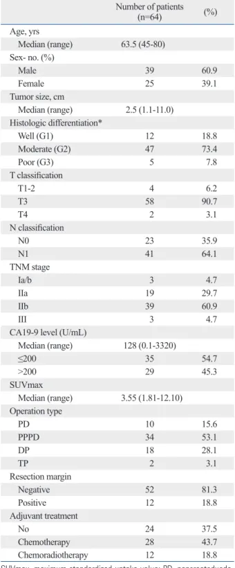
The clinical characteristics of the patient population are summarized in Table 1. Among the 64 patients studied, 34 (53.1%) underwent pylorus-preserving pancreatoduodenec- tomy, 18 (28.1%) distal pancreatectomy, 10 (15.6%) pan- creatoduodenectomy, and two (3.1) total pancreatectomy.
Forty patients had adjuvant treatment, 28 had chemothera- py, and 12 had chemoradiotherapy.
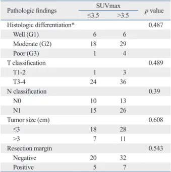
The median SUVmax of pancreatic cancers was 3.55 (range, 1.81-12.10). To compare patient characteristics de- scriptively according to the SUVmax, patients were divided into two groups using a cutoff of 3.5: a high SUVmax group (n=32; SUVmax >3.5) and a low SUVmax group (SUV- max ≤3.5). There were no significant differences in base- line characteristics and histologic findings between these two groups (Table 2).
Prognostic value of FDG PET and other parameters The mean OS and DFS of the 64 patients were 42.9 months (27.6-58.2; 95% CI) and 14.9 months (10.1-19.7; 95% CI), respectively. Survival analysis showed that the high SUV- max group had a significantly shorter OS than the corre- sponding low SUVmax group (p=0.011): 23.5 months vs.
45.4 months (Fig. 1). DFS was also significantly shorter in the high SUVmax group (p=0.002): 9.2 months vs. 26.1 months.
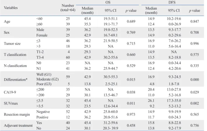
Univariate analysis of OS showed that patients with a higher SUVmax, CA19-9 over 200 ng/mL, or poor differen- tiation had significantly shorter survival (p=0.011, p=0.038, and p=0.015, respectively) (Table 3). Multivariate analysis of OS showed that higher SUVmax, like poor differentia- tion, was a significant poor prognostic factor (p=0.025 and p=0.043, respectively) (Table 4). Univariate analysis of DFS showed that a higher SUVmax was significantly correlated with shorter DFS (p=0.002), and CA19-9 over 200 ng/mL and poor differentiation were also correlated with signifi- cantly shorter DFS (p=0.029 and p=0.000, respectively).
Multivariate analysis of DFS showed that higher SUVmax
Table 1. Demographical and Baseline Characteristics
Number of patients(n=64) (%)
Age, yrs
Median (range) 63.5 (45-80) Sex- no. (%)
Male 39 60.9
Female 25 39.1
Tumor size, cm
Median (range) 2.5 (1.1-11.0) Histologic differentiation*
Well (G1) 12 18.8
Moderate (G2) 47 73.4
Poor (G3) 5 7.8
T classification
T1-2 4 6.2
T3 58 90.7
T4 2 3.1
N classification
N0 23 35.9
N1 41 64.1
TNM stage
Ia/b 3 4.7
IIa 19 29.7
IIb 39 60.9
III 3 4.7
CA19-9 level (U/mL)
Median (range) 128 (0.1-3320)
≤200 35 54.7
>200 29 45.3
SUVmax
Median (range) 3.55 (1.81-12.10) Operation type
PD 10 15.6
PPPD 34 53.1
DP 18 28.1
TP 2 3.1
Resection margin
Negative 52 81.3
Positive 12 18.8
Adjuvant treatment
No 24 37.5
Chemotherapy 28 43.7
Chemoradiotherapy 12 18.8
SUVmax, maximum standardized uptake value; PD, pancreatoduode- nectomy; PPPD, pylorus preserving pancreatoduodenectomy; DP, distal pancreatectomy; TP, total pancreatectomy; UICC, Union for International Cancer Control.
*UICC classification.
fulness of FDG PET in pancreatic cancer patients. In a prospective study, Kauhanen, et al.
9reported that FDG PET had a high diagnostic value in patients with pancreatic can- cer compared with computed tomography (CT) and mag- netic resonance imaging, with a sensitivity of 85%, speci- ficity of 94%, positive predictive value of 94%, and negative predictive value of 85%.
Beyond the conventional role of FDG PET as a diagnos- tic modality, it can predict treatment responses to chemo- therapy and/or radiotherapy
3,16,17and prognoses
10-15in pa- tients with pancreatic cancer. An accelerated rate of both glucose transport and glycolysis are characteristic biochem- ical features of malignant transformation. FDG is a glucose analogue that is actively transported via glucose transport- ers (GLUT) into cells and phosphorylated by hexokinase during the first step of the glycolytic pathway. However, unlike normal glucose, phosphorylated FDG cannot contin- ue in glycolysis and becomes trapped within the cell. Over- expression of GLUT-1
18,19and hexokinase-II
20has been re- ported in pancreatic adenocarcinomas and differences in the biologic aggressiveness of the tumor, represented as SUV, could explain the differences in disease-free survival and overall survival in patients with pancreatic cancer after curative resection.
Some researchers have shown the clinical value of SUV during pretreatment FDG PET in predicting prognosis in patients with pancreatic cancer. For all stages combined, survival time was significantly longer in patients with lower SUV than with higher SUV.
10,14Nakata, et al.
11reported that SUV could predict prognosis in patients with unresectable disease. In a study using dual time FDG PET, a retention However, a higher CA19-9 level did not show any signifi-
cant statistical power for either OS or DFS in multivariate analysis (p=0.325 and p=0.248, respectively).
DISCUSSION
Recently, FDG PET has been widely used in cancer pa- tients for diagnosis, staging, therapeutic monitoring, and re- staging, and previous studies have reported the clinical use- Table 2. Association between SUVmax and Histopathologic Findings
Pathologic findings SUVmax p value
≤3.5 >3.5
Histologic differentiation* 0.487
Well (G1) 6 6
Moderate (G2) 18 29
Poor (G3) 1 4
T classification 0.489
T1-2 1 3
T3-4 24 36
N classification 0.39
N0 10 13
N1 15 26
Tumor size (cm) 0.608
≤3 18 28
>3 7 11
Resection margin 0.543
Negative 20 32
Positive 5 7
SUVmax, maximum standardized uptake value; UICC, Union for Interna- tional Cancer Control.
*UICC classification.
Fig. 1. Overall survival (OS) and disease-free survival (DFS) in low SUVmax and high SUVmax groups. Tick marks on curves indicate drop-outs. (A) Cumulative survival rate was higher in the low SUVmax group (≤3.5; continuous line) than the high SUVmax group (>3.5; dotted line) (p=0.011). (B) Cumulative disease-free rate was also higher in the low SUVmax group (p=0.002). SUVmax, maximum standardized uptake value.
0.0 0.0
0.2 0.2
0.4 0.4
0.6 0.6
0.8 0.8
1.0 1.0
Cumulative survival Cumulative survival
0.00 20.00 40.00 60.00 0.00 20.00 40.00 60.00
OS (months) DFS (months)
SUVmax ≤3.5
SUVmax ≤3.5
SUVmax >3.5 SUVmax >3.5
B
A
3.5. In a comparison of various prognostic factors by multi- variate analysis, FDG PET proved to be an independent prognostic factor, where OS and DFS were significantly different above and below the cutoff value. Yet no consen- sus has been established on a cutoff value for SUV; differ- ent median values have been chosen as cutoff values, as in this study. Because these cutoff values vary greatly, ranging from 3.0
11to 7.0,
10an absolute cutoff is not recommended for further investigations; instead, it should be determined on the basis of the individual or larger group data.
For pancreatic cancer, many prognostic factors have been suggested: tumor size, location, TNM stage, tumor differ- index of more than 10% was suggested to be a significant
poor prognostic factor.
13For resectable pancreatic cancer, little information is available. SUV was significantly differ- ent between patients with resectable and unresectable dis- eases,
21and Sperti, et al.
12noted that a SUVmax of more than 4.0 was significantly related to poor prognosis after resec- tion in subpopulation analysis. Recently, Lee, et al.
15sug- gested a glucose-corrected SUVmax of pancreatic cancer as a prognostic marker for tumor recurrence after resection.
Our study also reinforced the prognostic value of FDG PET:
both OS and DFS were significantly correlated with SUV- max on baseline FDG PET scans with a SUVmax cutoff of Table 3. Univariate Analysis of OS and DFS
Variables Number
(total=64)
OS DFS
Median
(month) 95% CI p value Median
(month) 95% CI p value
Age <60 25 45.4 19.5-51.1
0.689 14.9 10.2-19.6
0.847
≥60 39 35.3 19.1-71.7 12.4 0.0-26.9
Sex Male 39 36.2 19.8-52.5
0.769 13.5 9.3-17.7
0.708
Female 25 42.9 16.7-69.1 14.9 0.2-29.6
Tumor size ≤3 46 36.2 21.9-50.5
0.715 16.9 7.6-26.2
0.996
>3 18 29.3 NA 11.0 5.6-16.4
T classification T1-2 4 29.3 NA 0.660 14.9 NA 0.575
T3-4 60 42.9 30.2-55.6 13.5 8.2-18.8
N classification N0 23 NA NA
0.529 16.9 0.0-34.4
0.335
N1 41 36.2 25.9-44.7 12.4 4.2-20.6
Differentiation* Well (G1)
Moderate (G2) 59 42.9 30.5-55.3 0.015 16.9 9.3-24.5 0.000
Poor (G3) 5 13.8 2.5-25.1 4.8 1.8-7.8
CA19-9 ≤200 35 NA NA 0.038 20.4 13.0-27.8 0.029
>200 29 30.1 13.5-46.7 11.0 5.2-16.8
SUVmax ≤3.5 32 45.4 NA 0.011 26.1 17.3-35.0 0.002
>3.5 32 23.5 12.6-34.4 9.2 5.2-13.2
Resection margin Negative 52 42.9 25.8-60.0
0.973 14.9 9.9-19.9
0.565
Positive 12 36.2 20.8-51.6 11.7 0.0-24.3
Adjuvant treatment Yes 40 45.4 31.2-59.6
0.458 15.8 8.8-22.8
0.756
No 24 30.1 20.3- 39.9 13.8 9.2-17.9
OS, overall survival; DFS, disease-free survival; SUVmax: maximum standardized uptake value; CA19-9, carbohydrate antigen 19-9; CI, confidence interval;
UICC, Union for International Cancer Control.
*UICC classification.
Table 4. Multivariate Analysis of OS and DFS
Variable OS DFS
Hazard ratio 95% CI p value Hazard ratio 95% CI p value
SUVmax >3.5 2.763 1.139-6.704 0.025 2.246 1.162-4.339 0.016
CA19-9 >200 U/mL 1.605 0.626-4.111 0.325 1.473 0.764-2.841 0.248
Differentiation, poor 4.515 1.047-19.460 0.043 11.294 3.530-36.141 0.000
T classification, T3 0.454 0.075-2.746 0.390 0.839 0.172-4.091 0.828
N classification, N1 1.208 0.465-3.137 0.698 1.031 0.524-2.030 0.929
Adjuvant treatment, no 1.063 0.409-2.765 0.900 1.047 0.533-2.060 0.893
OS, overall survival; DFS, disease-free survival; SUVmax, maximum standardized uptake value; CA19-9, carbohydrate antigen 19-9; CI, confidence interval.
hand, discrepancy with measured SUV between the two modalities may exist due to different attenuation correction methods, and a new cutoff value specific for PET/CT needs to be established for this reason.
There are several limitations in this study. Although we included a larger population with longer observation time than previous studies, the retrospective nature of the study with a still small number of cases and limited standard ref- erences limit the interpretation of the results. And as men- tioned above, PET-only scanners used in this study were somewhat out-of-date. Despite these limitations, our results suggested that SUVmax is a potent prognostic factor asso- ciated with DFS, as well as OS, in patients with resectable pancreatic cancer. Now, more therapeutic options are avail- able for pancreatic cancer with clinical evidence, and the prognostic role of FDG PET should evolve and be estab- lished with further controlled studies including a larger population.
In conclusion, SUV on FDG PET provided prognostic in- formation in patients with resectable pancreatic cancer and may therefore play an important role in risk stratification and treatment planning prior to undertaking surgical resection.
ACKNOWLEDGEMENTS
This work was supported by a research fund provided by the Yonsei Cancer Research Institute.
REFERENCES
1. Li D, Xie K, Wolff R, Abbruzzese JL. Pancreatic cancer. Lancet 2004;363:1049-57.
2. Yeo CJ, Cameron JL, Lillemoe KD, Sitzmann JV, Hruban RH, Goodman SN, et al. Pancreaticoduodenectomy for cancer of the head of the pancreas. 201 patients. Ann Surg 1995;221:721-31.
3. Bang S, Chung HW, Park SW, Chung JB, Yun M, Lee JD, et al.
The clinical usefulness of 18-fluorodeoxyglucose positron emis- sion tomography in the differential diagnosis, staging, and re- sponse evaluation after concurrent chemoradiotherapy for pancre- atic cancer. J Clin Gastroenterol 2006;40:923-9.
4. Humphris JL, Chang DK, Johns AL, Scarlett CJ, Pajic M, Jones MD, et al. The prognostic and predictive value of serum CA19.9 in pancreatic cancer. Ann Oncol 2012;23:1713-22.
5. Lee KJ, Yi SW, Chung MJ, Park SW, Song SY, Chung JB, et al.
Serum CA 19-9 and CEA levels as a prognostic factor in pancreat- ic adenocarcinoma. Yonsei Med J 2013;54:643-9.
6. Lim JE, Chien MW, Earle CC. Prognostic factors following cura- tive resection for pancreatic adenocarcinoma: a population-based, linked database analysis of 396 patients. Ann Surg 2003;237:74-85.
entiation, CA19-9 level, resection margin, neurovascular invasion, and adjuvant chemotherapy.
3-8Many tissue bio- markers have also been reported to be of significant prog- nostic value in pancreatic ductal adenocarcinoma.
22Recent comparative studies reported that conventional imaging modalities show limited accuracy in preoperative evalua- tion and have a tendency to understage primary pancreatic cancer.
23,24Because exact tumor stage and tissue biomark- ers can only be assessed by examining the surgical speci- men, such prognostic factors cannot be used to predict sur- vival outcomes before surgery. In contrast, FDG PET is a noninvasive, convenient, and feasible modality, which is now widely used in the management of a variety of can- cers. The SUV on FDG PET has several advantages over the other suggested prognostic factors mentioned above;
besides its nature of preoperative assessment and whole body imaging, FDG PET does not require additional proce- dures for prognosis prediction. Furthermore, SUV measure- ment is less time-consuming and easy to calculate in every- day practice.
Our results showed that tumor differentiation was also an independent prognostic factor. CA19-9 level was signifi- cantly correlated with survival on univariate analysis, but was not an independent factor on multivariate analysis. Un- like tumor differentiation and preoperative CA19-9, the other known factors did not show statistical significance in this study. In fact, there is still controversy as to which fac- tors can be used as independent predictors of prognosis and which have an influence on survival as well.
Curative resection is the best option for pancreatic can- cer, but life expectancy is still compromised by frequent tu- mor recurrence. Based on our study, FDG PET would play an important role in risk stratification and thus treatment planning. We suppose that patients with high SUV, who may have a more aggressive tumor, should undergo intensive treatment as well as close follow-up. Neoadjuvant chemo- radiation could also be an attractive option.
In recent years, integrated PET/CT scanners have rapidly
replaced PET-only scanners. In PET/CT, probably the most
relevant additional effect is that CT data frequently add
specificity to the FDG PET data by providing anatomic in-
formation and thus excluding physiologic or non-specific
uptakes.
25,26CT information also improves detectability of
small metastatic lung nodules or lymph nodes. Published
data demonstrated the superior performance of PET/CT
over PET in detection, differential diagnosis, and staging of
cancers,
27-30including pancreatic cancer.
9,31,32On the other
in oncologic questions: results of an interdisciplinary consensus conference. Schirmerreschaft der Deutschen Gesellschaft for Nuk- learmedizin]. Nuklearmedizin 1996;35:42-52.
20. Lyshchik A, Higashi T, Hara T, Nakamoto Y, Fujimoto K, Doi R, et al. Expression of glucose transporter-1, hexokinase-II, prolifer- ating cell nuclear antigen and survival of patients with pancreatic cancer. Cancer Invest 2007;25:154-62.
21. Wakabayashi H, Nishiyama Y, Otani T, Sano T, Yachida S, Okano K, et al. Role of 18F-fluorodeoxyglucose positron emission to- mography imaging in surgery for pancreatic cancer. World J Gas- troenterol 2008;14:64-9.
22. Jamieson NB, Carter CR, McKay CJ, Oien KA. Tissue biomark- ers for prognosis in pancreatic ductal adenocarcinoma: a system- atic review and meta-analysis. Clin Cancer Res 2011;17:3316-31.
23. Bley TA, Uhl M, Simon P, Mayerle J, Ghanem NA, Geml B, et al.
Diagnostic accuracy of MRI for preoperative staging of pancreatic carcinoma: tendency for understaging. In Vivo 2005;19:983-7.
24. Soriano A, Castells A, Ayuso C, Ayuso JR, de Caralt MT, Ginès MA, et al. Preoperative staging and tumor resectability assessment of pancreatic cancer: prospective study comparing endoscopic ul- trasonography, helical computed tomography, magnetic resonance imaging, and angiography. Am J Gastroenterol 2004;99:492-501.
25. Kapoor V, McCook BM, Torok FS. An introduction to PET-CT imaging. Radiographics 2004;24:523-43.
26. Hany TF, Steinert HC, Goerres GW, Buck A, von Schulthess GK.
PET diagnostic accuracy: improvement with in-line PET-CT sys- tem: initial results. Radiology 2002;225:575-81.
27. Mottaghy FM, Sunderkötter C, Schubert R, Wohlfart P, Blumstein NM, Neumaier B, et al. Direct comparison of [18F]FDG PET/CT with PET alone and with side-by-side PET and CT in patients with malignant melanoma. Eur J Nucl Med Mol Imaging 2007;
34:1355-64.
28. Halpern BS, Yeom K, Fueger BJ, Lufkin RB, Czernin J, Allen- Auerbach M. Evaluation of suspected local recurrence in head and neck cancer: a comparison between PET and PET/CT for biopsy proven lesions. Eur J Radiol 2007;62:199-204.
29. Freudenberg LS, Rosenbaum SJ, Beyer T, Bockisch A, Antoch G.
PET versus PET/CT dual-modality imaging in evaluation of lung cancer. Radiol Clin North Am 2007;45:639-44.
30. Kim JH, Czernin J, Allen-Auerbach MS, Halpern BS, Fueger BJ, Hecht JR, et al. Comparison between 18F-FDG PET, in-line PET/
CT, and software fusion for restaging of recurrent colorectal can- cer. J Nucl Med 2005;46:587-95.
31. Tang S, Huang G, Liu J, Liu T, Treven L, Song S, et al. Usefulness of 18F-FDG PET, combined FDG-PET/CT and EUS in diagnos- ing primary pancreatic carcinoma: a meta-analysis. Eur J Radiol 2011;78:142-50.
32. Buchs NC, Bühler L, Bucher P, Willi JP, Frossard JL, Roth AD, et al. Value of contrast-enhanced 18F-fluorodeoxyglucose positron emission tomography/computed tomography in detection and pre- surgical assessment of pancreatic cancer: a prospective study. J Gastroenterol Hepatol 2011;26:657-62.
7. Sohn TA, Yeo CJ, Cameron JL, Koniaris L, Kaushal S, Abrams RA, et al. Resected adenocarcinoma of the pancreas-616 patients:
results, outcomes, and prognostic indicators. J Gastrointest Surg 2000;4:567-79.
8. Ueda M, Endo I, Nakashima M, Minami Y, Takeda K, Matsuo K, et al. Prognostic factors after resection of pancreatic cancer. World J Surg 2009;33:104-10.
9. Kauhanen SP, Komar G, Seppänen MP, Dean KI, Minn HR, Ka- jander SA, et al. A prospective diagnostic accuracy study of 18F- fluorodeoxyglucose positron emission tomography/computed to- mography, multidetector row computed tomography, and magnetic resonance imaging in primary diagnosis and staging of pancreatic cancer. Ann Surg 2009;250:957-63.
10. Zimny M, Fass J, Bares R, Cremerius U, Sabri O, Buechin P, et al.
Fluorodeoxyglucose positron emission tomography and the prog- nosis of pancreatic carcinoma. Scand J Gastroenterol 2000;35:
883-8.
11. Nakata B, Nishimura S, Ishikawa T, Ohira M, Nishino H, Kawabe J, et al. Prognostic predictive value of 18F-fluorodeoxyglucose positron emission tomography for patients with pancreatic cancer.
Int J Oncol 2001;19:53-8.
12. Sperti C, Pasquali C, Chierichetti F, Ferronato A, Decet G, Pedraz- zoli S. 18-Fluorodeoxyglucose positron emission tomography in predicting survival of patients with pancreatic carcinoma. J Gas- trointest Surg 2003;7:953-9.
13. Lyshchik A, Higashi T, Nakamoto Y, Fujimoto K, Doi R, Imamura M, et al. Dual-phase 18F-fluoro-2-deoxy-D-glucose positron emission tomography as a prognostic parameter in patients with pancreatic cancer. Eur J Nucl Med Mol Imaging 2005;32:389-97.
14. Maemura K, Takao S, Shinchi H, Noma H, Mataki Y, Kurahara H, et al. Role of positron emission tomography in decisions on treat- ment strategies for pancreatic cancer. J Hepatobiliary Pancreat Surg 2006;13:435-41.
15. Lee SM, Kim TS, Lee JW, Kim SK, Park SJ, Han SS. Improved prognostic value of standardized uptake value corrected for blood glucose level in pancreatic cancer using F-18 FDG PET. Clin Nucl Med 2011;36:331-6.
16. Choi M, Heilbrun LK, Venkatramanamoorthy R, Lawhorn-Crews JM, Zalupski MM, Shields AF. Using 18F-fluorodeoxyglucose positron emission tomography to monitor clinical outcomes in pa- tients treated with neoadjuvant chemo-radiotherapy for locally ad- vanced pancreatic cancer. Am J Clin Oncol 2010;33:257-61.
17. Kuwatani M, Kawakami H, Eto K, Haba S, Shiga T, Tamaki N, et al. Modalities for evaluating chemotherapeutic efficacy and sur- vival time in patients with advanced pancreatic cancer: compari- son between FDG-PET, CT, and serum tumor markers. Intern Med 2009;48:867-75.
18. Higashi T, Saga T, Nakamoto Y, Ishimori T, Fujimoto K, Doi R, et al. Diagnosis of pancreatic cancer using fluorine-18 fluorodeoxyglu- cose positron emission tomography (FDG PET) --usefulness and limitations in “clinical reality”. Ann Nucl Med 2003;17:261-79.
19. Reske SN, Bares R, Büll U, Guhlmann A, Moser E, Wannenm- acher MF. [Clinical value of positron emission tomography (PET)