ORIGINAL ARTICLE
췌장암에 대한 예후 생물표지자로서의 MicroRNA-200c
백우현1,2,*, 송병준1,3,*, 김형우1, 김혜리1, 황진혁1
서울대학교 의과대학 분당서울대학교병원 내과학교실1, 인제대학교 일산백병원 내과2, 명지병원 내과3
MicroRNA-200c as a Prognostic Biomarker for Pancreatic Cancer
Woo Hyun Paik1,2,*, Byeong Jun Song1,3,*, Hyoung Woo Kim1, Hye Ree Kim1, and Jin-Hyeok Hwang1
Department of Internal Medicine, Seoul National University Bundang Hospital, Seoul National University College of Medicine, Seongnam1, Department of Internal Medicine, Inje University Ilsan Paik Hospital, Goyang2, Department of Internal Medicine, Myongji Hospital, Goyang3, Korea
Background/Aims: MicroRNA (miRNA) regulates messenger RNA stability and translation. In cancer biology, miRNA affects the growth and metastasis of cancer cells by controlling epithelial-mesenchymal transition (EMT). MiR-200 family (200a/200b/
200c/141) and miR-205 are associated with the regulation of EMT. We investigated the prognostic role of EMT-related miRNAs in pancreatic cancer.
Methods: We analyzed miR-200 family and miR-205 expression in tissue samples of 84 patients who underwent radical resection for pancreatic cancer.
Results: Patients were followed from the date of diagnosis until death or censoring. The mean overall survival was 25.0±2.0 months (2-140 months). The R0 resection rate was obtained in 84.5% (n=71) of patients. The relative expressions of miR-200a/200b/200c/141 and miR-205 were 266.9±57.3/18.5±2.2/0.7±0.1/27.2±6.6 folds and 0.1±0.1 compared with hu- man pancreatic ductal epithelial cells, respectively. Overall survival was longer in the low miR-200c expression group than in the high expression group (35 vs. 19 months, p=0.013). Multivariate analysis confirmed that patients with low miR-200c expression survived longer than the high expression group (hazard ratio, 1.771; 95% CI, 1.081-2.900; p=0.023). There was a trend toward longer disease-free survival in low miR-200c group without statistical significance (p=0.061).
Conclusions: The expression of miR-200c may be an important prognosis factor in pancreatic cancer, and it could be a novel therapeutic target of pancreatic cancer. (Korean J Gastroenterol 2015;66:215-220)
Key Words: Pancreatic neoplasms; MicroRNAs; Epithelial-mesenchymal transition; MIRN200 microRNA, human
Received July 2, 2015. Revised September 7, 2015. Accepted September 7, 2015.
CC This is an open access article distributed under the terms of the Creative Commons Attribution Non-Commercial License (http://creativecommons.org/licenses/
by-nc/4.0) which permits unrestricted non-commercial use, distribution, and reproduction in any medium, provided the original work is properly cited.
Copyright © 2015. Korean Society of Gastroenterology.
교신저자: 황진혁, 13620, 성남시 분당구 구미로 173번길 82, 서울대학교 의과대학 분당서울대학교병원 내과학교실
Correspondence to: Jin-Hyeok Hwang, Department of Internal Medicine, Seoul National University Bundang Hospital, Seoul National University College of Medicine, 82 Gumi-ro 173 Beon-gil, Bundang-gu, Seongnam 13620, Korea. Tel: +82-31-787-7017, Fax: +82-31-787-4051, E-mail: [email protected]
*These authors contributed equally to this work as co-first authors.
Financial support: This study was supported by grant no 02-2010-017 from SNUBH research fund and by A110931 from Korea Health Industry Development Institute research fund. Conflict of interest: None.
INTRODUCTION
MicroRNA (miRNA) is a small noncoding RNA molecule consisting of 21-25 nucleotides with a complementary base sequence at the 3’-UTR binding site of target genes, sup- pressing or accelerating protein synthesis of target genes.1,2
Since it was first identified from Caenorhabditis elegans in 1993, more than 700 different types of miRNA have been de- scribed in human cells.3,4 More than 30% of genes are regu- lated by miRNA, which is involved in differentiation, growth, and proliferation of cells by regulating messenger RNA translation.5,6 Aberrantly upregulated or downregulated ex-
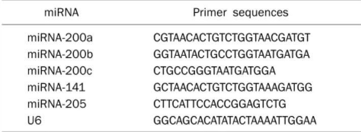
Table 1. The MicroRNA (miRNA) Primer Sequences for Quantitative Real-time PCR Analysis
miRNA Primer sequences
miRNA-200a CGTAACACTGTCTGGTAACGATGT miRNA-200b GGTAATACTGCCTGGTAATGATGA miRNA-200c CTGCCGGGTAATGATGGA miRNA-141 GCTAACACTGTCTGGTAAAGATGG miRNA-205 CTTCATTCCACCGGAGTCTG
U6 GGCAGCACATATACTAAAATTGGAA
pression of miRNA is observed in various cancers, suggesting it is important in carcinogenesis.7-12 In pancreatic cancer, ex- pression of miRNAs including miRNA-21, miRNA-221/222 and miRNA-181a/b/c is higher than in normal pancreatic tissues.13
Epithelial-mesenchymal transition (EMT) is a process by which cell-to-cell adhesion of epithelial cells disappears, and the epithelial cells gain migratory and invasive function like mesenchymal cells. This is closely related to metastasis and prognosis in gastrointestinal cancers (including colorectal cancer, hepatic cancer and pancreatic cancer) since it in- duces chemotherapy resistance.14-19 MiRNA-200 family and miRNA-205 regulate EMT by E-cadherin suppressing factors, ZEB1 and ZEB2 (SIP1).20 However, there are a few studies about EMT regulation by miRNA in pancreatic cancer.
Based on this background, the intent of this study is to elu- cidate the prognostic role of EMT-related miRNAs including miRNA-200 family (miRNA-200a/200b/200c/141) and miRNA-205 in resected pancreatic cancer patients.
SUBJECTS AND METHODS
1. Patients
Study subjects were drawn from among patients who were diagnosed with pancreatic cancer and underwent curative surgery in Seoul National University Bundang Hospital (Seoul, Korea) from 2003 through 2011. Patients who had palliative surgery or expired within 30 days after surgery due to surgical complications were excluded. This left 84 patients in the study. The study was approved by the human subjects committee of Seoul National University Bundang Hospital (IRB No. B-1103-124-302). Informed consent was waived by the board.
2. RNA extraction from pancreatic cancer tissue RNA from formalin-fixed, paraffin embedded pancreatic cancer tissue was extracted with a nucleic acid extraction kit (Ambion, Austin, TX, USA) and an RNeasyasylic Kit (Qiagen, Tokyo, Japan) according to the manufacturer’s instructions.21-24 The relative expression of miRNAs of pancreatic cancer tis- sues was measured with immortalized human pancreatic ductal epithelial (HPDE) cells as the expression reference.
HPDE cells were cultured in 5% fetal bovine serum (Gibco- BRL, Grand Island, NY, USA) and mixture of low glucose
(DMEM; Gibco-BRL) containing 750 ng/mL puromycin and medium M3 base (Incell Co., San Antonio, TX, USA), mixed in 3:1 ratio. After the cultured cell line was isolated by Trypsin-EDTA (Gibco-BRL) and collected with centrifugation (300×g, 5 min), RNA extraction was performed by manufac- tural protocol.
3. Quantitative real-time PCR
Isolated and quantified 5 μg of RNA was incubated in re- verse transcription premix (Maxime RT premix; Intron, Seoul, Korea) at 42oC for 50 minutes and at 95oC for 10 minutes to synthesize cDNA. With use of miScript (SYBR) Green PCR kit (Qiagen, Valencia, CA, USA), synthesized cDNA was mixed with 3 ng of cDNA, 10 L of SYBR Green master mix, 1 L of universal primer and 1 μL of each miRNA primer (Table 1) and titrated with diethylpyrocarbonate treated water, resulting in a total volume of 20 L. After incubation at 95oC for 15 mi- nutes, sequential incubation at 94oC for 15 seconds, at 55oC for 30 seconds and at 70oC for 30 seconds was repeated 45 times. The PCR results were analyzed by quantifying miRNA according to Ct values with 7500 Real-time PCR System (Applied Biosystems, Foster, CA, USA). U6 was used as an in- ternal control for quantification.
4. Statistics
The average values of continuous variables, standard er- ror of the means and median values were calculated and cat- egorical variables were measured. To evaluate the effects of miRNA on recurrence or metastasis, patients were dicotom- ized by the mean value of miRNA and Student’s t-test was performed. The relationship between miRNA expression and survival time after the diagnosis was evaluated with Pearson’s correlation analysis. Survival period and dis- ease-free survival period between the operation and re- currence were analyzed by Kaplan-Meier curves with log rank
Table 2. Baseline Characteristics of 84 Resected Pancreatic Cancer Patients
Characteristic Data
Age (yr) 64 (44-83)
Sex, male 52 (61.9)
Pathology, adenocarcinoma 79 (94.1)
R0 resection 71 (84.5)
pT stage
T1/T2 0 (0)/2 (2.4)
T3/T4 81 (96.4)/1 (1.2)
pN stage
N0/N1 36 (42.9)/48 (57.1)
AJCC stage
IB 1 (1.2)
IIA/IIB 37 (44.1)/45 (53.6)
III 1 (1.2)
Invasion
Angiolymphatic 39 (46.4)
Venous 17 (20.2)
Perineural 59 (70.2)
Adjuvant chemotherapy 43 (51.2)
Gemcitabine based 17 (20.2)
Adjuvant radiation therapy 45 (53.6)
Recurrence 70 (83.3)
Local recurrence 25 (29.8)
Distant metastasis 45 (53.6)
Values are presented as median (range) or n (%).
AJCC, American Joint Committee on Cancer.
Table 3. MicroRNA (miRNA) Expression Levels in Pancreatic Cancer Tissue Comparing with Immortalized Pancreatic Duct Epithelial Cells
miRNA HPDE cells Mean±SEM
miRNA-200a 1 266.9±57.3
miRNA-200b 1 18.5±2.2
miRNA-200c 1 0.7±0.1
miRNA-141 1 27.2±6.6
miRNA-205 1 0.1±0.1
HPDE, human pancreatic ductal epithelial; SEM, standard error of the mean.
test. We evaluated the prognostic factors affecting survival time by using univariate and multivariate analysis with Cox proportional hazards model. The prognostic factors of which p-values were less than 0.1 in univariate analysis were in- cluded in multivariate analysis. Null hypotheses of no differ- ence were rejected if p-values were less than 0.05, or, equiv- alently, if the 95% CIs of risk point estimates excluded 1. Most statistical analyses were carried out by IBM SPSS statistics ver- sion 20.0 (IBM Co., Armonk, NY, USA).
RESULTS
1. Baseline characteristics of enrolled patients There were 52 male and 32 female patients (Table 2). The mean age was 62.6±0.9 years. Pathologically, 79 patients (94.1%) had adenocarcinoma. Other cancers included one adenosquamous carcinoma, one mucoepidermoid carcino- ma, two undifferentiated carcinomas, and one anaplastic carcinoma. R0 resection was done in 71 patients (84.5%).
The pathologic stage of pancreatic cancer was mostly stage II, consisting of 37 patients with stage 2A (44.1%) and 45 pa-
tients with stage 2B (53.6%). Seventy patients (83.3%) expe- rienced recurrence during the follow-up period (25 in locore- gional and 45 in distant metastases). The mean time of re- currence was 16.0±2.2 months and the average overall sur- vival period was 25.0±2.3 months (2-140 months).
2. Expression of miRNA-200 family and miRNA-205 in pancreatic cancer
Relative expressions of miRNA-200a/200b/200c/141 and miRNA-205 in pancreatic cancer tissue were 266.9±
57.3 folds in miRNA-200a, 18.5±2.2 folds in miRNA-200b, 0.7±0.1 folds in miRNA-200c, 27.2±6.6 folds in miRNA-141 and 0.1±0.1 folds in miRNA-205 (Table 3). There were no sig- nificant differences in miRNA expression in patients in terms of recurrence or recurrence patterns (data not shown).
3. Survival and prognosis by expression of miRNA-200 family and miRNA-205 in pancreatic cancer The correlation between expressions of each miRNA and survival were evaluated. The miR-200c expression was in- versely correlated with survival period (r=−0.22, p=0.043), but the others were not associated (data not shown).
The subjects were dichotomized by the mean value of miRNA-200c; high miRNA-200c group (>0.65, n=29) and low miRNA-200c group (<0.65, n=55). The mean values of miRNA-200c were 1.5±0.2 in high miRNA-200c group and 0.3±0.03 in low miRNA-200c group. Overall survival was sig- nificantly longer in low miRNA-200c group (35 vs. 19 months, p=0.013) (Fig. 1A). By Cox proportional hazard model, survival was shorter in the high miRNA-200c group (hazard ratio, 1.771; 95% CI, 1.081-2.900; p=0.023) (Table 4).
When analyzing the correlation between miRNA-200a/
200b/200c/141 or miRNA-205 expression and disease free survival (DFS), there was a trend toward longer DFS in low
Fig. 1. Overall survival (p=0.013) (A) and disease-free survival (p=0.061) (B) according to the level of microRNA (miRNA)-200c.
Table 4. Prognostic Factors of Pancreatic Cancer
HR 95% CI p-value Univariate analysis
Age (>60 yr) 1.081 0.652-1.793 0.763
Sex (male) 1.286 0.794-2.085 0.307
Margin invasion (positive) 1.249 0.615-2.536 0.539
pN (positive) 0.955 0.593-1.539 0.850
Angiolymphatic invasion (positive) 1.314 0.819-2.108 0.258 Venous invasion (positive) 0.992 0.551-1.788 0.979 Perineural invasion (positive) 0.953 0.571-1.591 0.854 Post operative chemotherapy 0.866 0.540-1.390 0.552 Post operative gemcitabine 0.524 0.279-0.982 0.044 Post operative radiation therapy 1.036 0.645-1.664 0.883 High miRNA-200c (>0.65) 1.849 1.131-3.025 0.014 Multivariate analysis
Post operative gemcitabine 0.551 0.293-1.036 0.064 High miRNA-200c (>0.65) 1.771 1.081-2.900 0.023 HR, hazard ratio.
miRNA-200c group, however, it did not reach a statistical sig- nificance (24 vs. 12 months, p=0.061, Fig. 1B).
DISCUSSION
EMT is a process that is closely associated with metastasis and prognosis in gastrointestinal cancer including pancre- atic cancer.14,15 MiRNA-200 family and miRNA-205 are re- ported to regulate EMT and affected the prognosis in several cancers. However, there are only a few studies about these in pancreatic cancer. Based on this, we evalutated the the role of miRNA-200 family and miRNA-205 as an EMT-related prognostic marker in curatively resective pancreatic cancer.
In this study, the results of quantitative RT-PCR revealed
higher miRNA-200a/200b/141 and lower miRNA-200c/
205 expression in pancreatic cancer than in HPDE cells.
Moreover, the high miRNA-200c group, where the mean val- ue was over 1.5 fold of the reference, suffered a high re- currence rate and poor prognosis. In the present study, the ex- pression of miRNA-200c and miRNA-205 were suppressed in pancreatic cancers as in a previous study,25 and miRNA-200c was an independent prognostic factor. However, the overall survival was longer in the patient group with low miRNA-200c expression, unlike previous studies. These conflicting results suggest that roles of miRNA differ by various interactions be- tween miRNA and the surrounding environment. MiRNA and associated transcription factors function in a heterogeneous fashion in regulating genes in the context of tumor develop- ment, growth, metastasis and apoptosis. High levels of the miRNA-200 family are related to recurrence and poor prog- nosis in patients with ovarian cancer,26-28 although in other cancers that is not the case.29,30 Similarly, miRNA-200c in- hibits the metastatic ability of colon cancer cells by targeting ZEB1.31 However, another study observed that serum miRNA-200c levels were significantly higher in stage IV than in low stage colorectal cancer and high levels of miRNA-200c was associated with grave prognosis including lymph node and distant metastasis.32 Further study about the complex gene regulatory mechanism by miRNA-200c and related transcription factors is warranted.
This study is limited in that the results cannot reflect all pancreatic cancer patients because we included only pa- tients who had received surgical treatment and who were mostly stage II. Retrospective data collection produces errors
as medical records are not intended for research and can contain errors. As disease stage and lymph node metastasis are prognostic of pancreatic cancer, this may increase stat- istical error and reduce significance. Furthermore, the small study sample size might have been the reason that influenc- ing factors affecting survival in the univariate analysis lost statistical significance in the multivariate analysis.
The expression of miRNA was investigated by using ex- tracted RNA from specimens in this study. Further study about measuring miRNA expression by in situ hybridization analysis of cancer tissue is necessary.
In conclusion, this study found that miRNA-200a/200b/
141 was increased and miRNA-200c/205 was deceased in pancreatic cancer. Increased miRNA-200c expression is as- sociated with poor prognosis. Further studies on mecha- nisms of miRNA-200c in the progression and metastasis of pancreatic cancer will be required.
REFERENCES
1. Lai EC. Micro RNAs are complementary to 3' UTR sequence mo- tifs that mediate negative post-transcriptional regulation. Nat Genet 2002;30:363-364.
2. Bartel DP. MicroRNAs: target recognition and regulatory functions. Cell 2009;136:215-233.
3. Lee RC, Feinbaum RL, Ambros V. The C. elegans heterochronic gene lin-4 encodes small RNAs with antisense complementarity to lin-14. Cell 1993;75:843-854.
4. Ambros V. microRNAs: tiny regulators with great potential. Cell 2001;107:823-826.
5. Lu M, Zhang Q, Deng M, et al. An analysis of human microRNA and disease associations. PLoS One 2008;3:e3420.
6. Wienholds E, Plasterk RH. MicroRNA function in animal development. FEBS Lett 2005;579:5911-5922.
7. Krützfeldt J, Rajewsky N, Braich R, et al. Silencing of microRNAs in vivo with 'antagomirs'. Nature 2005;438:685-689.
8. Lu J, Getz G, Miska EA, et al. MicroRNA expression profiles classi- fy human cancers. Nature 2005;435:834-838.
9. Scherr M, Venturini L, Battmer K, et al. Lentivirus-mediated anta- gomir expression for specific inhibition of miRNA function.
Nucleic Acids Res 2007;35:e149.
10. He L, He X, Lowe SW, Hannon GJ. microRNAs join the p53 net- work--another piece in the tumour-suppression puzzle. Nat Rev Cancer 2007;7:819-822.
11. Calin GA, Croce CM. MicroRNA signatures in human cancers.
Nat Rev Cancer 2006;6:857-866.
12. Esquela-Kerscher A, Slack FJ. Oncomirs - microRNAs with a role in cancer. Nat Rev Cancer 2006;6:259-269.
13. Bloomston M, Frankel WL, Petrocca F, et al. MicroRNA ex- pression patterns to differentiate pancreatic adenocarcinoma from normal pancreas and chronic pancreatitis. JAMA 2007;
297:1901-1908.
14. Wells A, Yates C, Shepard CR. E-cadherin as an indicator of mes- enchymal to epithelial reverting transitions during the meta- static seeding of disseminated carcinomas. Clin Exp Metastasis 2008;25:621-628.
15. Thompson EW, Williams ED. EMT and MET in carcinoma--clinical observations, regulatory pathways and new models. Clin Exp Metastasis 2008;25:591-592.
16. Kajiyama H, Shibata K, Terauchi M, et al. Chemoresistance to pa- clitaxel induces epithelial-mesenchymal transition and enhan- ces metastatic potential for epithelial ovarian carcinoma cells.
Int J Oncol 2007;31:277-283.
17. Yang AD, Fan F, Camp ER, et al. Chronic oxaliplatin resistance in- duces epithelial-to-mesenchymal transition in colorectal cancer cell lines. Clin Cancer Res 2006;12:4147-4153.
18. Hiscox S, Jiang WG, Obermeier K, et al. Tamoxifen resistance in MCF7 cells promotes EMT-like behaviour and involves modu- lation of beta-catenin phosphorylation. Int J Cancer 2006;118:
290-301.
19. Hiscox S, Morgan L, Barrow D, Dutkowskil C, Wakeling A, Nicholson RI. Tamoxifen resistance in breast cancer cells is ac- companied by an enhanced motile and invasive phenotype: in- hibition by gefitinib ('Iressa', ZD1839). Clin Exp Metastasis 2004;21:201-212.
20. Gregory PA, Bert AG, Paterson EL, et al. The miR-200 family and miR-205 regulate epithelial to mesenchymal transition by tar- geting ZEB1 and SIP1. Nat Cell Biol 2008;10:593-601.
21. Abrahamsen HN, Steiniche T, Nexo E, Hamilton-Dutoit SJ, Sorensen BS. Towards quantitative mRNA analysis in paraf- fin-embedded tissues using real-time reverse transcriptase- polymerase chain reaction: a methodological study on lymph no- des from melanoma patients. J Mol Diagn 2003;5:34-41.
22. Godfrey TE, Kim SH, Chavira M, et al. Quantitative mRNA ex- pression analysis from formalin-fixed, paraffin-embedded tissues using 5' nuclease quantitative reverse transcription-polymerase chain reaction. J Mol Diagn 2000;2:84-91.
23. Doleshal M, Magotra AA, Choudhury B, Cannon BD, Labourier E, Szafranska AE. Evaluation and validation of total RNA extraction methods for microRNA expression analyses in formalin-fixed, paraffin-embedded tissues. J Mol Diagn 2008;10:203-211.
24. Zhang X, Chen J, Radcliffe T, Lebrun DP, Tron VA, Feilotter H. An array-based analysis of microRNA expression comparing match- ed frozen and formalin-fixed paraffin-embedded human tissue samples. J Mol Diagn 2008;10:513-519.
25. Yu J, Ohuchida K, Mizumoto K, et al. MicroRNA, hsa-miR-200c, is an independent prognostic factor in pancreatic cancer and its upregulation inhibits pancreatic cancer invasion but increases cell proliferation. Mol Cancer 2010;9:169.
26. Iorio MV, Visone R, Di Leva G, et al. MicroRNA signatures in hu- man ovarian cancer. Cancer Res 2007;67:8699-8707.
27. Nam EJ, Yoon H, Kim SW, et al. MicroRNA expression profiles in serous ovarian carcinoma. Clin Cancer Res 2008;14:2690-2695.
28. Helleman J, Jansen MP, Burger C, van der Burg ME, Berns EM.
Integrated genomics of chemotherapy resistant ovarian cancer:
a role for extracellular matrix, TGFbeta and regulating microRNAs.
Int J Biochem Cell Biol 2010;42:25-30.
29. Eitan R, Kushnir M, Lithwick-Yanai G, et al. Tumor microRNA ex- pression patterns associated with resistance to platinum based chemotherapy and survival in ovarian cancer patients. Gynecol Oncol 2009;114:253-259.
30. Leskelä S, Leandro-García LJ, Mendiola M, et al. The miR-200 family controls beta-tubulin III expression and is associated with paclitaxel-based treatment response and progression-free sur- vival in ovarian cancer patients. Endocr Relat Cancer 2010;18:
85-95.
31. Chen ML, Liang LS, Wang XK. miR-200c inhibits invasion and mi- gration in human colon cancer cells SW480/620 by targeting ZEB1. Clin Exp Metastasis 2012;29:457-469.
32. Toiyama Y, Hur K, Tanaka K, et al. Serum miR-200c is a novel prognostic and metastasis-predictive biomarker in patients with colorectal cancer. Ann Surg 2014;259:735-743.