691
Copyright © 2021 The Korean Society of Fisheries and Aquatic Science pISSN:0374-8111, eISSN:2287-8815
서 론
급성간췌장괴사병
(acute hepatopancreatic necrosis disease, AHPND)
은흰다리새우,
홍다리얼룩새우양식에서100%
폐 사를일으켜경제적손실을발생시키는매우심각한질병이다.
AHPND
에감염된새우는체색이옅어지고,
위장관이비어있으며
,
간췌장이흰색으로변하거나비정상적으로크기가줄어 드는등의증상이관찰된다(Tran et al., 2013; Han et al., 2015a;
Lee et al., 2015).
일반적으로
AHPND
의원인병원체는특정독소유전자(pirA, pirB)
를포함하고있으며,
병원성플라스미드(pVPA3-1)
을가 진Vibrio parahaemolyticus (Vp
AHPND)
로 알려져 있다(Han et al., 2015a).
중국(2009
년)
에서 첫 보고이후 베트남(2011
년),
말레이시아(2011
년),
태국(2012
년),
멕시코(2013
년),
필 리핀(2014
년),
미국(2017
년)
등여러국가로급속히확산되었 고(Lightner et al., 2012; Tran et al., 2013; Joshi et al., 2014;
흰다리새우(Penaeus vannamei)에서 급성간췌장괴사병(Acute Hepato- pancreatic Necrosis Disease)과 새우미포자충(Enterocytozoon
hepatopenaei)의 PCR 동시 진단법 개발
전혜진·이초롱·김범근·김수미
1·장광일
1·이강윤
2·권혜민
3·한지은*
경북대학교 수의학과, 1국립수산물품질관리원 수산방역과, 2군산대학교 해양과학대학, 3한국생명공학연구원 감염병연구센터
Development of a Simultaneous PCR Assay for Acute Hepatopancreatic Necrosis Disease (AHPND) and Enterocytozoon hepatopenaei (EHP) in Penaeus vannamei
Hye Jin Jeon, Chorong Lee, Bum Keun Kim, Sumi Kim1, Gwang Il Jang1, Gahngyoon Rhee2, Hyemin Kwon3 and Jee Eun Han*
College of Veterinary Medicine, Kyungpook National University, Daegu 41566, Korea
1Aquatic Disease Control Division, National Fishery Products Quality Management Service (NFQS), Busan 46083, Korea
2Kunsan National University Department of Ocean Science and Engineering, Gunsan 54150, Korea
3Infectious Disease Research Center, Korea Research Institute of Bioscience and Biotechnology, Daejeon 34141, Korea
Vibrio parahaemolyticus associated with acute hepatopancreatic necrosis disease ( Vp
AHPND) and Enterocytozoon hep- atopenaei (EHP) are the two most important pathogens in shrimp aquaculture and they have caused enormous losses to the shrimp industry worldwide. In ponds, the major target organ for the two pathogens is the hepatopancreas, and infection with EHP is a known potential risk factor for Vp
AHPNDinfection. This study aimed to develop a PCR (polymerase chain reaction)-based diagnostic method for simultaneously detecting Vp
AHPNDand EHP. The newly developed PCR diagnostic method could be used to test various samples, such as seawater, shrimp, and feces. The diagnostic method exhibited high sensitivity and specificity for both pathogens. This will help reduce the potential economic losses that may have been caused by the two major shrimp pathogens, Vp
AHPNDand EHP, and will allow for the efforts and time spent combatting them to be dedicated elsewhere.
Keywords: Acute hepatopancreatic necrosis disease, Diagnosis, Enterocytozoon hepatopenaei , Peneaus ( Litopenae- us ) vannamei , Shrimp
*Corresponding author: Tel: +82. 53. 950. 5972 Fax: +82. 53. 950. 5994 E-mail address: [email protected]
This is an Open Access article distributed under the terms of the Creative Commons Attribution Non-Commercial Licens (http://creativecommons.org/licenses/by-nc/3.0/) which permits unrestricted non-commercial use, distribution, and reproduction in any medium, provided the original work is properly cited.
Received 3 August 2021; Revised 11 September 2021; Accepted 23 September 2021 저자 직위: 전혜진(대학원생), 이초롱(박사후연구원), 김범근(대학생), 김수미 (주무관), 장광일(연구사), 이강윤(대학원생), 권혜민(대학원생), 한지은(교수) https://doi.org/10.5657/KFAS.2021.0691
Korean J Fish Aquat Sci 54(5), 691-697, October 2021
Nunan et al., 2014; Leobert et al., 2015; Dhar et al., 2019),
최 근에는국내새우양식장에서도보고된바가있다(Han et al., 2020a).
세계동물보건기구(OIE)
는2016
년부터AHPND
를관 리대상질병으로지정하여관리하고있으며,
국내에서도법정 전염병및 제1
종 살처분대상 질병으로 추가되었다(Aquatic Life Disease Control Act, 2021).
미포자충류는갑각류또는어류를포함하는다양한숙주의 세포내에서포자를형성하여기생할수있는단세포진핵생 물인세포내기생충이다
(Texier et al., 2010).
양식새우에서도 미포자충인Enterocytozoon hepatopenaei (EHP)
가보고되었 으며,
이는새우의간췌장을감염시켜소화,
흡수기능에영향 을끼치기때문에사료섭이저조,
성장지연을유발한다(Tang et al., 2015).
EHP
감염새우는 면역력감소로인해2
차질병감염에 취약 하게 되며(Newman, 2015),
현재까지Vp
AHPND를 포함한 다 양한 새우 병원균이EHP
와 복합감염으로 보고된 바 있다(Aranguren et al., 2017; Tang et al., 2017; Thamizhvanan et al., 2019; Han et al., 2020b; Jithendran et al., 2021). 2004
년 태국에서 성장지연을보이는홍다리얼룩새우에서 이름이알 려지지않은미포자충으로처음보고되었고(Chayaburakul et al., 2004), 2009
년태국의양식장에서특성화및명명된이후(Tourtip et al., 2009),
베트남(2010
년),
중국(2014
년),
인도(2016
년),
인도네시아(2016
년)
등여러아시아국가로급속하게 확산되고있다(Ha et al., 2010; Sritunyalucksana et al., 2014;
Rajendran et al., 2016; Tang et al., 2016).
최근에는남미에서 도EHP
감염이보고되었다(Tang et al., 2017). EHP
에감염된 새우는특이적인임상증상이관찰되지않고조직병리학적또 는PCR (polymerase chain reaction)
분석을통해서만진단이 가능하기때문에양식장에서의관리가매우어려워경제적으로 심각한문제를발생시킨다.
Vp
AHPND는세균이내뿜는독소가간췌장을괴사시키는반면,
EHP
는새우간췌장세포내에감염하여소화를방해하는등두질병의감염기작은다르나표적장기가간췌장으로동일하여복 합감염에대한문제가대두되고있다
(Han et al., 2020b).
실제 로인도새우양식장에서EHP
와Vibrio
의복함감염발병율이6%
이상으로 집계되었으며,
베트남과 태국의새우양식장에 서도Vp
AHPND와EHP
의복합감염발생빈도가높은것으로보 고되고있다(FAO, 2015; Babu et al., 2021).
또한,
실험실내에 서진행된연구에서EHP
에감염된새우는Vp
AHPND 감염에대 한감수성이증가하는것으로보고되고있다(Aranguren et al., 2017).
국내에서도수입된베트남흰다리새우에서Vp
AHPND와EHP
가동시에검출된바있다(Han et al., 2020b).
본연구에서는
Vp
AHPND와EHP
를동시에진단할수있는du- plex PCR
법을개발하였다.
개발된동시진단법은새우뿐만아 니라해수,
분변등의다양한종류의샘플에서적용가능성을알 아보기위해수행되었다.
재료 및 방법
샘플 수집 방법
Vp
AHPND와EHP
를동시에진단할수있는duplex PCR
법을 개발을위하여,
기존보고된PCR
방법으로진단된샘플을사 용하였다(Han et al., 2015a; Tang et al., 2015). AHPND
감염 샘플은양식장수집새우(N=4),
양식장물(N=3),
인공감염새 우(N=8), AHPND Vibrio parahaemolyticus (N=4), AHPND V. campbellii (N=1)
를, EHP
감염샘플은베트남수입새우(N=12) (Han et al., 2020b),
양식장수집새우(N=6),
양식장물(N=1),
새우분변(N=1)
을사용하였다. AHPND
인공감염을위 해22 L
의수조에1.0-1.5 g
의새우를10
5CFU (colony form- ing unit)/mL
의농도로감염시킨후,
폐사새우의간췌장샘플 을수집하였다. AHPND V. parahaemolyticus
와AHPND V.
campbellii
는TSB
+(tryptic soy broth+2% NaCl)
에서18
시간 이상진탕배양(28°C, 200 rpm)
후세균배양액(1 μL)
을PCR
분 석을위한template
로사용하였다.
동시감염샘플은Vp
AHPND와EHP
에대하여각각양성인DNA
샘플을혼합하여사용하였다.
수집된샘플은
DNeasy Blood & Tissue kit (Qiagen, Hilden, Germany)
를사용하여새우의간췌장(30 mg),
분변(30 mg),
해 수(300 μL)
에서각각DNA
를추출후PCR
분석에사용하였 으며,
진단분석에사용한샘플항목은Table 1
에나타내었다. Vp
AHPND와 EHP 동시진단 primer 제작
Vp
AHPND 진단primer
는 독소유전자(pirA, pirB)
를 포함하 는 병원성 플라스미드(pVPA3-1) (Genbank accession no.
KM067908)
의 염기서열을 바탕으로1-step PCR primer
인AHPND-729F/R
과2-step PCR primer
인AHPND-377F/R
을 새롭게제작하였다.
EHP
진단primer
는기존에제작된1-step PCR primer
를사 용하였고(Jaroenlak et al., 2016), 2-step PCR primer
는spore wall protein (Genbank accession no. KX258197)
의염기서열 을바탕으로EHP-203F/R
을새롭게제작하였다.
실험에사용한
primer
는Table 2
에나타내었다.
제작된prim- er
는Primer 3 (Version 0.4, primer design software)
를사용하 여설계하였으며, oligonucleotide
는Macrogen
에의뢰하여사 용전까지-20°C
에보관하였다.
Duplex PCR을 통한 Vp
AHPND와 EHP 동시진단
본 연구에서 개발된 동시진단primer
와Vp
AHPND, EHP
감 염샘플과의 반응여부는duplex PCR
분석으로 확인하였다.
1-step PCR amplification
을위하여,
수집된양성샘플의DNA,
AHPND-729F/R primer, EHP-SWP1F/1R primer
를 각각1
μL
씩PCR premix (K-2016; Bioneer, Daejeon, Korea)
에혼 합하였다. PCR
혼합물은95°C
에서5
분간pre-denaturation
시 킨후, 95°C
에서30
초denaturation, 58°C
에서45
초annealing,
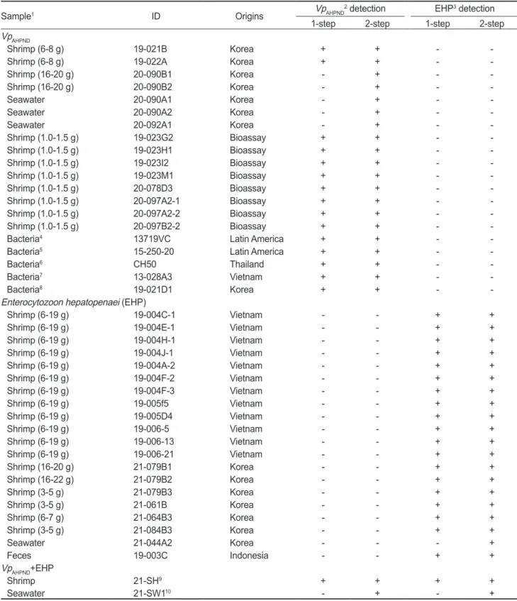
Table 1. Sample list and PCR detection of the duplex PCR assay
Sample1 ID Origins VpAHPND2 detection EHP3 detection
1-step 2-step 1-step 2-step
VpAHPND
Shrimp (6-8 g) 19-021B Korea + + - -
Shrimp (6-8 g) 19-022A Korea + + - -
Shrimp (16-20 g) 20-090B1 Korea - + - -
Shrimp (16-20 g) 20-090B2 Korea - + - -
Seawater 20-090A1 Korea - + - -
Seawater 20-090A2 Korea - + - -
Seawater 20-092A1 Korea - + - -
Shrimp (1.0-1.5 g) 19-023G2 Bioassay + + - -
Shrimp (1.0-1.5 g) 19-023H1 Bioassay + + - -
Shrimp (1.0-1.5 g) 19-023I2 Bioassay + + - -
Shrimp (1.0-1.5 g) 19-023M1 Bioassay + + - -
Shrimp (1.0-1.5 g) 20-078D3 Bioassay + + - -
Shrimp (1.0-1.5 g) 20-097A2-1 Bioassay + + - -
Shrimp (1.0-1.5 g) 20-097A2-2 Bioassay + + - -
Shrimp (1.0-1.5 g) 20-097B2-2 Bioassay + + - -
Bacteria4 13719VC Latin America + + - -
Bacteria5 15-250-20 Latin America + + - -
Bacteria6 CH50 Thailand + + - -
Bacteria7 13-028A3 Vietnam + + - -
Bacteria8 19-021D1 Korea + + - -
Enterocytozoon hepatopenaei (EHP)
Shrimp (6-19 g) 19-004C-1 Vietnam - - + +
Shrimp (6-19 g) 19-004E-1 Vietnam - - + +
Shrimp (6-19 g) 19-004H-1 Vietnam - - + +
Shrimp (6-19 g) 19-004J-1 Vietnam - - + +
Shrimp (6-19 g) 19-004A-2 Vietnam - - + +
Shrimp (6-19 g) 19-004F-2 Vietnam - - + +
Shrimp (6-19 g) 19-004F-3 Vietnam - - + +
Shrimp (6-19 g) 19-005f5 Vietnam - - + +
Shrimp (6-19 g) 19-005D4 Vietnam - - + +
Shrimp (6-19 g) 19-006-5 Vietnam - - + +
Shrimp (6-19 g) 19-006-13 Vietnam - - + +
Shrimp (6-19 g) 19-006-21 Vietnam - - + +
Shrimp (16-20 g) 21-079B1 Korea - - + +
Shrimp (16-22 g) 21-079B2 Korea - - + +
Shrimp (3-5 g) 21-079B3 Korea - - + +
Shrimp (3-5 g) 21-061B Korea - - + +
Shrimp (6-7 g) 21-064B3 Korea - - + +
Shrimp (3-5 g) 21-084B3 Korea - - + +
Seawater 21-044A2 Korea - - - +
Feces 19-003C Indonesia - - + +
VpAHPND+EHP
Shrimp 21-SH9 + + + +
Seawater 21-SW110 - + - +
1The species of shrimp in the samples are Penaeus vannamei. 2Vibrio parahaemolyticus causing acute hepatopancreatic necrosis disease.
3Enterocytozoon hepatopenaei. 4Bacteria species is V. campbellii, 2016, unpublished. 5Bacteria species is V. parahaemolyticus, 2015, un- published. 6Bacteria species is V. parahaemolyticus, 2019, unpublished. 7Bacteria species is V. parahaemolyticus, 2013 (Tran et al., 2013).
8Bacteria species is V. parahaemolyticus, 2019 (Han et al., 2020). 9EHP positive shrimp sample (19-004H1) and VpAHPND positive shrimp sample (20-078D3) mixed DNA. 10EHP positive shrimp sample (21-044A2) and VpAHPND positive shrimp sample (20-090A1) mixed DNA.
68°C
에서45
초extension
반응을30 cycles
수행한후68°C
에서5
분간final-extension
시켰다.
2-step PCR amplification
을 위하여1-step PCR product, AHPND-377F/R primer, EHP-203F/R primer
를각각1 μL
씩PCR premix (K-2016; Bioneer)
에혼합하였다. PCR
혼합물은95°C
에서5
분간pre-denaturation
시킨후, 95°C
에서30
초dena- turation, 64°C
에서30
초annealing, 68°C
에서20
초extension
반응을20 cycles
수행한후68°C
에서5
분간final-extension
시 켰다.
PCR
분석후, ethidium bromide
를함유한1.5% gel
에서PCR
산물을loading
하고gel doc (DAIHAN Scientific Co. Ltd., Seoul, Korea)
을이용해결과를확인하였다.
Vp
AHPND와 EHP 동시진단 primer의 특이도 및 민감 도 확인
Vp
AHPND와EHP
동시진단primer
가표적질병외다른병원체 에서는반응을일으키지않는다는것을확인하기위해특이도(specificity)
평가를실시하였다.
실험에는AHPND
를유발하지 않는V. parahaemolyticus (N=4), V. harveyi (N=6), V. camp- bellii (N=3)
와 흰반점바이러스(white spot syndrome virus, WSSV)
에감염된새우(N=7),
전염성피하및조혈괴사증(infec- tious hypodermal and hematopoietic necrosis virus, IHHNV)
에감염된새우(N=2),
무병새우(Penaeus vannamei) (N=1)
샘 플을사용하였다.
실험에사용된무병새우는제주도서귀포시 에위치한종묘장에서구입후,
흰다리새우의대표질병병원체(Vp
AHPND, EHP, IHHNV, WSSV)
를PCR
분석한결과음성으 로검출된샘플을사용하였다.
수집된샘플의DNA
추출,
세균 배양은‘Vp
AHPND와EHP
감염샘플수집방법’
과동일한방법으로진행되었다
.
Vp
AHPND와EHP
동시진단primer
의 민감도(sensitivity)
는Vp
AHPND와EHP
에각각감염된새우의DNA (19-004H1, 20- 078D3)
를10
배씩계단희석(10
-0~10
-6)
하여사용하였다.
신규Vp
AHPND 진단primer (729F/R, 377F/R), EHP (EHP-1F/1R, EHP-203F/R)
와기존진단에사용하는Vp
AHPND진단primer (VpPirA-284F/R, VpPirB-392F/R), EHP (EHP-1F/1R, EHP- 2F/2R)
의감도를비교하였다(Han et al., 2015a; Jaroenlak et al., 2016).
결 과
PCR분석 결과
양식장 수집 새우
(N=4),
양식장 물(N=3),
인공감염 새우(N=8), AHPND V. parahaemolyticus (N=4), AHPND V.
campbellii (N=1)
의총20
개Vp
AHPND샘플중,
양성률은1-step PCR
와2-step PCR
에서각각75% (N=15), 100% (N=20)
였 다.
베트남수입새우(N=12),
양식장수집새우(N=6),
양식장 물(N=1),
새우분변(N=1)
의총20
개의EHP
샘플중,
양성률 은1-step PCR
와2-step PCR
에서각각95% (N=19), 100%
(N=20)
였다.
제작된Vp
AHPND, EHP
동시감염 샘플(N=2)
의1-step PCR
와2-step PCR
에서각각50% (N=1), 100% (N=2)
였으며,
분석에사용된primer
간교차반응은일어나지않았다. PCR
분석결과는Table 1
에나타내었다.
특이도, 민감도 테스트
특이도 검사를 위해 사용된
AHPND
를 유발하지 않는V.
parahaemolyticus (N=4), V. harveyi (N=6), V. campbellii
Table 2. The new duplex PCR primer sequences and PCR condition used in this studyDiagnosis Primers Sequence (5’ to 3’) Amplicon size (bp) Reference
EHP
1-step 1F TTGCAGAGTGTTGTTAAGGGTTT
514 Jaroenlak et al. (2016)
1R CACGATGTGTCTTTGCAATTTTC
2-step 203F TGATCATCAACGCAAAACTGT
203 In this study
203R CACGATGTGTCTTTGCAATTT
VpAHPND
1-step 729F GGCTGGAAAGTGGCTAAATC
729
In this study
729R ATGCACATCAGAATCGGTGA
2-step 377F CGCCAAATGAGCCAGATATT
377R ATGCACATCAGAATCGGTGA 377
PCR condition
1-step
1 cycle 95°C, 5 min
In this study 35 cycles 95°C, 30 sec/58°C, 40 sec/68°C, 45 sec
1 cycle 68°C 5 min
2-step
1 cycle 95°C, 5 min
35 cycles 95°C, 30 sec/64°C, 30 sec/68°C, 20 sec 1 cycle 68°C 5 min
EHP, Enterocytozoom hepatopenaei; VpAHPND, Vibrio parahaemolyticus associated with AHPND.
(N=3)
와흰반점바이러스(WSSV)
에감염된새우(N=7),
전염 성피하및조혈괴사증(IHHNV)
에감염된새우(N=2),
무병새우(P. vannamei) (N=1)
샘플모두에서교차반응은일어나지않았 다.
특이도분석결과는Table 3
에나타내었다.
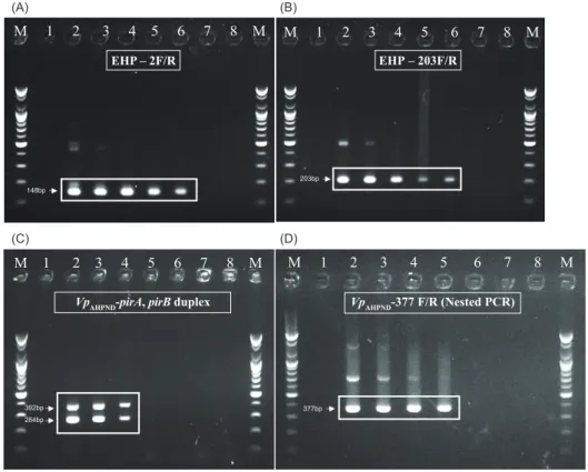
민감도테스트에서는기존
Vp
AHPND진단primer (Han et al., 2015a), EHP (Jaroenlak et al., 2016)
와신규개발된primer
의 감도를비교하였다.
신규Vp
AHPND진단primer
의검출한계는 양성DNA
의10
-3배희석샘플까지검출되었고,
기존Vp
AHPNDprimer
보다10
배민감하였다.
신규EHP primer
의검출한계 는양성DNA
의10
-4배희석샘플까지검출되었고,
기존EHP primer
와동일한감도로검출되었다. PCR
분석결과는Fig. 1
에나타내었다.
고 찰
이번연구에서는표적장기가동일한두가지병원체
Vp
AHPND 와EHP
의동시진단을위한새로운nested duplex PCR
방법을 개발했다.
실질적으로양식장에서두질병에의한경제적,
노동 적손실을최소화하기위해서는빠르고정확한스크리닝을통 해이두병원체(Vp
AHPND, EHP)
를초기단계에서진압하여질 병전파를최소화하는것이필요하다.
현재까지Vp
AHPND진단을 위해pirA
및pirB
독소유전자를표적으로하는PCR, real-time PCR
등의여러분자생물학적진단법이개발되었고(Han et al.,
Fig. 1. Result of sensitivity test. DNAs of 19-004H1 positive for Enterocytozoon hepatopenaei (EHP), and 20-078D3 positive for Vibrio parahaemolyticus causing acute hepatopancreatic necrosis disease (VpAHPND) were used. EHP PCR assay using (A), previous EHP primer (148bp); (B), new EHP primer (203bp); VpAHPND PCR assay using (C), Previous VpAHPND duplex primer, targeting pirA (284bp) and pirB (392bp); (D), New VpAHPND primer (377bp); Lane M, 1kb-plus DNA ladder; Lane 1, negative control; Lane 2, original DNA; Lane 3, 10-1 dilution; Lane 4, 10-2 dilution; Lane 5, 10-3 dilution; Lane 5, 10-4 dilution; Lane 6, 10-5 dilution; Lane7, 10-6 dilution.(B) (A)
M 1 2 3 4 5 6 7 8 M
EHP – 2F/R EHP – 203F/R
M 1 2 3 4 5 6 7 8 M
203bp 148bp
(D) (C)
M 1 2 3 4 5 6 7 8 M VpAHPND-pirA, pirB duplex
284bp 392bp
M 1 2 3 4 5 6 7 8 M VpAHPND-377 F/R (Nested PCR)
377bp
Table 3. PCR result of the specificity test
Type Sample
Number
detectionEHP VpAHPND detection 1-step 2-step 1-step 2-step
WSSV1 7 - - - -
IHHNV2 2 - - - -
Vibrio parahaemolyticus3 4 - - - -
V. harveyi4 6 - - - -
V. campbellii5 3 - - - -
SPF shrimp6 1 - - - -
1White spot syndrome virus. 2Infectious hypodermal and hema- topoietic necrosis virus. 3non-AHPND vibrio parahaemolyticus.
4non-AHPND vibrio harveyi, 5non-AHPND vibrio campbellii.
6Specific pathogen free Penaeus vannamei.
2015a, Han et al., 2015b), EHP
또한PCR, real-time PCR, In situ hybridization, LAMP
등여러분자생물학적진단법이개 발되었다(Sritunyalucksana et al., 2014; Tang et al., 2015; Ja- roenlak et al., 2016; Makesh et al., 2018).
이두질병을야기하 는병원체(Vp
AHPND, EHP)
는필드에서복합감염이빈번하게보 고되고있지만(Han et al., 2020b),
이를동시에진단할수있는PCR
법은아직개발되지않았다.
이번연구에서개발된
nested duplex PCR
법은새우양식장에 서주요한두가지질병인AHPND
와EHP
의복합감염과단일 감염을모두진단할수있고,
기존의진단primer
보다높은감도 를나타내기때문에초기감염단계에서질병을진단할수있다 는장점이있다.
또한Vp
AHPND와EHP
에만특이적으로반응하 기때문에위양성검출에의한결과의오류를줄일수있다.
그 리고양식장새우샘플뿐만아니라실험실내인공감염샘플에 서도검출이용이하며,
분리된세균이나환경시료(
물과분변)
에서도적용가능하다.
따라서,
신속하고정확한질병진단을통 해양식장내에서보다효율적인질병관리가가능할것으로판 단된다.
추가적으로,
개발된PCR
법의활용과객관적인데이터 확보를위해,
보다다양한샘플수집및분석,
그리고Pir
독소의 정량화등의후속연구가필요하다고생각한다.
사 사
이논문은국립수산물품질관리원의수산생물 방역기술개발 및프로그램운영과제
(R2021071)
와정부(
과학기술정보통신 부,
교육부)
의 재원으로 한국연구재단의지원을받아수행된 기초연구 사업입니다(No. NRF-2018R1C1B5086350, NRF- 2019R1C1C1006212, NRF-2021R1I1A1A01040303).
References
Aquatic Life Disease Control Act. 2021. Enforcement date 30.
June, 2021. Act No.486, 30. June, 2021., partial amendment.
Retrieved form https://www.law.go.kr/lsSc.do?menuId=1&
subMenuId=15&tabMenuId=81&eventGubun=060114#un defined on Jul 27, 2021.
Aranguren LF, Han JE and Tang KF. 2017. Enterocytozoon
hepatopenaei (EHP) is a risk factor for acute hepatopancre-
atic necrosis disease (AHPND) and septic hepatopancreatic necrosis (SHPN) in the Pacific white shrimp Penaeus van-namei. Aquaculture 471, 37-42. https://doi.org/10.1016/j.
aquaculture.2016.12.038.
Babu B, Sathiyaraj G, Mandal A, Kandan S, Biju N, Palanisamy S, You SG, Nisha R G and Prabhu N M. 2021. Surveillance of disease incidence in shrimp farms located in the east coastal region of India and in vitro antibacterial efficacy of probiot- ics against Vibrio parahaemolyticus. J Invertebr Pathol 179, 107536. https://doi.org/10.1016/j.jip.2021.107536.
Chayaburakul K, Nash G, Pratanpipat P, Sriurairatana S and
Withyachumnarnkul B. 2004. Multiple pathogens found in growth-retarded black tiger shrimp Penaeus monodon cul- tivated in Thailand. Dis Aquat Org 60, 89-96. https://doi.
org/10.3354/dao060089.
Dhar AK, Piamsomboon P, Aranguren Caro LF, Kanrar S, Ad- ami R Jr and Juan YS. 2019. First report of acute hepatopan- creatic necrosis disease (AHPND) occurring in the USA. Dis Aquat Org 132, 241-247. https://doi.org/10.3354/dao03330.
FAO (Food and Agriculture Organization of the United Na- tions). 2015. Highlights of the first international technical seminar/workshop: “EMS/AHPND: Government, scientist and farmer responses” (June 2015, Panama City, Panama) under the FAO Project. TCP/INT/3502. 2015, FAO, Rome, Italy.
Ha NTH, Ha DT, Thuy NT and Lien VTK. 2010. Enterocytozo-
on hepatopenaei has been detected parasitizing tiger shrimp Penaeus monodon cultured in Vietnam and showing white
feces syndrome. J Agric Sci 12, 45-50.Han JE, Tang KF, Tran LH and Lightner DV. 2015a. Photorhab-
dus insect-related (Pir) toxin-like genes in a plasmid of Vib- rio parahaemolyticus, the causative agent of acute hepato-
pancreatic necrosis disease (AHPND) of shrimp. Dis Aquat Org 113, 33-40. https://doi.org/10.3354/dao02830.Han JE, Tang KF, Pantoja CR, White BL and Lightner DV.
2015b. qPCR assay for detecting and quantifying a viru- lence plasmid in acute hepatopancreatic necrosis disease (AHPND) due to pathogenic Vibrio parahaemolyticus.
Aquaculture 442, 12-15. https://doi.org/10.1016/j.aquacul- ture.2015.02.024.
Han JE, Choi SK, Han SH, Lee SC, Jeon HJ, Lee C, Kim KY, Lee YS, Park SC, Rhee GY, Park SY, Kim JS, Park S, Kim JH and Lee KJ. 2020a. Genomic and histopathological characteristics of Vibrio parahaemolyticus isolated from an acute hepatopancreatic necrosis disease outbreak in Pacific white shrimp Penaeus vannamei cultured in Korea. Aqua- culture 524, 735284. https://doi.org/10.1016/j.aquacul- ture.2020.735284.
Han JE, Lee SC, Park SC, Jeon HJ, Kim KY, Lee YS, Park S, Han SH, Kim JH and Choi SK. 2020b. Molecular detection of Enterocytozoon hepatopenaei and Vibrio parahaemolyti-
cus-associated acute hepatopancreatic necrosis disease in
Southeast Asian Penaeus vannamei shrimp imported into Korea. Aquaculture 517, 734812. https://doi.org/10.1016/j.aquaculture.2019.734812.
Jaroenlak P, Sanguanrut P, Williams BA, Stentiford GD, Fle- gel TW, Sritunyalucksana K and Itsathitphaisarn O. 2016.
A nested PCR assay to avoid false positive detection of the microsporidian Enterocytozoon hepatopenaei (EHP) in environmental samples in shrimp farms. PLoS One 11, e0166320. https://doi.org/10.1371/journal.pone.0166320.
Jithendran KP, Navaneeth Krishnan A, Jagadeesan V, Anan- daraja R, Ezhil Praveena P, Anushya S, Amarnath BC and
Bhuvaneswari T. 2021. Co-infection of infectious myone- crosis virus and Enterocytozoon hepatopenaei in Penaeus
vannamei farms in the east coast of India. Aquac Res 52,
4701-4710. https://doi.org/10.1111/are.15304.Joshi J, Srisala J, Truong VH, Chen I-T, Nuangsaeng B, Suthienkul O, Lo CF, Flegel TW, Sritunyalucksana K and Thitamadee S. 2014. Variation in Vibrio parahaemolyticus isolates from a single Thai shrimp farm experiencing an out- break of acute hepatopancreatic necrosis disease (AHPND).
Aquaculture 428-429, 297-302. https://doi.org/10.1016/j.
aquaculture.2014.03.030.
Lee CT, Chen IT, Yang YT, Ko TP, Huang YT, Huang JY, Huang MF, Lin SJ, Chen CY, Lin SS, Lighter DV, Wang HC, Wang AHJ, Wang HC, Hor LI and Lo CF. 2015. The opportunistic marine pathogen Vibrio parahaemolyticus becomes virulent by acquiring a plasmid that expresses a deadly toxin. Proc Natl Acad Sci 112, 10798-10803. https://doi.org/10.1073/
pnas.1503129112.
Leobert D, Cabillon NAR, Catedral DD, Amar EC, Usero RC, Monotilla WD, Calpe AT, Fernandez DDG and Saloma CP.
2015. Acute hepatopancreatic necrosis disease (AHPND) outbreaks in Penaeus vannamei and P. monodon cultured in the Philippines. Dis Aquat Org 116, 251-254. https://doi.
org/10.3354/dao02919.
Lightner DV, Redman RM, Pantoja CR, Noble BL and Tran LH.
2012. Early mortality syndrome affects shrimp in Asia. Glob Aquac Advocate, 40.
Makesh M. 2018. Visual loop-mediated isothermal amplifica- tion (LAMP) for the rapid diagnosis of Enterocytozoon hep-
atopenaei (EHP) infection. Parasitol Res 117, 1485-1493.
https://doi.org/10.1007/s00436-018-5828-4.
Newman SG. 2015. Microsporidian impacts shrimp production- industry efforts address control, not eradication. Glob Aquac Advocate, 33-35.
Nunan L, Lightner D, Pantoja C and Gomez-Jimenez S. 2014.
Detection of acute hepatopancreatic necrosis disease (AHPND) in Mexico. Dis Aquat Org 111, 81-86. https://doi.
org/10.3354/dao02776.
Rajendran KV, Shivam S, Praveena PE, Rajan JJS, Kumar TS, Avunje S and Vijayan KK. 2016. Emergence of Enterocyto-
zoon hepatopenaei (EHP) in farmed Penaeus (Litopenaeus) vannamei in India. Aquaculture 454, 272-280. https://doi.
org/10.1016/j.aquaculture.2015.12.034.
Snowden KF. 2004. Zoonotic microsporidia from animals and arthropods with a discussion of human infections. In: Op- portunistic infections: toxoplasma, sarcocystis, and micro- sporidia. Lindsay DS and Weiss LM, eds. Kluwer Academic Publishers, New York, NY, U.S.A., 123-134. https://doi.
org/10.1007/978-1-4020-7846-0_8.
Sritunyalucksana K, Sanguanrut P, Salachan PV, Thita- madee S and Flegel TW. 2014. Urgent appeal to control spread of the shrimp microsporidian parasite Enterocyto-
zoon hepatopenaei (EHP). Retrieved from https://enaca.
org/?id=101&title=urgent-appeal-to-control-spread-of-the- shrimp-microsporidian-parasite-enterocytozoon-hepatope- naei-ehp on Mar 21, 2021.
Tang KF, Pantoja CR, Redman RM, Han JE, Tran LH and Lightner DV. 2015. Development of in situ hybridiza- tion and PCR assays for the detection of Enterocytozoon
hepatopenaei (EHP), a microsporidian parasite infecting
penaeid shrimp. J Invertebr Pathol 130, 37-41. https://doi.org/10.1016/j.jip.2015.06.009.
Tang KF, Han JE, Aranguren LF, White-Noble B, Schmid MM, Piamsomboon P, Risdiana E and Hanggono B. 2016. Dense populations of the microsporidian Enterocytozoon hepa-
topenaei (EHP) in feces of Penaeus vannamei exhibiting
white feces syndrome and pathways of their transmission to healthy shrimp. J Invertebr Pathol 140, 1-7. https://doi.org/10.1016/j.jip.2016.08.004.
Tang KF, Aranguren LF, Piamsomboon P, Han JE, Maskaykina IY and Schmidt MM. 2017. Detection of the microsporid- ian Enterocytozoon hepatopenaei (EHP) and Taura syn- drome virus in Penaeus vannamei cultured in Venezuela.
Aquaculture 480, 17-21. https://doi.org/10.1016/j.aquacul- ture.2017.07.043.
Texier C, Vidau C, Viguès B, El Alaoui H and Delbac F. 2010.
Microsporidia: a model for minimal parasite-host inter- actions. Curr Opin Microbiol 13, 443-449. https://doi.
org/10.1016/j.mib.2010.05.005.
Thamizhvanan S, Sivakumar S, Santhosh Kumar S, Vinoth Ku- mar D, Suryakodi S, Balaji K, Rajkumar T, Vimal S, Abdul Majeed S, Taju G and Sahul Hameed AS. 2019. Multiple infections caused by white spot syndrome virus and Entero-
cytozoon hepatopenaei in pond-reared Penaeus vannamei in
India and multiplex PCR for their simultaneous detection. J Fish Dis 42, 447-454. https://doi.org/10.1111/jfd.12956.Tourtip S, Wongtripop S, Stentiford GD, Bateman KS, Sriurai- ratana S, Chavadej J, Sritunyalucksana K and Withyachum- narnkul B. 2009. Enterocytozoon hepatopenaei sp. nov.
(Microsporida: Enterocytozoonidae), a parasite of the black tiger shrimp Penaeus monodon (Decapoda: Penaeidae):
Fine structure and phylogenetic relationships. J Invertebr Pathol 102, 21-29. https://doi.org/10.1016/j.jip.2009.06.004.
Tran LH, Nunan L, Redman RM, Mohney LL, Pantoja CR, Fitzsimmons K and Lightner DV. 2013. Determination of the infectious nature of the agent of acute hepatopancreatic necrosis syndrome affecting penaeid shrimp. Dis Aquat Org 105, 45-55. https://doi.org/10.3354/dao02621.