Introduction
Open wedge high tibial osteotomy (OWHTO) is a commonly performed procedure for knees exhibiting medial compartment osteoarthritis with varus malalignment1), and favorable surgical outcomes have been reported in the majority of clinical followup studies2,3). However, this procedure also carries the risk of com
plications such as nonunion, infection, tibial plateau fracture and patellofemoral problems48).
Among these complications, postoperative patellofemoral problems such as patella baja and anterior knee pain have been considered as potential shortcomings of OWHTO because this procedure inevitably lowers the tibial tubercle position creating a potential risk for patella baja and increases patellofemoral pres
sure2,4,912). Therefore, measures against these unfavorable patel
lofemoral mechanics are essential to improve the outcomes of OWHTO.
Lateral retinacular release is a popular arthroscopic procedure performed to improve the patellofemoral alignment and relieve the excessive joint pressure8). Therefore, concomitant lateral re
lease (LR) at the time of OWHTO may effectively improve patel
lofemoral mechanics leading to reduced anterior knee symptoms and improved surgical outcome and patient satisfaction.
There have been a few studies that examined the effect of con
comitant LR on the postoperative patellofemoral mechanics and
The Effect of Concomitant Arthroscopic Lateral
Retinacular Release on Postoperative Patellar Position and Orientation in Open Wedge High Tibial Osteotomy
Kazuhiro Murayama, MD
1, Hiroshi Nakayama, MD
1, Tomohiko Murakami, MD
2, Shinichi Yoshiya, MD
1, Shuhei Otsuki, MD
2, and Toshiya Tachibana, MD
11Department of Orthopaedic Surgery, Hyogo College of Medicine, Nishinomiya; 2Department of Orthopedic Surgery, Osaka Medical College, Osaka, Japan
Purpose: The purpose of this study was to evaluate the effect of concomitant arthroscopic lateral release (LR) in open wedge high tibial osteotomy (OWHTO) by comparing the pre and postoperative radiological parameters of patellar position and orientation.
Materials and Methods: The study was comprised of 19 knees undergoing OWHTO and concomitant LR and 18 knees undergoing OWHTO alone.
Radiological parameters for patellar position and orientation included the CatonDeschamps index (CDI), BlackburnePeel index (BPI), patellar tilting angle (PTA), patellar lateral shift (PLS), and patellofemoral distance (PFD), which were evaluated in the preoperative period and at one year after surgery.
Results: Patellar height was significantly reduced after surgery as indicated by the decrease in BPI (p=0.03) in the OWHTO/LR group, and decrease in CDI (p=0.03) and BPI (p=0.04) in the OWHTO alone group. PTA and PLS were significantly reduced after the combined OWHTO/LR procedure (p=0.04 and p=0.04, respectively). By contrast, no significant changes were detected when isolated OWHTO was performed.
Conclusions: OWHTO induced a postoperative decrease in patellar height in both groups. Regarding the change in patellofemoral alignment, concomitant LR in OWHTO significantly decreased lateral patellar tilt and shift, while no significant difference in those parameters were noted in the OWHTO alone knees.
Keywords: Knee, Osteotomy, Open wedge, Arthroscopy, Lateral retinacular release, Patellar position pISSN 2234-0726 · eISSN 2234-2451
Knee Surgery & Related Research
Received January 13, 2018; Revised (1st) April 19, 2018;
(2nd) July 9, 2018; Accepted July 23, 2018 Correspondence to: Kazuhiro Murayama, MD
Department of Orthopaedic Surgery, Hyogo College of Medicine, 11 Mukogawacho, Nishinomiya, Hyogo 6638501, Japan
Tel: +81798456452, Fax: +81798456453 Email: [email protected]
241
This is an Open Access article distributed under the terms of the Creative Commons Attribution NonCommercial License (http://creativecommons.org/licenses/bync/4.0/) which permits unrestricted noncommercial use, distribution, and reproduction in any medium, provided the original work is properly cited.
Copyright © 2018 KOREAN KNEE SOCIETY www.jksrr.org
outcomes of high tibial osteotomy (HTO). Christodoulou et al.9) compared the results of HTO with and without LR and reported that concomitant LR could afford better postoperative patello
femoral alignment, range of motion, and clinical score compared to isolated HTO without LR. However, their HTO procedure was a Coventrytype closed wedge osteotomy. Saito et al.13) reported that anterior advancement of distal fragment associated with LR could improve patellofemoral alignment; however, their series also dealt with subjects with LR combined with closed wedge HTO. To date, the efficacy of LR for the improvement of patello
femoral mechanics in OWHTO has not been reported.
Therefore, the primary purpose of this study was to evaluate the effect of concomitant arthroscopic LR in OWHTO on postopera
tive patellar position and orientation based on the comparison of pre and postoperative radiological parameters. It was hypoth
esized that concomitant LR would improve the postoperative patellofemoral mechanics as evidenced by radiological findings.
Materials and Methods
The subjects of this study were composed of a consecutive series of 19 knees undergoing OWHTO concomitant with LR (OWH
TO/LR group) and 18 knees undergoing OWHTO alone without LR (OWHTO alone group). The patients and/or their families were informed that data from the case would be submitted for publication and gave their consent. The surgeries were performed by two surgeons (HN and SO). During the period from January 2013 to March 2014, one surgeon (HN) performed OWHTO concomitant with LR in all cases when the opening wedge width was more than 10 mm, while the other surgeon (SO) performed isolated HTO for all knees regardless of the wedge width.
All knees underwent OWHTO for medial compartment osteo
arthritis with varus deformity after failure of supervised conser
vative treatment. OWHTO was not indicated for knees with sub
stantial patellofemoral osteoarthritis and symptoms, rheumatoid arthritis, flexion contracture of >10°, and total range of motion
<120°. In order to make a comparison between knees undergo
ing OWHTO with comparable amount of correction, knees with a wedge width of less than 10 mm were excluded from the study population. In addition, knees with other concomitant proce
dures such as anterior cruciate ligament reconstruction were also excluded.
At surgery, the lateral patellar retinaculum was released with a radiofrequency device under arthroscopic view. The release area was between the anterolateral portal and the proximal pole of the patella as in other studies reported in the literature14,15).
A TomoFix plate (DePuy Synthes, Zuchwil, Switzerland) was used to attain solid fixation at the osteotomy site, and a βTCP spacer was inserted into the open gap.
Postoperatively, the same rehabilitation protocol was followed in both groups. Patients were allowed range of motion and straight leg raising exercises from the 1st postoperative day. Touch
down weight bearing was started at 1 week after surgery with full weight bearing allowed at 2 weeks after surgery.
Radiological examination included a bilateral standing antero
posterior whole leg radiograph, standing anteroposterior and lateral views, and an axial view. The radiographs obtained pre
operatively and at 1 year after surgery were used for the analysis, and pre and postoperative results were compared. Examined radiological parameters were as follows: coronal limb alignment (anatomical femorotibial angle), posterior slope of the proxi
mal tibia (tibial slope)16), CatonDeschamps index (CDI) and
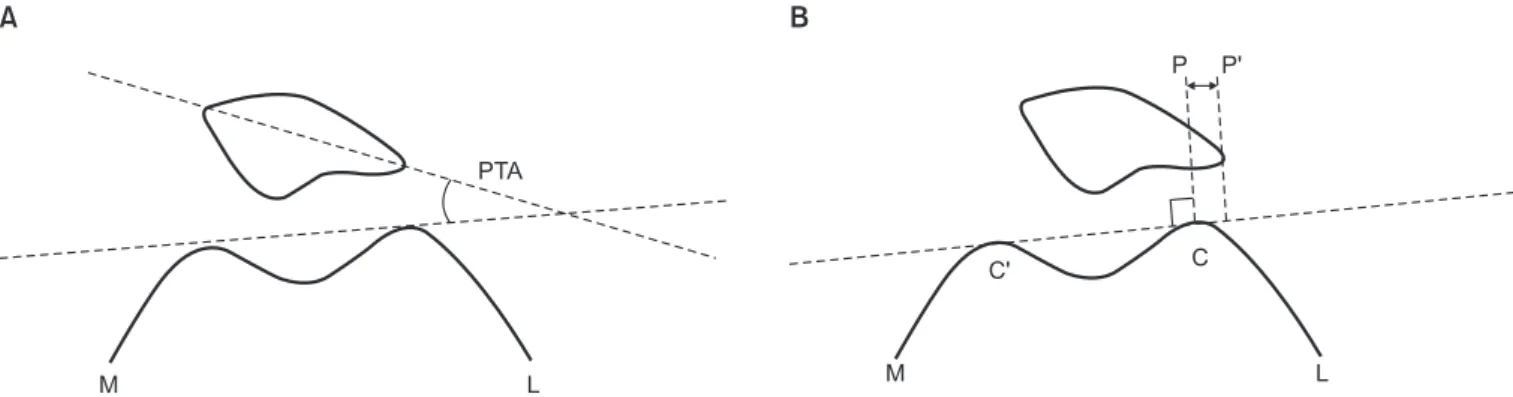
PTA
M L
C' C
M L
P P'
A B
Fig. 1. (A) Measurement of patellar tilting angle (PTA). PTA was defined as the angle between a line intersecting the widest bony structure of the patella and a line tangent to the anterior surface of the femoral condyles on a skyline view. (B) Measurement of patellar lateral shift (PLS). PLS was de
fined as the ratio of PP’ to CC’. CC’: distance between the summits of the condyles on the medial and lateral sides, PP’: distance between the summit of the lateral condyle and the point where a line drawn from the lateral edge of the patella runs perpendicular to a line that passes through the sum
mits of lateral (L) and medial (M) condyles.
BlackburnePeel index (BPI) on the lateral radiograph to assess the patellar height in reference to the joint level13), patellar tilting angle (PTA) and patellar lateral shift (PLS) (Fig. 1)2), and medial and lateral patellofemoral joint distances (MPFD and LPFD) (Fig. 2) as parameters for patellofemoral alignment measured on the 30° axial view2).
MannWhitney Utest was used to statistically analyze the dif
ference in numerical variables between the groups as well as pre
and postoperative radiological results. The difference in gender distribution between the groups was assessed using the Fisher exact test.
This study was performed after obtaining Institutional Review Board approval from our institution.
Results
Preoperative demographic and clinical characteristics in both groups are comparatively shown in Table 1. All demographic and clinical characteristics were comparable without significant intergroup difference except for age (52.0 years and 63.1 years on average for the OWHTO/LR group and OWHTO alone group, respectively).
The mean opening height was 12.8 mm and 11.8 mm in the OWHTO/LR and OWHTO alone groups, respectively. Regard
ing intra and postoperative complications, there was no intra
operative fracture on the lateral side of tibia requiring additional procedure or modification of the postoperative protocol. Delayed union at the osteotomy site was not encountered in the study subjects. One patient in the OWHTO/LR group sustained super
ficial wound infection which was resolved by local management.
Pre and postoperative values measured for each of the radio
logical parameters in both groups are comparatively shown in Table 2. In the OWHTO/LR group, patellar height significantly decreased in the assessment of BPI, while a significant postopera
tive reduction in CDI was detected in the OWHTO alone group.
Regarding patellar shift and tilt, in the OWHTO/LR group, both PTA and PLS values significantly decreased after surgery indicat
ing significant postoperative reductions in lateral patellar shift and tilt. Although an increase in LPFD and a decrease in M
PFD were observed after surgery, these postoperative changes did not reach the level of statistical significance. By contrast, in the OWHTO alone group, there were no significant changes in the parameters for patellar tilt and shift after surgery. In addition, al
though not statistically significant, an increase in LPFD was ob
served after combined LR and OWHTO, while the correspond
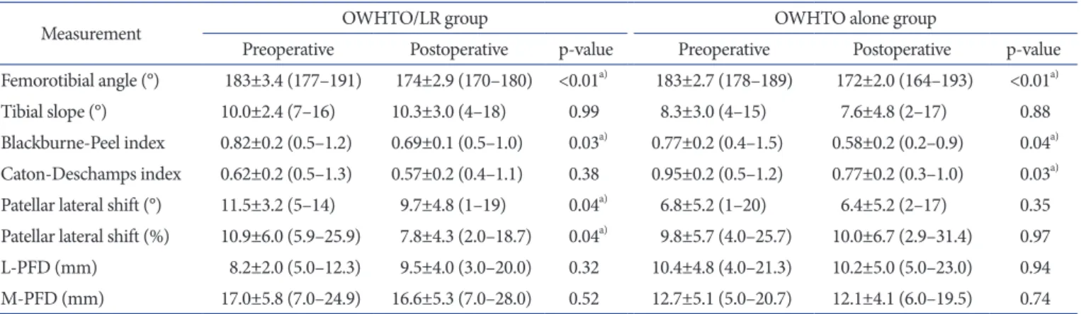
M-PFD L-PFD
M L
Fig. 2. Measurement of patellofemoral distance (PFD). Distance from the lateral (medial) edge of the lateral trochlea to the lateral (medial) edge of the patella was measured on a skyline view and defined as lateral PFD (LPFD) and medial PFD (MPFD), respectively. M: medial, L: lat
eral.
Table 1. Preoperative Demographics and Clinical Characteristics
Variable OWHTO/LR group (n=19) OWHTO alone group (n=18) pvalue
Age (yr) 52.0±9.1 (32–72) 63.1±7.1 (53–74) 0.04a)
Sex (male), no. 13 13 0.08b)
Preoperative FTA (°) 183±3.4 (177–191) 183±2.7 (178–189) 0.81
Tibial slope (°) 10.0±2.4 (7–16) 8.3±3.0 (4–15) 0.06
Average opening wedge value (mm) 12.8±2.2 (10–16) 11.8±1.8 (10–15) 0.32
Height (m) 1.66±0.09 (1.49–1.83) 1.65±0.09 (1.47–1.76) 0.93
Weight (kg) 65.7±11.6 (48.7–85.2) 68.8±9.5 (53.6–85.2) 0.59
BMI (kg/m2) 23.8±3.0 (19.3–29.5) 25.3±3.1 (24.0–30.8) 0.22
Values are presented as mean±standard deviation (range).
OWHTO: open wedge high tibial osteotomy, LR: lateral release, FTA: femorotibial angle, BMI: body mass index.
a)Statistically significant difference (p<0.05).
b)Fisher exact test.
ing distance was reduced after isolated OWHTO. The increase in PFD induced by concomitant LR may result in reduction of patellofemoral joint pressure.
Discussion
Although OWHTO has been reported to provide consistently satisfactory clinical outcome, the adverse effect of wedge opening proximal to the tibial tubercle on postoperative patellofemoral mechanics has been raised as a concern57,17). Since arthroscopic LR has been widely utilized with the intent of improving patel
lofemoral mechanics and symptoms8), LR concomitant with OWHTO may potentially reduce the risk of patellofemoral com
plication after OWHTO.
In the present study, postoperative changes in patellar posi
tion and orientation were comparatively examined in knees undergoing OWHTO with and without concomitant LR. The comparison of pre and postoperative radiological parameters showed postoperative reduction in patellar height in both groups.
Regarding the patellar position on the axial plane as indicated by patellar shift and tilt, concomitant LR effectively reduced lateral patellar shift and tilt, while OWHTO alone did not change the alignment postoperatively.
The effect of OWHTO on postoperative patellar position and tracking has been investigated in both clinical and biomechanical studies. The majority of these studies dealt with the problem of patellar height reduction with resultant increase in patellofemoral pressure potentially leading to patellofemoral osteoarthritis2,4,1822). There have been a few reports examining the change in patellar tilt and shift after OWHTO. Bito et al.2) examined the patellofem
oral alignment after OWHTO and showed a significant decrease in lateral patellar tilt with no appreciable change in patellar shift.
Based on the study results, they stated that OWHTO may nega
tively affect the congruency of the patellofemoral joint. Gaasbeek et al.20) compared dynamic patellar tracking after OWHTO and closed wedge HTO by measuring threedimensional patellar tracking in a cadaveric biomechanical study. Their study showed increased medial patellar tilt and reduced medial patellar rotation after HTO and these changes were more pronounced in OWH
TO, while no significant change was observed in mediolateral patellar translation.
In the present study, both lateral patellar tilt and shift were re
duced after surgery by concomitant LR at the time of OWHTO.
These changes were not observed after isolated OWHTO without LR. In addition, although not statistically significant, an increase in lateral patellofemoral distance was observed after combined LR and OWHTO, while the corresponding distance was rather reduced after isolated OWHTO. In the majority of our patient population undergoing OWHTO, the patella is laterally tilted or shifted with narrowing of the lateral patellofemoral joint space.
The present study results have shown decreased lateral patellar tilt and shift following combined OWHTO and LR. Therefore, it is thought that concomitant LR may effectively relieve the pressure in the lateral patellofemoral joint and potentially prevent anterior knee pain and progressive patellofemoral osteoarthritis following OWHTO especially for larger wedge size. Longino et al.6) pro
posed a combined tibial tubercle osteotomy with OWHTO and showed that concomitant tibial tubercle osteotomy could prevent the unfavorable patellofemoral mechanics after surgery. Although concomitant procedures to the tibial tubercle can be another op
Table 2. Preoperative and Postoperative Patellar Positions in Two Groups
Measurement OWHTO/LR group OWHTO alone group
Preoperative Postoperative pvalue Preoperative Postoperative pvalue
Femorotibial angle (°) 183±3.4 (177–191) 174±2.9 (170–180) <0.01a) 183±2.7 (178–189) 172±2.0 (164–193) <0.01a)
Tibial slope (°) 10.0±2.4 (7–16) 10.3±3.0 (4–18) 0.99 8.3±3.0 (4–15) 7.6±4.8 (2–17) 0.88
BlackburnePeel index 0.82±0.2 (0.5–1.2) 0.69±0.1 (0.5–1.0) 0.03a) 0.77±0.2 (0.4–1.5) 0.58±0.2 (0.2–0.9) 0.04a) CatonDeschamps index 0.62±0.2 (0.5–1.3) 0.57±0.2 (0.4–1.1) 0.38 0.95±0.2 (0.5–1.2) 0.77±0.2 (0.3–1.0) 0.03a) Patellar lateral shift (°) 11.5±3.2 (5–14) 9.7±4.8 (1–19) 0.04a) 6.8±5.2 (1–20) 6.4±5.2 (2–17) 0.35 Patellar lateral shift (%) 10.9±6.0 (5.9–25.9) 7.8±4.3 (2.0–18.7) 0.04a) 9.8±5.7 (4.0–25.7) 10.0±6.7 (2.9–31.4) 0.97 LPFD (mm) 8.2±2.0 (5.0–12.3) 9.5±4.0 (3.0–20.0) 0.32 10.4±4.8 (4.0–21.3) 10.2±5.0 (5.0–23.0) 0.94 MPFD (mm) 17.0±5.8 (7.0–24.9) 16.6±5.3 (7.0–28.0) 0.52 12.7±5.1 (5.0–20.7) 12.1±4.1 (6.0–19.5) 0.74 Values are presented as mean±standard deviation (range).
OWHTO: open wedge high tibial osteotomy, LR: lateral release, LPFD: lateral patellofemoral joint distance, MPFD: medial patellofemoral joint distance.
a)Statistically significant difference (p<0.05).
tion to cope with the patellofemoral problem after OWHTO, an additional bony procedure may add to surgical invasion. LR can be concomitantly performed with minimal surgical morbidity, and the authors feel supplementation with LR can be considered especially when performing OWHTO for knees with some patel
lofemoral malalignment or arthritic changes.
Since each of the two involved surgeons (HN and SO) per
formed HTO with and without LR in this study, indication for LR was not specifically determined based on the study results. In ad
dition, there are other study limitations such as small sample size, no priori power analysis performed, short followup period, ret
rospective analysis of two surgical procedures performed by two surgeons (HN and SO), no randomization for the two procedures (a concern for selection bias), and the lack of clinical results. A prospective comparative study including clinical evaluation for a larger number of patients with a longer followup period would be required to critically examine the effect of concomitant LR on the surgical outcome of OWHTO.
Conclusions
OWHTO performed for varus osteoarthritis knees induced a postoperative decrease in patellar height in both patient groups with and without concomitant LR. Regarding the change in patellofemoral alignment, however, concomitant LR in OWHTO significantly decreased lateral patellar tilt and shift, while no significant difference in those parameters was noted in the HTO alone knees.
Conflict of Interest
No potential conflict of interest relevant to this article was re
ported.
References
1. Backstein D, Meisami B, Gross AE. Patella baja after the modified CoventryMaquet high tibial osteotomy. J Knee Surg. 2003;16:2038.
2. Bito H, Takeuchi R, Kumagai K, Aratake M, Saito I, Hayashi R, Sasaki Y, Saito T. Opening wedge high tibial osteotomy affects both the lateral patellar tilt and patellar height. Knee Surg Sports Traumatol Arthrosc. 2010;18:95560.
3. Bode G, Ogon P, Pestka J, Zwingmann J, Feucht M, Sudkamp N, Niemeyer P. Clinical outcome and return to work follow
ing singlestage combined autologous chondrocyte implan
tation and high tibial osteotomy. Int Orthop. 2015;39:689
96.
4. Brouwer RW, BiermaZeinstra SM, van Koeveringe AJ, Ver
haar JA. Patellar height and the inclination of the tibial pla
teau after high tibial osteotomy: the open versus the closed
wedge technique. J Bone Joint Surg Br. 2005;87:122732.
5. Chae DJ, Shetty GM, Wang KH, Montalban AS Jr, Kim JI, Nha KW. Early complications of medial opening wedge high tibial osteotomy using autologous tricortical iliac bone graft and Tplate fixation. Knee. 2011;18:27884.
6. Longino PD, Birmingham TB, Schultz WJ, Moyer RF, Giffin JR. Combined tibial tubercle osteotomy with medial opening wedge high tibial osteotomy minimizes changes in patellar height: a prospective cohort study with historical controls.
Am J Sports Med. 2013;41:284957.
7. Matthews LS, Goldstein SA, Malvitz TA, Katz BP, Kaufer H.
Proximal tibial osteotomy. Factors that influence the dura
tion of satisfactory function. Clin Orthop Relat Res. 1988;
(229):193200.
8. OgilvieHarris DJ, Jackson RW. The arthroscopic treatment of chondromalacia patellae. J Bone Joint Surg Br. 1984;66:
6605.
9. Christodoulou NA, Tsaknis RN, Sdrenias CV, Galanis KG, Mavrogenis AF. Improvement of proximal tibial osteotomy results by lateral retinacular release. Clin Orthop Relat Res.
2005;441:3405.
10. ElAzab H, Glabgly P, Paul J, Imhoff AB, Hinterwimmer S. Patellar height and posterior tibial slope after open and closedwedge high tibial osteotomy: a radiological study on 100 patients. Am J Sports Med. 2010;38:3239.
11. Kaper BP, Bourne RB, Rorabeck CH, Macdonald SJ. Patellar infera after high tibial osteotomy. J Arthroplasty. 2001;16:
16873.
12. Stoffel K, Willers C, Korshid O, Kuster M. Patellofemoral contact pressure following high tibial osteotomy: a cadaveric study. Knee Surg Sports Traumatol Arthrosc. 2007;15:1094
100.
13. Saito T, Takeuchi R, Ara Y, Yoshida T, Koshino T. High tibial osteotomy with anterior advancement of distal fragment for medial and patellofemoral compartmental osteoarthritis of the knee. Knee. 2002;9:12732.
14. Ford DH, Post WR. Open or arthroscopic lateral release.
Indications, techniques, and rehabilitation. Clin Sports Med.
1997;16:2949.
15. Elkousy H. Complications in brief: arthroscopic lateral re
lease. Clin Orthop Relat Res. 2012;470:294953.
16. ElAzab H, Halawa A, Anetzberger H, Imhoff AB, Hinter
wimmer S. The effect of closed and openwedge high tibial osteotomy on tibial slope: a retrospective radiological review of 120 cases. J Bone Joint Surg Br. 2008;90:11937.
17. Gaasbeek RD, Sonneveld H, van Heerwaarden RJ, Jacobs WC, Wymenga AB. Distal tuberosity osteotomy in open wedge high tibial osteotomy can prevent patella infera: a new technique. Knee. 2004;11:45761.
18. Phillips CL, Silver DA, Schranz PJ, Mandalia V. The mea
surement of patellar height: a review of the methods of im
aging. J Bone Joint Surg Br. 2010;92:104553.
19. Flahiff CM, Kraus VB, Huebner JL, Setton LA. Cartilage mechanics in the guinea pig model of osteoarthritis studied
with an osmotic loading method. Osteoarthritis Cartilage.
2004;12:3838.
20. Gaasbeek R, Welsing R, Barink M, Verdonschot N, van Kampen A. The influence of open and closed high tibial osteotomy on dynamic patellar tracking: a biomechanical study. Knee Surg Sports Traumatol Arthrosc. 2007;15:978
84.
21. Fan JC. Open wedge high tibial osteotomy: cause of patellar descent. J Orthop Surg Res. 2012;7:3.
22. Agarwala S, Sobti A, Agrawal P. A report of nonunion at me
dial wedge high tibial osteotomy site and its management. J Nat Sci Biol Med. 2015;6(Suppl 1):S1602.