에스트로겐성 물질에 의해 자극된 인간 유방암 세포 증식에 대한 다이드제인, 바이칼레인, 헤스페리딘 및 우르솔산의 억제 효과
이미남·이수열·이현재·석정호·이충재# 충남대학교 의학전문대학원 약리학교실
(Received March 14, 2010; Revised March 29, 2010; Accepted March 29, 2010)
Anti-proliferative Effects of Daidzein, Baicalein, Hesperidin and Ursolic Acid on Human Breast Cancer Cells Stimulated by Estrogenic Compounds
Mi Nam Lee, Su Yel Lee, Hyun Jae Lee, Jeong Ho Seok and Choong Jae Lee#
Department of Pharmacology, School of Medicine, Chungnam National University, Daejeon 301-131, Korea
Abstract
— We investigated whether three flavonoids including daidzein, baicalein, hesperidin and ursolic acid, a triterpene acid, affect proliferation of MCF-7 human breast cancer cells stimulated by estrogenic compounds. Ursolic acid and baicalein inhibited proliferation of MCF-7 cells induced by PhIP, a food-derived carcinogen with estrogenic activity. Daidzein and hes- peridin inhibited estradiol-induced proliferation of MCF-7 cells. These compounds should be further investigated for the pos- sible involvement in signaling pathway after estrogen receptor binding in breast cancer cells.
Keywords □
flavonoids, triterpenoids, MCF-7, cytotoxicity
인체에발병할수있는다양한악성종양병변중
,
유방암은 악성종양으로인한사망및후유장애의주요인으로자리잡고있 다.
1,2)유방암의치료를위하여다수의항암제들이개발되어현재임상에서활용되고있지만여전히더효능이향상되고부작 용이적은새로운유방암치료제의개발이절실히요구되고있 는실정이다
.
잘알려진바와같이,
에스트로겐성물질은여성생 식과관련된다양한세포의증식,
분화및정상적기능유지에 있어서중요한역할을하고있으며에스트로겐활성을발현하는 물질중생리적으로가장중요한물질은17beta-estradiol
로서,
이물질은유방암의발병에있어서도주위험요인으로작용하고 있다
.
3)동시에,
인체악성종양의다양한병인중식이,
특히육식은주요한병인으로작용할수있음이역학적연구에의해알 려져있으며
,
우육,
계육,
생선등의고온가열조리과정에서생 성되는다수의복소환아민성화합물(heterocyclic amines)
중2- amino-1-methyl-6-phenylimidazo [4,5-b] pyridine(PhIP)
는돌연 변이원성및발암성을나타냄으로써전립선,
대장,
유선조직등에악성종양생성을유발할수있음이보고되어있다
.
4)PhIP
는 특히암컷흰쥐에서대장조직보다는유선조직에우선적으로 악성종양을 유발하며,
기존에 알려진 것과 같이 유전독성(genotoxicity)
을발현함과동시에에스트로겐수용체를경유하는 세포신호전달경로를활성화시킬수있음이보고되어있다.
3,4)한편
,
식이중채식(
과일의과육,
과피및야채)
을통하여인체에 흡수되는플라보노이드및트리터페노이드계물질은복수의기 전을경유하여항암작용을나타낼가능성이많은연구자들에의 해 보고되어 왔다.
5-9) 즉, daidzein
및balcalein
을 포함하여hesperidin
과같은플라보노이드는항염증,
항산화,
특정한암세포주에대한항증식
(
항암)
활성을발현하는것으로알려져있다.
5-12)Pecking
등과Cluzan
등은hesperidin
을포함한플라보노이드성 물질들이유방암의치료후에관찰되는림프부종을효과적으로 조절할수있음을보고하였고,
8,9)So
등은hesperidin
및다양한플라보노이드성물질들이인간유방암세포주의증식을억제하 고
,
유방암의발병과정을지연시킬수있음을보고하였다.
6)Choi
등은
hesperidin
의aglycone form
인hesperetin
이인간유방암세포의세포주기를
G1-phase
에서정체시킴으로써항증식효과 를발현할가능성을제시하였다.
5)또한,
트리터페노이드인ursolic acid
는인간의간암세포,
10)전립선암세포,
11)난소암세포12)등#본논문에관한문의는저자에게로
(
전화) 042-580-8255 (
팩스) 042-585-6627 (E-mail) [email protected]
종설
에대해항증식효과를발현함이보고되어있다
.
그러나,
상기한천연물들이
PhIP
등에스트로겐성활성을지닌물질에의해야기되는유방암세포의증식에어떠한영향을미칠수있는지에 대해서는지금까지검증된바가없었다
.
따라서,
본연구에서는, daidzein, baicalein, hesperidin
등3
종의플라보노이드와ursolic acid
와같은트리터페노이드가17beta-estradiol
또는PhIP
에의해자극된
MCF-7
인간유방암세포의증식에어떠한영향을미치는지를알아보고자하였다
.
실험방법
세포주및시약
인간유방암세포인
MCF-7
세포주는American Type Culture Collection
사(Manassas, VA, U.S.A.)
에서구입하였다. 2-Amino- 1-methyl-6-phenylimidazo [4,5-b] pyridine(PhIP)
은Toronto Research Chemicals(North York, ON, Canada)
에서, Sulforhod- amine B(SRB), trypsin-EDTA, Trizma base, 17beta-estradiol, 5% charcoal stripped-fetal bovine serum(CS-FBS), trichloro- acetic acid(TCA), hesperidin(purity: 90.0%), baicalein(purity:
95.0%), daidzein(purity: 95.0%), ursolic acid(purity: 90.0%), L-glutamine, sodium bicarbonate, dimethyl sulfoxide(DMSO)
등은
Sigma-Aldrich
사(St. Louis, MO, U.S.A.)
에서, penicillin-G, streptomycin, fetal bovine serum(FBS), Dulbecco's Modified Eagle's Medium(DMEM), RPMI 1640
은GIBCO-BRL
사(Grand Island, NY, U.S.A.)
에서각각구입하였고,
기타시약들은일급시약등급이상의것들을사용하였다
.
인간유방암세포의배양
MCF-7
세포는습도가충분히유지되고95%
공기, 5% CO
2를 함유하는37
oC
조건의 배양기 내에서L-glutamine(2 mM), sodium bicarbonate(44 mM), penicillin G(100 U/m
l), strepto- mycin(100
µg/m
l), FBS(10%, V/V)
등이첨가된DMEM
배양액 을이용하여배양되었으며, 1
주에2
회빈도로subculture
하였고배양액은
2
일마다1
회씩교체해주었다.
유방암세포의증식에미치는각약물의영향검증
PhIP
및17beta-estradiol
처리에의한MCF-7
세포의증식유 발효능과,
각약물에의한증식저해(
억제)
효능을정확하게검 증하기 위하여direct cell counting
방법을적용하였다.
즉, 6 well plate
의각well
에1×10
4개의세포를함유하는DMEM
배 양액(FBS 10%
함유) 1 m
l를가하여37
oC, 5% CO
2존재하에 서72
시간동안배양하였다.
세포에약물을처리하기이전에,
세포에가해지는모든에스트로겐성영향을차단하기위하여
phenol red
가제거된DMEM
배양액에5% charcoal-stripped FBS
를첨가한배양액으로교체하고다시
72
시간동안배양한후, 10 nM
의
17beta-estradiol
또는100 nM
의PhIP
단독처리혹은0.1~
100
µM
농도의hesperidin, baicalein, daidzein, ursolic acid
를 각각전처리후10 nM
의17beta-estradiol
또는100 nM
의PhIP
를처리하였다
.
이러한약물처리방법을적용하여48
시간마다 약물을함유한배양액을교체해주는방법으로총6
일간(48
시간×3
회)
배양한후,
각well
에 존재하는세포의수를hemacy- tometer
를이용하여직접계수하였다.
통계처리
모든측정결과는
Mean±S.E.M.
으로환산된후,
약물처리군 의측정치는대조군측정치의백분율로나타냈다.
통계처리는one-way ANOVA
및unpaired Student's
t-test
로 하였으며, p<0.05
인경우에통계적으로유의성이있는것으로판정하였다.
실험결과
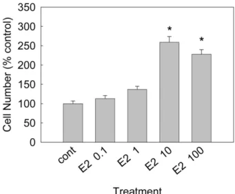
MCF-7세포에대한 17beta-estradiol의증식유발(자극)효과
Fig. 1
에서볼수있는것처럼, 17beta-estradiol(E2
로약칭)
은0.1~10 nM
범위에서농도의존적으로MCF-7
세포의증식을유 발함을관찰하였다.
처리농도별세포수는대조군, E2 0.1 nM, E2 1 nM, E2 10 nM, E2 100 nM
처리군에서 각각100±7%, 113±8%, 137±8%, 259±15%, 228±12%
이었다. E2 10 nM
처리군과
E2 100 nM
처리군에서세포증식유발효과가거의유사하므로이후의실험에서
E2
를처리하여세포증식을유발하고Fig. 1 −
Effect of 17beta-estradiol on proliferation of MCF-7 cells.
Each bar represents a mean±S.E.M. of three independent
experiments in comparison with that of the control set at
100%. The number of cells in control well was
145,000±10,150 (E2; 17beta-estradiol). * Significantly
different from control (p<0.05).
자할때에는
10 nM
농도로처리하기로하였다.
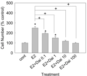
17beta-estradiol로유발된세포증식에대한daizein의억제 효과
Fig. 2
에서볼수있는것처럼, daidzein
은0.1~100
µM
범위에서
10 nM
의E2
로유발된유방암세포의증식을농도의존적으로억제함을관찰하였다
.
처리농도별세포수는대조군, E2 10 nM
단독
, E2 10 nM+daidzein 0.1
µM, E2 10 nM+daidzein 1
µM, E2 10 nM+daidzein 10
µM, E2 10 nM+daidzein 100
µM
처리 군에서각각100±5%, 250±14%, 195±11%, 150±14%, 118±
9%, 98±7%
이었다.
17beta-estradiol로유발된세포증식에대한hesperidin의억 제효과
Fig. 3
에서볼수있는것처럼, hesperidin
도역시0.1~100
µM
범위에서
10 nM
의E2
로유발된유방암세포의증식을농도의존적으로억제함을관찰하였다
.
처리농도별세포수는대조군, E2 10 nM
단독, E2 10 nM+hesperidin 0.1
µM, E2 10 nM+
hesperidin 1
µM, E2 10 nM+hesperidin 10
µM, E2 10 nM+
hesperidin 100
µM
처리군에서 각각100±5%, 250±14%, 230±9%, 210±11%, 182±7%, 155±8%
이었다.
MCF-7세포에대한PhIP의증식유발(자극)효과
Fig. 4
에서볼수있는것처럼, PhIP
는100 nM
과1
µM
농도에서
MCF-7
세포의증식을유발함을관찰하였다.
처리농도별세포수는 대조군
, PhIP 1 nM, PhIP 10 nM, PhIP 100 nM, PhIP 1,000 nM(1
µM)
처리군에서 각각100±5%, 101±8%, 110±9%, 185±6%, 178±10%
이었다. PhIP 100 nM
처리군과1
µM
처리군에서세포증식유발효과가거의유사하므로이후Fig. 4 −
Effect of PhIP on proliferation of MCF-7 cells. Each bar represents a mean±S.E.M. of three independent experi- ments in comparison with that of the control set at 100%.
The number of cells in control well was 135,000±6,750. * Significantly different from control (p<0.05).
Fig. 2 −
Effect of daidzein on 17beta-estradiol-induced proliferation of MCF-7 cells. Each bar represents a mean±S.E.M. of three independent experiments in comparison with that of the control set at 100%. The number of cells in control well was 128,000±6,400 (E2; 17beta-estradiol, Dai; daidzein). * Significantly different from control (p<0.05). + Significantly different from 17beta-estradiol-only treatment (p<0.05).
Fig. 3 −
Effect of hesperidin on 17beta-estradiol-induced proliferation of MCF-7 cells. Each bar represents a mean±S.E.M. of three independent experiments in comparison with that of the control set at 100%. The number of cells in control well was 114,000±5,700 (E2; 17beta-estradiol, Hes; hesperidin).
* Significantly different from control (p<0.05). + Sig-
nificantly different from 17beta-estradiol-only treatment
(p<0.05).
의실험에서
PhIP
를처리하여세포증식을유발하고자할때에는
100 nM
농도로처리하기로하였다.
PhIP로유발된세포증식에대한ursolic acid의억제효과
Fig. 5
에서볼수있는것처럼, ursolic acid
는1~100
µM
범위에서
100 nM
의PhIP
로유발된유방암세포의증식을농도의존적으로억제함을관찰하였다
.
처리농도별세포수는대조군, PhIP 100 nM
단독, PhIP 100 nM+ursolic acid 1
µM, PhIP 100 nM +ursolic acid 10
µM, PhIP 100 nM+ursolic acid 100
µM
처리군에서각각
100±7%, 181±7%, 175±4%, 106±10%, 32±9%
이었다
.
PhIP로유발된세포증식에대한baicalein의억제효과
Fig. 6
에서볼수있는것처럼, baicalein
은1~100
µM
범위에 서100 nM
의PhIP
로유발된유방암세포의증식을농도의존적으로억제함을관찰하였다
.
처리농도별세포수는대조군, PhIP 100 nM
단독, PhIP 100 nM+baicalein 1
µM, PhIP 100 nM+
baicalein 10
µM, PhIP 100 nM+baicalein 100
µM
처리군에서각각
100±7%, 181±7%, 169±8%, 102±6%, 33±5%
이었다.
고 찰
잘알려져있는것처럼
,
유방암세포에대한에스트로겐의주작용은
nuclear receptor superfamily
에속하는에스트로겐수용체
-alpha(ER-alpha)
와에스트로겐수용체-beta(ER-beta)
에의해 매개된다. Gene knockout technic
을이용한연구에의해밝혀진바
, ER-alpha
는정상적인유선의발육을포함하여여성생식계통의발달을위해필수적이며
, ER-beta
유전자를녹아웃시킬경 우에는유선의정상적발달에는외관상지장이없는것처럼보 이나,
유선말단부의정상적분화에문제가생길수있음이보고되어있다
.
3)대부분의유방암에서ER-beta
수용체의존재가확 인되나,
유방암발병에있어서의명확한역할에대해서는아직알려진바가없다
. ER-alpha
는유방암의진행에도중요한역할을하지만
,
유방암의개시과정(initiation)
에서도중요한역할을 할가능성이제시되었다.
본연구에서사용된PhIP
는유방암세포에존재하는
ER-alpha
수용체를자극함으로써에스트로겐에의해조절되는유전자의활성화및세포의증식을야기하는것 으로알려져있다
.
4)한편,
플라보노이드는복수의기전을경유하여인간암세포의증식을억제할수있는것으로보고되어있
다
.
5,13-16)플라바논화합물인hesperidin
을포함한다수의플라보노이드들은발암원
(carcinogen)
이생체내에서결정적인발암개시부위에접근하는과정을차단하거나
,
발암원의독성을무 독화시키는등의기전을경유하여발암원에의한새로운악성종 양의형성을예방할수있는것으로알려져있다.
16)Park
등은hesperidin
이caspase-3
의활성화를통하여대장암세포에대하 여세포자멸사를유도함을보고하였다.
13)Yeh
등은hesperidin
이matrix metalloproteinase-9
의유전자발현억제기전을경유하여간암세포의거동에영향을줄가능성이있다고보고하였으 며
,
14)hesperidin
은폐암세포15)에대해서도항증식효과를발현 Fig. 6 −Effect of baicalein on PhIP-induced proliferation of MCF-7
cells. Each bar represents a mean±S.E.M. of three independent experiments in comparison with that of the control set at 100%. The number of cells in control well was 115,000±8,050 (P; PhIP, Bai; baicalein). * Significantly different from control (p<0.05). + Significantly different from PhIP-only treatment (p<0.05).
Fig. 5 −
Effect of ursolic acid on PhIP-induced proliferation of MCF-
7 cells. Each bar represents a mean±S.E.M. of three
independent experiments in comparison with that of the
control set at 100%. The number of cells in control well
was 155,000±10,850 (P; PhIP, Urs; ursolic acid). * Sig-
nificantly different from control (p<0.05). + Significantly
different from PhIP-only treatment (p<0.05).
함이보고되어 있다
. Fukutake
등은생약인황금(Scutellariae Radix)
에서유래된플라보노이드성분인baicalin
등이흰쥐의직장에서
cyclooxygenase-1
을억제하는기전을경유하여효과적인 발암예방약물로작용할가능성을제시17)하였으며, Chuang
등은
baicalin
및baicalein
이암세포의세포자멸사를유도할수있 는TGF(transforming growth factor) beta-1
유전자의발현을촉 진시킴을보고하였다.
18)또한, ursolic acid
와같은triterpenoids
는다양한기전을경유하여인체악성종양세포의증식을억제
함이보고되어있다
.
10-12)그러나,
서론에서언급한바와같이이러한천연물들이
PhIP
등육식섭취를통하여인체에유입될수있는에스트로겐성활성을지닌물질에의해유도되는유방암세 포의증식에어떠한영향을미칠수있는지에대해서는지금까 지검증된바가없었다
. Daidzein
및hesperidin
은각각에스트로겐 수용체에대하여
selective estrogen receptor modulator (SERM)
로서 작용할 가능성19,20)이 제시되었으므로17beta-
estradiol
에의해자극된암세포의증식에대한영향이검증되었으며
, baicalein
과ursolic acid
는PhIP
에의해발암과정이자극 될수있는또다른주요인체악성종양인전립선암세포의증식을억제할수있는것으로보고되어있으므로
,
21,22)PhIP
에의해자극된유방암세포의증식에대한영향이검증되었다
.
실험결 과에서알수있듯이, 17beta-estradiol
및PhIP
는각각MCF-7
세포의증식을유발하였으며
, daidzein
및hesperidin
은17beta-
estradiol
에의해자극된암세포의증식을유의하게억제하였고,
baicalein
과ursolic acid
는PhIP
에의해자극된암세포의증식을유의하게억제하였다
.
이러한결과는일상적인식생활중육식을통하여인체에흡수되는
PhIP
등발암물질의작용을,
식이내 용물중채소,
과일등에함유된플라보노이드및트리터페노이드등이억제할가능성을일부나마시사하는연구결과라고생각 된다
.
따라서,
향후의 연구를 통하여daidzein, hesperidin, baicalein
및ursolic acid
등이PhIP
에의한수용체자극단계혹은여타의신호전달과정에서어떠한영향을미침으로써세포증 식억제작용을발현하는지를탐색함으로써상기의물질들이효 과적인발암예방약물로응용될가능성을지속적으로규명해나 아가야할것으로생각된다
.
감사의 말씀
This study was financially supported by research fund of Chungnam National University in 2008.
참고문헌
1) Muñoz, M., Estévez, L. G., Alvarez. I., Fernández, Y., Margelí, M., Tusquets, I., Seguí, M. A. and Lluch, A. : Evaluation of
international treatment guidelines and prognostic tests for the treatment of early breast cancer. Cancer Treat. Rev.
34(8), 701 (2008).
2) Long, B. J., Grigoryev, D. N., Nnane, I. P., Liu, Y., Ling, Y. Z.
and Brodie, A. M. : Antiandrogenic effects of novel androgen synthesis inhibitors on hormone-dependent prostate cancer.
Cancer Res.
60(23), 6630 (2000).
3 Lauber, S. N., Ali, S. and Gooderham, N. J. : The cooked food derived carcinogen 2-amino-1-methyl-6-phenylimidazo[4,5-b]
pyridine is a potent oestrogen: a mechanistic basis for its tissue-specific carcinogenicity. Carcinogenesis
25(12), 2509 (2004).
4) Lauber, S. N. and Gooderham, N. J. : The cooked meat derived genotoxic carcinogen 2-amino-3-methylimidazo[4,5-b]pyridine has potent hormone-like activity: mechanistic support for a role in breast cancer. Cancer Res.
67(19), 9597 (2007).
5) Choi, E. J. : Hesperetin induced G1-phase cell cycle arrest in human breast cancer MCF-7 cells: involvement of CDK4 and p21. Nutr. Cancer
59(1), 115 (2007).
6) So, F. V., Guthrie, N., Chambers, A. F., Moussa, M. and Carroll, K. K. : Inhibition of human breast cancer cell proliferation and delay of mammary tumorigenesis by flavonoids and citrus juices. Nutr. Cancer
26(2), 167 (1996).
7) Franke, A. A., Cooney, R. V., Custer, L. J., Mordan, L. J. and Tanaka, Y. : Inhibition of neoplastic transformation and bioavailability of dietary flavonoid agents. Adv. Exp. Med. Biol.
439
, 237 (1998).
8) Pecking, A. P., Février, B., Wargon, C. and Pillion, G. : Efficacy of Daflon 500 mg in the treatment of lymphedema (secondary to conventional therapy of breast cancer). Angiology
48(1), 93 (1997).
9) Cluzan, R. V., Alliot, F., Ghabboun, S. and Pascot, M. : Treatment of secondary lymphedema of the upper limb with CYCLO 3 FORT. Lymphology
29(1), 29 (1996).
10) Yan, S. L., Huang, C. Y., Wu, S. T. and Yin, M. C. : Oleanolic acid and ursolic acid induce apoptosis in four human liver cancer cell lines. Toxicol. In Vitro 2009 Dec 22. [Epub ahead of print].
11) Deeb, D., Gao, X., Jiang, H., Janic, B., Arbab, A. S., Rojanasakul, Y., Dulchavsky, S. A. and Gautam, S. C. : Oleanane triterpenoid CDDO-Me inhibits growth and induces apoptosis in prostate cancer cells through a ROS-dependent mechanism. Biochem. Pharmacol.
79(3), 350 (2010).
12) Petronelli, A,, Saulle, E., Pasquini, L., Petrucci, E., Mariani, G.,
Biffoni, M., Ferretti, G., Scambia, G., Benedetti-Panici, P.,
Greggi, S., Cognetti, F., Russo, M. A., Sporn, M. and Testa,
U. : High sensitivity of ovarian cancer cells to the synthetic
triterpenoid CDDO-Imidazolide. Cancer Lett.
282(2), 214
(2009).
13) Park, H. J., Kim, M. J., Ha, E. and Chung, J. H. : Apoptotic effect of hesperidin through caspase3 activation in human colon cancer cells, SNU-C4. Phytomedicine
15, 147 (2008).
14) Yeh, M. H., Kao, S. T., Hung, C. M., Liu, C. J., Lee, K. H. and Yeh, C. C. : Hesperidin inhibited acetaldehyde-induced matrix metalloproteinase-9 gene expression in human hepatocellular carcinoma cells. Toxicol. Lett.
184(3), 204 (2009).
15) Kamaraj, S., Ramakrishnan, G., Anandakumar, P., Jagan, S. and Devaki, T. : Antioxidant and anticancer efficacy of hesperidin in benzo(a)pyrene induced lung carcinogenesis in mice. Invest.
New Drugs
27(3), 214 (2009).
16) Benavente-García, O. and Castillo, J. : Update on uses and properties of citrus flavonoids: new findings in anticancer, cardiovascular, and anti-inflammatory activity. J. Agric. Food Chem.
56(15), 6185 (2008).
17) Fukutake, M., Yokota, S., Kawamura, H., Iizuka, A., Amagaya, S., Fukuda, K. and Komatsu, Y. : Inhibitory effect of Coptidis Rhizoma and Scutellariae Radix on azoxymethane-induced aberrant crypt foci formation in rat colon. Biol. Pharm. Bull.
21
(8), 814 (1998).
18) Chuang, H. N., Wang, J. Y., Chiu, J. H., Tsai, T. H., Yeh, S. F., Fu, S. L., Lui, W. Y. and Wu, C. W. : Enhancing effects of
Scutellaria baicalensis and some of its constituents on TGF- beta1 gene expression in RAW 264.7 murine macrophage cell line. Planta Med.
71(5), 440 (2005).
19) Martin, D., Song, J., Mark, C. and Eyster, K. : Understanding the cardiovascular actions of soy isoflavones: potential novel targets for antihypertensive drug development. Cardiovasc.
Hematol. Disord. Drug Targets.
8(4), 297 (2008).
20) Scambia, G., Ranelletti, F. O., Benedetti, Panici P., Piantelli, M., Rumi, C., Battaglia, F., Larocca, L. M., Capelli, A. and Mancuso, S. : Type-II estrogen binding sites in a lymphoblastoid cell line and growth-inhibitory effect of estrogen, anti-estrogen and bioflavonoids. Int. J. Cancer.
46(6), 1112 (1990).
21) Miocinovic, R., McCabe, N. P., Keck, R. W., Jankun, J., Hampton, J. A. and Selman, S. H. : In vivo and in vitro effect of baicalein on human prostate cancer cells. Int. J. Oncol.
26(1), 241 (2005).
22) Kassi, E., Papoutsi, Z., Pratsinis, H., Aligiannis, N., Manoussakis, M. and Moutsatsou, P. : Ursolic acid, a naturally occurring triterpenoid, demonstrates anticancer activity on human prostate cancer cells. J. Cancer Res. Clin. Oncol.
133