Original Article: Bioactive Materials
Anticancer Activities of the Methanolic Extract from Lemon Leaves in Human Breast Cancer Stem Cells
Jeong Yong Moon · Linh Thi Thao Nguyen · Ho Bong Hyun · Ahmed Osman · Minwhan Cho · Suyeong Han · Dong-Sun Lee* · Kwang Seok Ahn*
인간 유방암 줄기세포에서 레몬잎 메탄올 추출물의 항암 효능
문정용 · Linh Thi Thao Nguyen · 현호봉 · Ahmed Osman · 조민환 · 한수영 · 이동선* · 안광석*
Received: 16 April 2015 / Accepted: 6 May 2015 / Published Online: 30 September 2015
© The Korean Society for Applied Biological Chemistry 2015
Abstract The anticancer activity of a methanolic extract from lemon leaves (MLL) was assessed in MCF-7-SC human breast cancer stem cells. MLL induced apoptosis in MCF-7-SC, as evidenced by increased apoptotic body formation, sub-G1 cell population, annexin V-positive cells, Bax/Bcl-2 ratio, as well as proteolytic activation of caspase-9 and caspase-3, and degradation of poly (ADP-ribose) polymerase (PARP) protein. Concomitantly, MLL induced the formation of acidic vesicular organelles, increased LC3-II accumulation, and reduced the activation of Akt, mTOR, and p70S6K, suggesting that MLL initiates an autophagic progression in MCF-7-SC via the Akt/mTOR pathway. Epithelial- mesenchymal transition (EMT), a critical step in the acquisition of the metastatic state, is an attractive target for therapeutic interventions directed against tumor metastasis. At low concentrations, MLL induced anti-metastatic effects on MCF-7-SC by inhibiting the EMT process. Exposure to MLL also led to an increase in the epithelial marker E-cadherin, but decreased protein levels of the mesenchymal markers Snail and Slug. Collectively, this study provides evidence that lemon leaves possess cytotoxicity and anti- metastatic properties. Therefore, MLL may prove to be beneficial as a medicinal plant for alternative novel anticancer drugs and nutraceutical products.
Keywords apoptosis · autophagy · cancer stem cells · epithelial- mesenchymal transition · lemon leaf
서 론
암 줄기세포(cancer stem cells; CSCs)는 일반 암세포들에 비해 J. Y. Moon and L. T. T. Nguyen contributed equally.
J. Y. Moon
Subtropical Horticulture Research Institute, Jeju National University, Jeju 690-756, Republic of Korea
L. T. T. Nguyen · H. B. Hyun
Faculty of Biotechnology, College of Applied Life Sciences, SARI, Jeju National University, Jeju 690-756, Republic of Korea
A. Osman
Faculty of Advanced Convergence Technology & Science, Jeju National University, Jeju, 690-756, Republic of Korea
M. Cho
Seoul Global High School, 40 Sungkyunkwan-ro 13 gil, Jongno-gu, Seoul 110-521, Republic of Korea
S. Han
Cheongshim international academy, 324-213 Misari-ro, Sorak-myeon, Gapyeng-gun, Gyeonggi-do 477-855, Republic of Korea
D.-S. Lee
Faculty of Biotechnology, College of Applied Life Sciences, SARI, Jeju National University, Jeju 690-756, Republic of Korea
Subtropical Horticulture Research Institute, Jeju National University, Jeju 690-756, Republic of Korea
*Corresponding author (D.-S. Lee: [email protected]) K. S. Ahn
Department of Oriental Pathology, College of Oriental Medicine, Kyung Hee University, Seoul 130-701, Republic of Korea
*Corresponding author (K. S. Ahn: [email protected])
This is an Open Access article distributed under the terms of the Creative Commons Attribution Non-Commercial License (http://creativecommons.
org/licenses/by-nc/3.0/) which permits unrestricted non-commercial use, distribution, and reproduction in any medium, provided the original work is properly cited.
높은 생존율과 자가 재생 능력을 지닌 세포들로서, 일반적인 약 물이나 방사선 치료 방법으로는 완벽하게 제거하기 힘들기 때 문에 최근 CSCs에 대한 연구가 활발히 진행되고 있다(Siegel 등, 2013). 유방암 줄기세포가 그 대표적인 예이며, 이들은 유 방암의 진행과 전이에 있어서 매우 중요한 원인 중 하나로 밝 혀진 바 있다(Reya 등, 2001; Al-Hajj 등, 2003). 따라서, 유방 암 줄기세포를 표적으로 한 항암 치료방법이 효과적인 방법으 로 부상하고 있다. 다양한 종류의 유방암 줄기세포들이 보고되 고 있으며(Charafe-Jauffret 등, 2009; Han과 Crowe, 2009; Ali 등, 2011; Kotiyal과 Bhattacharya, 2014), 이들 중에서 MCF- 7-SC 세포는 CD44high/CD24low 표면 마커 단백질들의 발현특성 을 이용하여 인간 유방암 MCF-7 세포로부터 분리되었다(Phuc 등, 2010). 또한 MCF-7-SC 세포에 대한 이전 연구에서 anoikis 저항성, mammosphere 형성, 세포 전이와 약물에 대한 내성 등 과 같은 줄기세포의 특성들이 확인된 바 있다(Nguyen 등, 2014; Tran 등, 2014).
암세포를 이용한 새로운 항암제 개발 시 활용되는 중요한 현 상으로서 세포자멸(apoptosis)과 자식작용(autophagy)을 들 수 있 다. 세포예정사(programed cell death)라고도 불리는 apoptosis는 노화, 항상성 유지, 발달과정에서 일반적으로 발생하며 세포 내 에서 두 종류의 apoptosis 경로가 존재한다. 세포가 지닌 죽음 수용체에 의해 매개되는 외인성 경로(extrinsic pathway)와 미토 콘드리아 매개 내인성 경로(intrinsic pathway)가 있으며, 이러한 경로들을 통해 궁극적으로 caspase들의 활성화에 의해 세포들을 죽음에 이르게 한다. Autophagy는 진화적으로 잘 보존된 기작 중의 하나이며, 리소좀(lysosome)을 통하여 세포 내에서 불필요 한 구성물질이나 손상된 세포 소기관들을 분해하는 과정을 말 한다. Autophagy는 autophagosome으로 알려진 이중막 소포체 를 형성하고, 성숙되면 lysosome과 융합하여 autophagolysosome 을 형성한다. 이렇게 형성된 autophagolysosome은 포집된 불필 요한 구성물 또는 세포 소기관들을 가수분해효소를 이용하여 분 해시킨다. 단백질 kinase mTOR (mammalian target of rapamycin) 은 단백질 합성을 조절하기 위하여 세포의 에너지 상태를 감지 함으로써 autophagy의 주요 조절자 역할을 수행한다(Cuyàs 등, 2014). mTOR은 PI3K (phosphoinositide 3-kinase)와 Akt라 알 려진 PKB (protein kinase B) 신호경로의 하위 단백질로서 세 포의 성장과 분화, 생존을 촉진하는 반면 apoptotic signal을 저 해한다(Brunet 등, 2001; Hanada 등, 2004; Endo 등, 2006).
그러므로, 활성화된 PI3K/Akt/mTOR 신호경로는 apoptosis와 autophagy를 억제하여 세포의 생존을 증가시키는 역할을 한다.
EMT (epithelial-mesenchymal transition)는 정상적 배아발달 과정에서 발생하는 하나의 현상이지만, 암세포에서는 세포의 침 윤과 이동에 주요한 과정이며, 전이성 암세포에서 특히 많이 발 생된다(Lamouille 등, 2014). 암세포가 EMT 과정을 거치게 되 면 섬유모세포와 같은 표현형을 지니게 되며 원래의 epithelial 성질을 잃으면서 세포의 이동이 쉬운 형태로 변하게 된다. 다 양한 전사 조절인자들이 EMT 조절에 관여하는데, 특히 zinc finger transcription factor인 Snail과 Slug는 EMT의 주요 조절 인자로 알려져 있다(Thiery 등, 2009). Snail과 Slug는 E- cadherin의 프로모터에 부착하여 E-cadherin의 전사를 억제하며, 이것은 EMT 과정의 특징 중 하나로 알려져 있다(Bolós 등, 2003; Damian 등, 2008). 따라서, Snail과 Slug의 발현양을 비 교분석함으로써 후보물질들의 암전이 억제효능 여부를 평가할 수 있다(Park 등, 2008).
레몬(Citrus limon)은 주로 주스로 생산되기 위해 세계적으로 많은 곳에서 재배되고 있다. 레몬 주스에 대한 항암 활성 및 항 산화 활성은 이미 알려져 있으나, 레몬 잎에 대한 유효 성분 및 생리 활성에 대한 연구는 미비한 실정이다. 따라서, 본 연구에 서는 레몬 잎 메탄올 추출물(methanolic extract from lemon leaves, MLL)을 이용하여 유방암 줄기 세포인 MCF-7-SC에 대 한 항암 활성을 조사하였다.
재료 및 방법
추출물 제조. 실험에 사용된 레몬잎은 제주도에서 자생하고 있 는 유레카 품종으로, 5−6월에 샘플을 채취한 뒤, 온풍건조 한 시료 9 kg을 믹서기를 이용하여 분쇄하였다. 80% 메탄올 240 L 를 이용하여 3일 동안 실온에서 추출하고 여과지로 여과하여 40oC에서 감압농축기를 이용하여 농축 한 뒤 동결 건조하였다 (1.375 kg). 건조된 시료는 −20oC에서 보관하며 실험에 사용하 였다.
시약. RPMI 1640 medium, F-12K medium, Dulbecco’s Modified Eagle Medium (DMEM) medium, bovine serum albumin, trypsin/ethylenediaminetetraacetic acid (EDTA), fetal bovine serum (FBS), and antibiotic-antimycotic는 Invitrogen (USA)에서 구입 하여 사용하였으며, Hoechst 33342, 3-(4,5-dimethylthiazol-2-yl)- 2,5-diphenyltetrazolium bromide (MTT), propidium iodide (PI), RNase A, β-Actin 항체는 Sigma Chemical Co. (USA)에서 구 입하여 사용하였다. PARP, caspase-9, caspase-3, Bax, Bcl-2, p- Akt, Akt, p-mTOR, mTOR, p-p70S6K, Slug, Vimentin, E-cadherin 항체는 Cell Signaling (USA)에서 구입하였으며, Snail 항체는 Santa Cruz Biotechnology (USA)에서 구입하여 사용하였다.
기체 크로마토그래피-질량(GC-MS) 분석. GC-MS 분석은 Shimadzu사의 QP-2010(GC-MS; Model QP-2010, Shimadzu Co., Japan)을 이용하여 수행하였다. 분석조건은 오븐온도 40oC 에서 2분간 유지한 뒤 분당 5oC 승온하여 200oC에서 1분간 유 지하였다. GC 컬럼은 Rtx-5MS (30 m length, 0.25µm diameter, 0.25µm thickness)를 사용하였고, GC-MS 분석을 통하여 검출 된 피크의 화합물 동정은 Willey 9 library database를 이용하여 수행하였으며, library와 유사도가 80% 이상인 화합물만 동정하 였다.
세포 배양. MCF-7-SC는 RPMI-1640, MCF-7 세포는 DMEM 과 F-12K를 1:1로 혼합하여 배양하였다. 배지는 10% heat- inactivated FBS와 1% antibiotics를 첨가하여 37oC, 5% CO2 인큐베이터에서 배양하였다.
세포 생존율 측정. 96 well plate에 MCF-7-SC와 MCF-7 세포 를 3×10⁴ cells/mL로 준비한 뒤 MLL을 농도 별로 처리하여 24, 48시간 배양하였다. 24, 48시간 후 MTT 시약을 처리하고 4시간 동안 CO2 인큐베이터에서 반응시킨 후 배양액을 제거하 고 150µL의 dimethyl sulfoxide를 처리한 후 microplate reader (Sunrise, Tecan, Austria)를 이용하여 570 nm에서 흡광도를 측 정하였다.
세포 형태 관찰. MCF-7-SC 세포를 3×10⁴ cells/mL로 6-well plate에 준비한 뒤 MLL을 농도 별로 처리하여 24시간 배양하 였다. 24시간 후 Hoechst 33342와 acridine orange (AO)를 10 µM되게 처리한 뒤 형광현미경을 이용하여 관찰하였다(Olympus, UK).
세포 주기 분석. 세포주기를 관찰하기 위하여 MLL이 처리된 세포를 일정 시간 배양 뒤 70% 에탄올로 고정하였다. 고정 된 세포를 2 mM EDTA-phosphate buffered saline (PBS)로 세척 한 뒤 RNase A (25µg/mL)가 포함된 PI (40 µg/mL) 시약으로 염색하여 FACSCalibur flow cytometer (BD Biosciences, USA) 를 이용해서 세포주기를 측정하였다.
Annexin V 측정. Annexin V-FITC Apoptosis Detection Kit I을 이용하여 MLL이 처리된 세포에서 apoptosis를 확인하기 위 하여 phosphatidylserine의 이동을 측정하였다. MCF-7-SC 세포 에 MLL을 24시간 처리한 뒤 PBS로 세척하고 annexin V와 PI가 혼합된 binding 버퍼를 이용하여 상온에서 15분간 염색시 킨 뒤 FACSCalibur flow cytometer (BD Biosciences)를 이용 해서 annexin V positive 세포들을 측정하였다.
Acridine orange (AO) 염색. MLL에 의한 autophagy 현상을 측정하기 위하여 10µM의 AO를 세포에 처리한 뒤 15분간 CO2 인큐베이터에서 반응시킨다. 세포를 PBS로 세척 한 뒤 FACSCalibur flow cytometer (BD Biosciences, USA)를 이용 해서 세포들을 측정하였다.
Wound healing 측정. MCF-7-SC 세포를 6-well plate에 준비 한 뒤 세포가 거의 가득 차있는 상태에서 플라스틱 팁을 이용 하여 wound를 형성한 뒤, MLL을 처리하여 48시간 동안 CO2 인큐베이터에서 배양하였다. 48시간 뒤, 현미경(Olympus, UK) 을 통해 세포의 이동 정도를 비교하였다.
Reverse transcription-polymerase chain reaction (RT-PCR).
MCF-7-SC세포의 total RNA는 TRIzol reagent (Invitrogen)를 이용하여 추출하였다. 추출된 RNA 1µg을 reverse transcription system (Promega, USA)을 이용하여 cDNA를 합성한 뒤, 다음 과 같은 유전자들을 증폭하였다(Snail, forward 5'-GAGGACAG TGGGAAAGGCTC-3', reverse 5'-TGGCTTCGGATGTGCATC TT-3'; Slug, forward 5'-GAACTCACACGGAGAAG-3', reverse 5'-ACACAGCAGCCAGATTCCTC-3'; Vimentin, forward 5'- AATGGCTCGTCACCTTCGTGAAT-3', reverse 5'-CAGATTAG TTTCCCTCAGGTTCAG-3'; E-cadherin, forward 5’-GGAAGT CAGTTCAGACTCCAGCC-3', reverse 5'-AGGCCTTTTGACT GTAATCACACC-3'; GAPDH, forward 5’-GAGAAGGCTGGG GCTCATTT-3', reverse 5'-AGTGATGGCATGGACTGTGG-3').
Western blot 분석. MLL이 처리된 MCF-7-SC 세포를 수확 후 RIPA buffer를 이용하여 4oC에서 30분간 용해하였다.
Western blotting은 Moon 등(2009)의 보고에 따라 수행하였다.
5% skim milk로 blocking한 후, primary 항체를 1:1,000으로 사용하여 4oC에서 overnight 반응하였고, secondary 항체는 1:5,000으로 상온에서 1시간 동안 반응시켰다. ECL kit (iNtRON, Korea)를 이용하여 단백질 밴드를 검출하였다.
통계분석. 결과분석은 3회 반복수행 평균값과 표준편차로 나타 내었으며, 유의성 검증은 version 14의 Statistical Package for the Social Sciences를 이용하여 나타내었다(유의수준 *p <0.05,
**p <0.01, ***p <0.001).
결과 및 고찰
MLL에 의한 유방암 세포 증식 억제 효능. 유방암 줄기 세포 는 종양을 형성할 수 있는 능력 및 다약제 내성의 특징을 갖는 다(Al-Hajj 등, 2003; Ponti 등, 2005; Wright 등, 2008). 따라
서 우리는 우선 유방암줄기세포 MCF-7-SC에 대해서 MLL의 항증식 효과를 측정하고 이를 유방암 MCF-7에 대한 항증식 효 과와 비교하였다. Figs. 1A와 B에 나타난 바와 같이, MLL의 항증식 효과는 시간의존적으로 증가하였으며, 24시간 처리 후 MCF-7 세포와 MCF-7-SC의 IC50값은 각각, 996.2과 913.8 µg/mL였으며, 48시간 처리후의 IC50값은 MCF-7세포는 824.3, MCF-7-SC는 755.0µg/mL로 나타났다. Song 등(2015)이 보고 한 온주밀감 잎 메탄올 추출물의 위암세포주 AGS에 대한 IC50
값과 비교 시에는 레몬 잎 메탄올 추출물의 IC50값이 다소 높 게 나타났지만, Gali 등(2011)이 보고한 Argemone Mexicana L.
잎 메탄올 추출물의 유방암세포주 MCF-7과 자궁경부암 HeLa 세포주에 대한 IC50값과 비교했을 때는 큰 차이가 없음을 알 수 있었다. 위의 결과를 토대로 레몬잎 메탄올 추출물이 유방암세 포 MCF-7 뿐만 아니라, 항암제에 대해서 내성이 있다고 보고 된 유방암줄기세포 MCF-7-SC에서도 항증식 효과가 있음을 나 타내고 있다.
MLL에 의한 apoptosis 유도. MCF-7-SC에 대한 MLL의 항증 식 효과가 apoptosis에의해 기인한 것인지를 확인하기 위하여 다음과 같은 실험들을 수행하였다. Hoechst 33342 염색을 통하 여 MCF-7-SC에서 MLL 농도 의존적으로 DNA의 절편화와 응 축을 확인 할 수 있었으며(Fig. 2A), Annexin V/PI 염색을 통 해 annexin V-positive/PI-negative 세포들의 증가를 확인 할 수 있었다(Fig. 2B). 또한, Fig. 2C에서 보는 바와 같이, MLL 처 리에 따라 sub-G1기가 8.71%에서 26.02%로 증가되는 것을 확 인하였으며, Western blot을 통해 apoptosis 관련 단백질인 caspase-9과 -3의 활성화 및 PARP의 절단과 Bax/Bcl-2 ratio 증가를 관찰할 수 있었다(Fig. 2D). 결론적으로, MLL의 처리에 의해 MCF-7-SC에서 apoptosis가 유도되는 것을 확인할 수 하 였다.
Akt/mTOR 경로를 통한 autophagy 유도. Autophagy는 진화적 으로 잘 보존된 세포내 분해 경로이지만, 과다한 발생은 다른 형 Fig. 1 MLL inhibits breast cancer cell proliferation. MCF-7-SC and MCF-7 cells were seeded and treated with the indicated concentrations of MLL for (A) 24 h and (B) 48 h. Cell viability was then determined by the MTT assay. Data correspond to the mean ± standard deviation (SD) from three independent experiments.
Fig. 2 MLL induces apoptotic cell death in MCF-7-SC. Cells were seeded, incubated for 24 h, and incubated with the indicated concentrations of MLL for an additional 24 h. (A) Treated cells were fixed and stained with 10µM Hoechst 33342 and observed under a fluorescent microscope. (B) Treated cells were harvested and stained with PI and/or annexin V according to the manufacturer’s instructions. The results shown are representative of three independent cytometric analyses. (C) Treated cells were harvested, fixed with 70% cold ethanol, stained with PI, washed, and analyzed by flow cytometry. The results shown are representative of three experiments. (D) Cells were lysed following incubation with MLL at the indicated concentrations for 24 h. Cellular proteins were separated by SDS-PAGE and probed with appropriate primary antibodies. β-Actin served as an internal control. *p <0.05, **p <0.01, ***p <0.001. Densitometry values for PCR experiments were estimated by the Image J software (National Institutes of Health, USA).
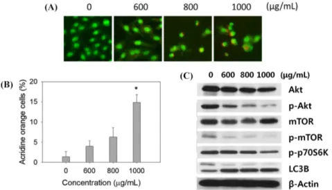
Fig. 3 MLL induces autophagic cell death in MCF-7-SC. Cells were seeded, incubated for 24 h, and incubated with the indicated concentrations of MLL for an additional 24 h. (A) Acidic vesicular organelles (AVOs) were examined by incubating cells with 10µM acridine orange (AO) for 5–10 min followed by fluorescence microscopy. The results shown are representative of three experiments. (B) Quantification of AVO-positive cells by flow cytometry. (C) Cells were lysed following incubation with MLL at the indicated concentrations for 24 h. Cellular proteins were separated by SDS- PAGE and probed with appropriate primary antibodies. β-Actin served as an internal control. *p <0.05, **p <0.01, ***p <0.001. Densitometry values for PCR experiments were estimated by the Image J software (National Institutes of Health, USA).
태의 세포 죽음을 유도할 수 있다(Kuma 등, 2004; Mizushima와 Komatsu, 2011). 따라서, MLL 처리에 따라 MCF-7-SC에서 autophagy가 유도되는지 알아보았다. 세포 내에서 autophagy가 유도될 때 발생하는 주요 현상 중의 하나인 acidic vesicular
organelles (AVOs) 형성 여부를 AO 염색을 통해 확인한 결과, Fig. 3A에서와 같이 MLL 처리에 따라 많은 수의 AVOs가 형성 되는 것을 현미경으로 확인할 수 있었다. 또한, flow cytometry 를 이용하여 AVOs의 형성을 정량적으로 측정한 결과, MLL Fig. 4 MLL interrupts the EMT in MCF-7-SC. (A) MCF-7-SC were incubated with sub-lethal concentrations of MLL. Cell migration was then measured by wound healing assays. Data represent the means ± SD of at least three replicates relative to the control. (B) After 48 h incubation with MLL, mRNA levels of EMT markers (E-cadherin, Vimentin, Snail, and Slug) were analyzed by RT-PCR. GAPDH served as the loading control. Data represent the mean ± SD of at least three replicates relative to the control. (C) MCF-7-SC were treated with the indicated concentrations of MLL for 48 h, and the expression of EMT markers was analyzed by Western blotting. β-Actin served as the loading control. This figure is representative of three replicates. *p <0.05, **p <0.01, ***p <0.001. Densitometry values for PCR experiments were estimated by the Image J software (National Institutes of Health,USA).
1000µg/mL 처리군에서 대조군과 비교하여 1.4에서 14.8%로 증 가됨을 확인하였다(Fig. 3B). 또한, autophagy의 중요한 마커인 LC3I에서 LC3II로의 전환 및 Akt, mTOR, p70S6K의 인산화 억제를 Western blot으로 확인함으로써 MLL에 의한 autophagy 유도가 일어나고 있음을 거듭 확인할 수 있었다(Fig. 3C). 이러 한 결과들은 MLL 처리에 의해 MCF-7-SC에서 autophagy와 apoptosis의 상호작용이 존재할 수 있음을 시사하고 있으며, 이 에 대해 더 많은 연구가 필요할 것으로 사료된다.
Snail과 Slug를 통한 EMT 억제 효능. 암의 전이는 암세포가 원래의 종양부위에서 다른 조직으로 확산되는 과정이다. EMT 과정에서 암세포는 epithelial 극성을 잃고 침윤성과 이동능력을 획득하며 줄기 세포와 같은 특성을 가지게 되어 새로운 종양을 발생시킨다(Herreros-Villanueva 등, 2013). MCF-7-SC에서 MLL 의 항전이 효능을 알아보기 위하여 wound healing assay를 수 행해 본 결과, MLL 처리에 따라 세포의 이동능력이 감소하였 으며(Fig. 4A), 예상한 바와 같이, epithelial 마커인 E-cadherin 의 mRNA 발현은 증가되었다(Fig. 4B). 또한 EMT 조절에 중 요한 역할을 하는 mesenchymal 마커인 Vimentin, Snail, Slug 의 발현을 확인해본 결과, mRNA의 변화는 없었지만 단백질의
발현이 현저히 감소되는 것을 확인 할 수 있었다(Fig. 4B, C).
종합적으로, 이러한 결과들을 토대로 MLL이 EMT 과정을 억 제할 수 있는 잠재력을 가지며 MCF-7-SC의 전이를 예방하는 데 중요한 역할을 한다고 사료된다.
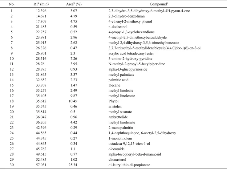
GC-MS를 이용한 MLL의 성분 분석. GC-MS를 이용하여 MLL의 성분을 분석한 결과, 총 29개의 화합물을 검출할 수 있 었으며(Table 1), 주요 화합물로는 di-lauryl thio-di-propionate (25.34%), phytol (10.45%), methyl linoleate (9.87%), 3-amino- 2-hydroxy-pyridine (7.26%), 2,3-dihydro-benzofuran (4.79%), 4- ethenyl-2-methoxy phenol(4.75%), N-methyl-2-propyl-5-butylpiperidine (3.95%), methyl palmitate (3.37%), 2,3-dihydro-3,5-dihydroxy- 6-methyl-4H-pyran-4-one (3.07%), 4-methyl-2,5-dimethoxybenzaldehyde (2.96%), methyl 2,4-dihydroxy-3,5,6-trimethylbenzoate (2.62%), methyl linoleate (2.49%), acrylic acid tetradecyl ester (2.30%), palmitic acid (2.23%)가 차지하고 있었다. 이전의 연구결과에서 4-ethenyl-2-methoxy phenol, phytol, methyl palmitate, palmitic acid 등과 같은 화합물들은 항증식, 항염증, 항종양 활성이 있다고 보고되었다(Harada 등, 2002; Tewtrakul, 2005; Yuenyongsawad와 Tewtrakul, 2005; El-Demerdash, 2011; Ravikumar 등, 2012).
Table 1 Compounds from methanolic extract of lemon leaves (MLL) identified by GC-MS
No. RTa (min) Areab (%) Compoundc
1 12.396 3.07 2,3-dihydro-3,5-dihydroxy-6-methyl-4H-pyran-4-one
2 14.671 4.79 2,3-dihydro-benzofuran
3 17.309 4.75 4-ethenyl-2-methoxy phenol
4 21.483 0.59 n-dodecanol
5 22.757 0.52 4-propyl-1,3-cyclohexandione
6 23.981 2.96 4-methyl-2,5-dimethoxybenzaldehyde
7 25.913 2.62 methyl 2,4-dihydroxy-3,5,6-trimethylbenzoate
8 26.326 0.47 3,7,7-trimethyl-5-methylidenebicyclo[4.4.0]dec-1(6)-en-3-ol
9 26.801 2.30 acrylic acid tetradecanyl ester
10 28.516 7.26 3-amino-2-hydroxy-pyridine
11 28.760 3.95 N-methyl-2-propyl-5-butylpiperidine
12 28.895 0.93 alpha-D-glucopyranoside
13 31.865 3.37 methyl palmitate
14 32.652 2.23 palmitic acid
15 33.708 1.47 Decane
16 35.257 2.49 methyl linoleate
17 35.405 9.87 methyl linolenate
18 35.612 10.450 Phytol
19 35.745 0.46 aristolen
20 35.814 0.50 methyl stearate
21 36.047 0.96 ambrettolide
22 36.205 4.42 methyl linolenate
23 42.396 0.29 2-monopalmitin
24 44.565 0.44 1,4-naphthoquinone, 6-acetyl-2,5-dihydroxy
25 44.745 0.27 1-monolinolein
26 44.863 0.34 octadeca-9,12,15-trien-1-ol
27 45.762 1.10 oleoamide
28 49.615 0.77 alpha-tocopheryl-beta-d-mannosid
29 52.485 1.02 clionasterol
30 57.031 25.340 di-lauryl thio-di-propionate
aRetention indices relative to C7-C30 n-alkanes calculated on an Rtx-5MS capillary column.
bRelative peak area percentage (peak area relative to total peak area percentage).
cCompounds tentatively identified based on retention index and elution order, as well as the fragmentation pattern described in the literature.
따라서 MLL의 항증식 및 항전이 활성은 이러한 화합물들로부 터 기인한 것이라고 사료된다. 한편MLL이 포함하고 있는 다양 한 생리활성 물질들의 상호 작용이 항암 효능에 있어서 상승 또는 상쇄 효과를 유발할 수 있기 때문에, 이러한 생리활성 물 질들의 상호 작용 효과와 이에 따른 항암치료제로서의 가능성 에 대한 지속적인 연구가 필요하다. 결론적으로, MLL은 apoptosis와 Akt/mTOR 활성 억제를 통한 autophagy를 유도하 여 MCF-7-SC의 증식을 저해하였다. 또한, MLL은 MCF-7-SC 에서 EMT 과정을 저해함으로써 항전이 활성을 나타냈다. 이러 한 결과들을 토대로 레몬잎 추출물이 CSCs 표적 치료를 통하 여 보다 효과적인 유방암 치료에 기여할 수 있는 소재가 될 것 이라고 사료된다.
초 록
본 연구에서는 레몬 잎 메탄올 추출물(MLL)의 인간 유방암 줄 기 세포인 MCF-7-SC에 대한 항암 활성을 조사하였다. MLL이 MCF-7-SC에서 apoptosis를 유도하였으며, 이를 apoptotic body 의 형성, sub-G1 phase 및 annexin V-positive 세포와 Bax/Bcl- 2 ratio의 증가, caspase-9과 caspase-3의 활성화 및 PARP의 절 단을 통하여 확인하였다. 동시에 MCF-7-SC에서 MLL은 acidic vesicular organelles의 형성, LC3-II의 축적 증가, Akt/mTOR/
p70S6K의 활성 억제 등을 통하여 autophagy를 유도하였다.
Epithelial-mesenchymal transition (EMT)는 세포가 전이 상태를 획득하기 위한 중요한 과정이며, 이 기작은 암세포가 전이되는 것을 억제함에 있어서 중요한 표적이 된다. 낮은 농도에서의 MLL은 epithelial 마커 단백질인 E-cadherin이 증가와 mesenchymal 마커 단백질인 Snail과 Slug의 발현 감소를 통해 EMT 과정을 저해함으로써 MCF-7-SC에서 항전이 활성을 나타내었다. 본 연 구에서는 레몬 잎 메탄올 추출물이 농도 의존적으로 유방암 줄 기세포에 대해 세포 독성과 항전이 활성을 나타내고 있으며, 따 라서 레몬잎은 항암 소재로서의 개발 가능성이 높은 식물이라 고 사료된다.
Keywords 레몬 잎 · 암 줄기 세포 · apoptosis · autophagy · epithelial-mesenchymal transition
Acknowledgments This research was supported by the Basic Science Research Program through the National Research Foundation of Korea (NRF) funded by the Ministry of Education (2014047495) and by the Ministry of Education (2013R1A1A2064103).
References
Al-Hajj M, Wicha MS, Benito-Hernandez A, Morrison SJ, and Clarke MF (2003) Prospective identification of tumorigenic breast cancer cells. Proc Natl Acad Sci USA 100, 3983–8.
Ali HR, Dawson SJ, Blows FM, Provenzano E, Pharoah PD, and Caldas C (2011) Cancer stem cell markers in breast cancer: pathological, clinical and prognostic significance. Breast Cancer Res 13, R118.
Bolós V, Peinado H, Pérez-Moreno MA, Fraga MF, Esteller M, and Cano A (2003) The transcription factor Slug represses E-cadherin expression and induces epithelial to mesenchymal transitions: a comparison with Snail and E47 repressors. J Cell Sci 116, 499–511.
Brunet A, Datta SR, and Greenberg ME (2001) Transcription-dependent and - independent control of neuronal survival by the PI3K-Akt signaling
pathway. Curr Opin Neurobiol 11, 297–305.
Charafe-Jauffret E, Ginestier C, and Birnbaum D (2009) Breast cancer stem cells: tools and models to rely on. BCM Cancer 9, 1449–53.
Cuyàs E, Corominas-Faja B, Joven J, and Menendez JA (2014) Cell cycle regulation by the nutrient-sensing mammalian target of rapamycin (mTOR) pathway. Methods Mol Biol 1170, 113–44.
El-Demerdash E (2011) Anti-inflammatory and antifibrotic effects of methyl palmitate. Toxicol Appl Pharmacol 254, 238–44.
Endo H, Nito C, Kamada H, Nishi T, and Chan PH (2006) Akt/GSK3beta survival signaling is involved in acute brain injury after subarachnoid hemorrhage in rats. J Cereb Blood Flow Metab 26, 1479–89.
Gali K, Ramakrishnan G, Kothai R, and Jaykar B (2011) in-vitro Anti-Cancer activity of Methanolic extract of leaves of Argemone mexicana Linn. Int J Pharm Tech Res 3, 1329–33.
Hanada M, Feng J, and Hemmings BA (2004) Structure, regulation and function of PKB/AKT: A major therapeutic target. Biochim Biophys Acta 1697, 3–16.
Han JS and Crowe DL (2009) Tumor initiating cancer stem cells from human breast cancer cell lines. Int J Oncol 34, 1449–53.
Harada H, Yamashita U, Kurihara H, Fukushi E, Kawabata J, and Kamei Y (2002) Antitumor activity of palmitic acid found as a selective cytotoxic substance in a marine red alga. Anticancer Res 22, 2587–90.
Herreros-Villanueva M, Zhang JS, Koenig A, Abel EV, Smyrk TC, Bamlet WR et al. (2013) SOX2 promotes dedifferentiation and imparts stem cell-like features to pancreatic cancer cells. Oncogenesis 2, e61.
Kotiyal S and Bhattacharya S (2014) Breast cancer stem cells, EMT and therapeutic targets. Biochem Biophys Res Commun 453, 112–6.
Kuma A, Hatano M, Matsui M, Yamamoto A, Nakaya H, Yoshimori T et al.
(2004) The role of autophagy during the early neonatal starvation period.
Nature 432, 1032–6.
Lamouille S, Xu J, and Derynck R (2014) Molecular mechanisms of epithelial-mesenchymal transition. Nat Rev Mol Cell Biol 15, 178–96.
transition through beta-catenin-T-cell factor-4-dependent expression of transforming growth factor-beta3. Mol Biol Cell 19, 4875–87.
Mizushima N and Komatsu M (2011) Autophagy: renovation of cells and tissues. Cell 147, 728–41.
Moon JY, Kim H, Cho M, Chang WY, Kim CT, and Cho SK (2009) Induction of apoptosis in SNU-16 human gastric cancer cells by the chloroform fraction of an extract of dangyuja (Citrus grandis) leaves. J Korean Soc Appl Biol Chem 52, 168–75.
Nguyen LTT, Song YW, Tran TA, Kim KS, and Cho SK (2014) Induction of apoptosis in anoikis-resistant breast cancer stem cells by supercritical CO2 extracts from Citrus hassaku Hort ex Tanaka. J Korean Soc Appl Biol Chem 57, 469−72.
Park SH1, Cheung LW, Wong AS, and Leung PC (2008) Estrogen regulates Snail and Slug in the down-regulation of E-cadherin and induces metastatic potential of ovarian cancer cells through estrogen receptor alpha. Mol Endocrinol 22, 2085−98.
Phuc PV, Khuong TTT, Dong LV, Kiet TD, Giang TT, and Ngoc PK (2010) Isolation and characterization of breast cancer stem cells from malignant tumours in Vietnamese women. Journal of Cell and Animal Biology 4, 163–9.
Ponti D, Costa A, Zaffaroni N, Pratesi G, Petrangolini G, Coradini D et al.
(2005) Isolation and in vitro propagation of tumorigenic breast cancer cells with stem/progenitor cell properties. Cancer Res 65, 5506–11.
Ravikumar VR, Gopal V, and Sudha T (2012) Analysis of phytochemical constituents of stem bark extracts of zanthoxylum tetraspermum wight &
arn. Res J Pharm Biol Chem Sci 3, 391–402.
Reya T, Morrison SJ, Clarke MF, and Weissman IL (2001) Stem cells, cancer, and cancer stem cells. Nature 414, 105–11.
Siegel R, Naishadham D, and Jemal A (2013) Cancer statistics. CA Cancer J Clin 63, 11–30.
Song YW, Shrestha S, Gyawali R, Lee DS, and Cho SK (2015) Citrus unshiu leaf extract containing phytol as a major compound induces autophagic cell death in human gastric adenocarcinoma AGS cells. J Korean Soc Appl Biol Chem 58, 257–65.
Tewtrakul S, Yuenyongsawad S, Kummes S, and Atsawajaruwan L (2005) Chemical components and biological activities of volatile oil of Kaempferia galangal L. Songklanakarin J Sci Techol 27, 503–7.
Thiery JP, Acloque H, Huang RY, and Nieto MA (2009) Epithelialme- senchymal transitions in development and disease Cell 139, 871–90.
Tran TA, Ahn KS, Song YW, Moon JY, Cho M, Lim Y et al. (2014) Mechanism of 2',3'-dimethoxyflavanone-induced apoptosis in breast cancer stem cells: roles of ubiquitination of caspase-8 and LC3. Arch Biochem Biophys 562, 92–102.
Wright MH, Calcagno AM, Salcido CD, Carlson MD, Ambudkar SV, and Varticovski L (2008) Brca1 breast tumors contain distinct CD44+/CD24
and CD133+ cells with cancer stem cell characteristics. Breast Cancer Res 10, R10.
Yuenyongsawad S and Tewtrakul S (2005) Essential oil components and biological activities of Coleus parvifolius leaves. Songklanakarin J Sci Technol 27, 497−501.