1-Furan-2-yl-3-pyridin-2-yl-propenone의 TNF-α 유도성 MCP-1과 IL-8의 발현 억제를 통한 장 상피세포 염증 억제효과
김경진·김종태·이응석·이종숙·김정애# 영남대학교 약학대학
(Received August 8, 2008; Revised September 29, 2008)
1-Furan-2-yl-3-Pyridine-2-yl-Propenone Inhibits TNF-α-induced Intestinal Inflammation via Suppression of MCP-1 and IL-8 Expressions
in HT-29 Human Colon Epithelial Cells
Kyoung-Jin Kim, Jong Tae Kim, Eung-Seok Lee, Jong Suk Lee and Jung-Ae Kim
#College of Pharmacy, Yeungnam University, Gyeongsan 712-749, Korea
Abstract— Previously, we have shown that 1-furan-2-yl-3-pyridin-2-yl-propenone (FPP-3) has an anti-inflammatory activ- ity in a rat paw-edema model. In the present study, we investigated an inhibitory effect of FPP-3 on the tumor necrosis factor (TNF)-α-induced inflammatory cytokine response in HT-29 human colon epithelial cells. Treatment with FPP-3 significantly prevented the TNF-α-induced attachment of leukocytes to HT-29 colon epithelial cells, which is one of the pathologic hall- marks in colon inflammation. The effect of FPP-3 was markedly superior than that of 5-aminosalicylic acid (5-ASA), a com- monly used drug for the treatment of inflammatory bowel disease (IBD). The pretreatment with FPP-3 inhibited TNF-α- induced monocyte chemoattractant protein (MCP)-1, interleukin (IL)-8 mRNA expressions. In addition, FPP-3 significantly suppressed TNF-α-induced nuclear factor (NF)-κB transcription activity. These results demonstrate that FPP-3 modulates intestinal inflammation via suppressing the NF-κB dependent expressions of MCP-1 and IL-8, and suggest that FPP-3 may be a valuable agent for the treatment of IBD.
Keywords □1-furan-2-yl-3-pyridin-2-yl-propenone, inflammatory bowel disease, monocyte chemoattractant protein-1, interleukin-8, 5-aminosalicylic acid
염증성장질환(Inflammatory bowel disease, IBD)은출혈성 설사, 극심한하복부통증, 체중감소등을주요증세로한소화 기계의만성적염증질환이다. 주요발병원인은아직명확하게
밝혀지지않았지만, nuclear factor(NF)-κB의활성화에의한염 증관련 chemokine들의증가가 IBD의유발이나지속에중요한 역할을한다는것이최근의연구에의하여잘밝혀졌다.1)
Monocyte chemoattractant protein(MCP)-1과 interleukin
(IL)-8은염증부위로각종면역관련세포들의부착을유도하는
주요 chemokine들로서, IBD의염증병변을악화시키는것으로
알려져있다. MCP-1은단핵구와 T 림프구를염증부위로유인 하는역할을하는 β chemokines의일종으로, IBD 환자와동물
모델에서얻어진장점막조직에서그발현이증가함이확인되었 다. IL-8은α chemokine의일종으로 MCP-1과마찬가지로단핵 구와 T 림프구, macrophage 등을염증부위로유인하는역할을
한다. 특히 IBD 환자의장점막조직에서발현이증가하며, 그발
현의정도는 IBD 질환의염증정도와밀접한상관관계가있는
것으로알려져있다.2)
NF-κB는염증상황에서중요한역할을하는전사인자로, 염증 관련유전자의발현유도를매개하는것으로널리알려져있다.
특히 NF-κB는 IBD에서중요한역할을하는것으로알려진염
증성 cytokine인 tumor necrosis factor(TNF)-α에의해증가하 는 MCP-1과 IL-8의 mRNA 발현을유도하는것으로알려져있 다.3)따라서 NF-κB 활성억제작용을통한 MCP-1과 IL-8의발
현을억제하는물질의개발은 IBD 환자의염증증상을치료하는
데크게기여할수있을것이다.
1-Furan-2-yl-3-pyridin-2-yl-propenone(FPP-3)는 propenone
#본논문에관한문의는저자에게로
(전화) 053-810-2816 (팩스) 053-810-4654 (E-mail) [email protected]
유도체로서 이미 COX/5-LOX dual inhibitor 작용과 anti- invasive 활성이보고된바 있다.4,5) 본연구에서는 TNF-α로
유도되어진 in vitro 장염증모델에서 FPP-3의 NF-κB 활성 억제 작용을통한 MCP-1과 IL-8의발현억제정도를조사하
였다.
실험재료 및 방법
시약
실험에 사용한 fetal bovine serum(FBS), penicillin/strepto- mycin(PS), RPMI1640 배지는 Hyclone사(South Logan, UT, USA)에서 구입하였다. 4-(2-hydroxyethyl)-1-piperazineethane- sulfonic acid(HEPES), sodium pyruvate, sodium bicarbonate, 3-[4,5-dimethylthiazol-2-yl]-2,5-diphenyltetrazolium bromide(MTT), 2,7-bis(2-carboxyethyl)-5(6)-carboxyfluorescein acetoxymethyl ester(BCECF-AM)은 Sigma사(St. Louis, MO, USA)에서 구입하였다. TNF-α는 Biosourse사(Camarillo, CA, USA)에서구입하였다. Trizol reagent는 Invitrogen사(Carlsbad, CA, USA)에서구입하였고, Taq DNA polymerase는 Takara사 (Otsu, Shiga, Japan)에서구입하였다. Dual-Luciferase Reporter Assay Kit는 Promega사(Madison, WI, USA)에서구입하였고, GeneJammer transfection reagent은 Stratagene사(La Jolla, CA, USA)에서구입하였다.
세포배양(Cell culture)
사람대장상피세포주인 HT-29 세포와사람단핵구세포주인
U937 세포를염증성장질환의세포모델로사용하였다. 두세포
모두 37oC, 5% CO2의 배양조건 하에서 10% fetal bovine serum(FBS), 1% penicillin/streptomycin(PS), 10 mM 4-(2- hydroxyethyl)-1-piperazineethanesulfonic acid(HEPES), 1 mM sodium pyruvate, 1.5 g/l sodium bicarbonate을 함유한
RPMI1640 배지를사용하여배양하였다. 2~3일에한번씩배지 를교체해주었으며, 배양 flask에 80% 이상자라게되면 1 : 5의
비율로계대하였다.
세포독성실험(MTT assay)
HT-29 세포를 96 well plate에 4×10⁴cells/cm2 의농도로배 양한뒤 FBS는없이 1% PS만을함유한배지에서 FPP-3를 1, 10, 20µM 농도로 1시간동안전처리하였다그다음 10 ng/ml TNF-α를처리하여 37oC에서 3시간동안반응시킨후 3-[4,5- dimethylthiazol-2-yl]-2,5-diphenyltetrazolium bromide(MTT)
시약을첨가하였다. 4시간의반응후배지를모두제거하고 DMSO
를첨가하여남아있는 formazan 결정을모두용해시킨후 540 nm에서흡광도를측정하였다.
부착능실험(Adhesion assay)
HT-29 세포를 48 well plate에 2×10⁵cells/cm2 의농도로배
양하여 1% PS만함유된 FPP-3를 1, 10, 20µM 농도로 1시간 동안전처리하였다. 그다음 10 ng/ml TNF-α를처리하여 37oC
에서 3시간동안반응시키고반응이끝나면화합물이처리된 HT- 29 세포의배지를제거하고 PBS로 1회씻어주었다. 그후 37oC
에서 30분동안 10µg/ml BCECF-AM로처리하여 반응시킨 U937 세포를 HT-29 세포와 37oC에서 1시간동안반응시킨후,
부착되지않은 U937 세포를제거하기위하여 PBS로 1회씻어 주었다. 세포용해를위하여 0.1% Triton X-100 in 0.1 M Tris를
처리하여 30분동안 실온에서 반응시킨 후, Fluostar optima microplate reader(BMG Labtechnologies, Germany)을사용하
여형광을측정하여정량하였다.
RT-PCR
Trizol reagent를이용하여 total cellular RNA를추출한다음, Ready-To-Go T-primed First Strand Kit를 이용하여 cDNA를 합성하였다. 그 후 0.5 U Taq DNA polymerase를 사용하여 MCP-1, IL-8, GAPDH primer로 PCR을 수행하였으며 그 sequence는 아래와 같다. human MCP-1(5'-ATG AAA GTC TCT GCC GCC CTT-3', 3'-TGG GTT TGA GGC TTC TGA ACT-5'); IL-8(5'-ATG ACT TCC AAG CTG GCC GTG GCT-3', 3'-CTC TTC AAA AAC TTC TCC CGA CTC T-5');
GAPDH(5'-GGT GAA GGT CGG AGT CAA CG-3', 3'-CCA GTA GGT ACT GTT GAA AC-5'). PCR 산물은 0.5µg/ml
ethidium bromide가함유된 2% agarose gel에전기영동을수행
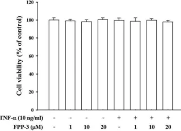
Fig. 1 −The effects of FPP-3 and TNF-α on the viability of HT-29 cells. Cells were treated with various concentrations of FPP-3 for 1 h, and then the cells were further incubated in the absence or presence of TNF-α (10 ng/ml) for 3 h. The cell viability was measured by MTT assay. The data represent the means±SEM of three independent experi- ments.
한 후 gel documentation system(UVP, Cambridge, UK)를 이
용하여각각의밴드를관찰하였다.
Reporter gene 발광효소 실험(Reporter gene luciferase assay)
50~60% 자란 HT-29 세포를 PBS로 2회씻은후, PS을함유
하지않고 10% FBS를함유한 RPMI1640 배지 6.5 ml와 trans- fection mixture 700µl를넣고 37oC 조건하에서 3시간동안반
응시켰다. FBS와 PS가없는 700µl의 RPMI1640 배지에 35µl
GeneJammer transfection reagent와 0.24µg/ml NF-κB luciferase construct(firefly luciferase) 그리고 0.2µg/ml pRL-TK(renilla luciferase)를넣어실온에서 7분간반응시켜 transfection mixture
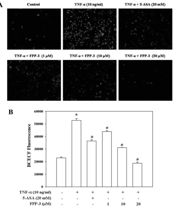
Fig. 2 −FPP-3 inhibits U937 monocyte adhesion to HT-29 cells. The cells were pretreated with various concentrations of FPP-3 for 1 h, followed by 10 ng/ml of TNF-α for 3 h. Then, BCECF/AM-prelabeled U937 cells were co-incubated with HT-29 cells for 30 min. Non- adhering U937 cells were removed, and fluorescence-emitting from the remaining cells (A) was imaged by inverted fluorescence microscopy (TE2000-U; Nikon, Japan). In a different set of experiment (B), the fluorescence from the remaining cells was measured by a fluorescence-detecting microplate reader (Fluostar optima; BMG Labtech GmbH, Germany) with excitation at 485 nm and emission at 520 nm. Data are shown as means±SEM from three independent experiments. *P<0.05, compared to untreated control and #P<0.05, compared to TNF-α-treated group.
을만든후사용하였다. 3시간 transfection 반응후, 10% FBS
와 1% PS를함유한 RPMI1640 배지를 7 ml넣어주고 37oC에
서 24시간배양시켰다. NF-κB가 transfection 된세포를 24 well plate에 1×10⁵ cells/cm2이되게배양하고 FPP-3를 1시간동안
전처리한후, 10 ng/ml TNF-α를처리하고 3시간동안 37oC에서 반응시켰다. PBS로 2회씻은후 24시간동안 -70oC에서 freezing
시키고, 그후세포들을모은다음 kit에포함되어있는 LAS, stop
& Glow buffer를 첨가하여 TD20/20 luminometer(Turner Biosystems, CA, USA)을이용하여측정하였다.
결과 및 고찰
본연구실에서앞서선행된 FPP-3에대한연구의결과, 20µM
의농도에서세포독성이적으면서유효한효과가있음을확인
하였으므로,5)이를기초로하여본실험에서는 FPP-3의 1, 10, 20µM의 cell viability을측정하였다. 그결과, FPP-3를단독으
로처리한군과 TNF-α를같이처리한군에서모두세포독성이
없음을확인하였다(Fig. 1).
TNF-α에의해유도된장상피세포에 U937이부착하는현상 에대한억제능력을조사한결과, Fig. 2과같이 TNF-α에의해 유도된장상피세포와 U937의부착이 FPP-3에의해농도의존
적으로억제되는것으로나타났다. 특히현재임상적으로염증 성장질환치료에많이쓰이고있는약물인 sulfasalazine의활성 대사체인 5-ASA의유효농도인 20 mM과비교할때, TNF-α가
처리되지않은대조군의수준으로억제하는 FPP-3의유효농도
는 10µM~20µM인것으로나타났다. 이는 FPP-3가 5-ASA보
다약 1000배낮은농도에서도더우수한효과를보인다는것을
의미하는것으로 FPP-3가염증성장질환과관련된염증반응을
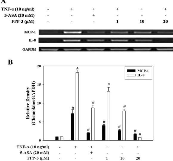
Fig. 3 −FPP-3 suppresses the TNF-α-induced MCP-1 and IL-8 mRNA expression. The cells were pretreated with various concentrations of FPP-3 for 1 h, followed by 10 ng/ml of TNF-α for 3 h. The MCP-1 and IL-8 mRNA expression level was measured by RT-PCR. The PCR products of MCP-1, IL-8, and GAPDH were separated in 2% agarose gel (A). The expressions of MCP-1 and IL-8 mRNA were statistically analyzed relative to GAPDH by densitometry (B). Data are shown as means±SEM from three independent experiments.
*P<0.05, compared to untreated control and #P<0.05, compared to TNF-α-treated group.
효과적으로억제할수있음을암시하고있다.
다음으로 U937의장상피세포부착을매개하는 MCP-1과 IL- 8의발현증가에대한 FPP-3의영향을조사하였다. 그결과, Fig.
3와 같이 FPP-3가 TNF-α에 의해 유도된 MCP-1과 IL-8의
mRNA 발현증가를농도의존적으로억제하는것으로나타났다.
특히 IL-8은염증성장질환에서특이적으로높게발현되는것으
로알려져있는데, FPP-3를 20µM 농도로처리하였을때 IL-8
의 mRNA 발현이 TNF-α가처리되지않은대조군과유사한수
준으로까지억제함을확인하였다.
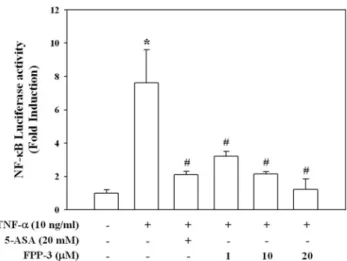
뿐만아니라 MCP-1과 IL-8의발현조절에공통적으로관련된 전사인자인 NF-κB에대한 FPP-3의영향을조사하기위해 NF-
κB luciferase reporter gene constructs가 transfection된장상피 세포에 TNF-α를처리하였을때, NF-κB의활성이유의적으로증 가되었고, 이러한증가는 FPP-3가처리된실험군에서농도의존적
으로억제됨을확인하였다(Fig. 4). 또한 Fig. 3의결과와비교해 보면 MCP-1의경우, 본실험의결과와거의일치하는경향을보
였으나, IL-8의경우에는그발현억제경향이 NF-κB의활성억
제경향과약간의차이를보였다. 이는 IL-8의발현조절이 NF-
κB를경유한경로를통해서뿐만아니라 NF-κB에비의존적인경
로를통해서도이루어질수있기때문인것6)으로판단된다.
결 론
FPP-3 화합물이 TNF-α로유도되는소장상피세포의염증상 태에미치는영향을조사한결과, TNF-α에의해유도되어진단
핵구의장상피세포부착현상을 FPP-3가농도의존적으로억제
함을확인하였다. 또한 TNF-α에의한단핵구의부착현상과깊은
관련이있으며, 소장의염증병변을악화시키는주요 chemokine
인 MCP-1과 IL-8의발현증가및이들의발현조절인자인 NF-κB
의활성화도 FPP-3에의해농도의존적으로억제됨을확인하였
다. 특히임상적으로널리쓰이는약물인 sulfasalazine의활성대 사체인 5-ASA의농도와비교하였을때, FPP-3는 1000배낮은농
도에서우수한효과를나타내었다. 위의결과로미루어봤을때, FPP-3는 TNF-α로유도된장상피세포의염증을강력하게억제 함으로염증성장질환치료제개발의선도물질이될수있을것이다.
감사의 말씀
이연구는 2007년도정부(과학기술부)의재원으로한국과학재 단의지원을받아수행된연구임(No. R11-2007-040-02004-0).
참고문헌
1) Ito, R., Shin-Ya, M., Kishida, T., Urano, A., Takada, R., Sakagami, J., Imanishi, J., Kita, M., Ueda, Y., Iwakura, Y., Kataoka, K., Okanoue, T. and Mazda, O. : Interferon-gamma is causatively involved in experimental inflammatory bowel disease in mice. Clin. Exp. Immunol. 46, 330 (2006).
2) Banks, C., Bateman, A., Payne, R., Johnson, P. and Sheron, N. : Chemokine expression in IBD. Mucosal chemokine expression is unselectively increased in both ulcerative colitis and Crohn's disease. J. Pathol. 199, 28 (2003).
3) Dijkstra, G., Moshage, H. and Jansen, P. L. : Blockade of NF- kappaB activation and donation of nitric oxide: new treatment options in inflammatory bowel disease. Scand J. Gastroenterol.
Suppl. 236, 37 (2002).
4) Lee, E. S., Park, B. C., Paek, S. H., Lee, Y. S., Basnet, A., Jin, D. Q., Choi, H. G., Yong, C. S. and Kim, J. A. : Potent analgesic and anti-inflammatory activities of 1-furan-2-yl-3-pyridin-2-yl- propenone with gastric ulcer sparing effect. Biol. Pharm. Bull.
29, 361 (2006).
5) Park, B. C., Thapa, D., Lee, Y. S., Kwak, M. K., Lee, E. S., Choi, H. G., Yong, C. S. and Kim, J. A. : 1-Furan-2-yl-3-pyridin- 2-yl-propenone inhibits the invasion and migration of HT1080 human fibrosarcoma cells through the inhibition of proMMP-2 activation and down regulation of MMP-9 and MT1-MMP.
Eur. J. Pharmacol. 567, 193 (2007).
6) Moon, Y., Yang, H. and Park, S. H. :Hypo-responsiveness of interleukin-8 production in human embryonic epithelial intestine 407 cells independent of NF-kappaB pathway: new lessons from endotoxin and ribotoxic deoxynivalenol. Toxicol.
Appl. Pharmacol. 15, 231 (2008).
Fig. 4 −FPP-3 suppresses the TNF-α-induced NF-κB gene trans- cription activity. The NF-κB gene transcription activity was measured by a luciferase assay kit (Dual Luciferase Activity assay system; Promega) in the cells transfected with NF-κB plasmid in the presence of GeneJammer (Stratagene). Data are means±SEM from three independent experiments. *P<0.05, compared to untreated control and
#P<0.05, compared to TNF-α-treated group.