TNF-α 자극에 의한 U937 단핵구 세포의 HT29 대장 상피 세포 부착에 대한 Berberine의 PPARγ가 아닌 NF-κB 경로를 통한 억제 효과
박수영·이광익·김일엽·김정애# 영남대학교 약학대학
(Received September 8, 2009; Revised October 20, 2009; Accepted October 20, 2009)
Inhibitory Effect of Berberine on TNF- α -induced U937 Monocytic Cell Adhesion to HT29 Human Colon Epithelial Cells is Mediated through NF- κ B Rather than PPAR γ
Su-Young Park, Gwang Ik Lee, Il-Yeob Kim and Jung-Ae Kim
#College of Pharmacy, Yeungnam University, Gyeongsan 712-749, Korea
Abstract
— Berberine, an isoquinoline alkaloid, has a wide range of pharmacological effects, including anti-inflammation.
It has been reported that berberine inhibits experimental colitis through inhibition of IL-8, and that inhibitory effect of ber- berine on inflammatory cytokine expression is mediated through peroxisome proliferator activated receptor (PPAR)-
γ. In this study, we examined the effects and action mechanism of berberine on the tumor necrosis factor (TNF)-
α-induced mono- cyte adhesion to HT29 human colonic epithelial cells, which is commonly used as an in vitro model of inflammatory bowel disease (IBD). Berberine significantly inhibited the TNF-
α-induced monocyte adhesion to HT29, which is similar to the effect of PDTC, a nuclear factor (NF)-
κB inhibitor. However, ciglitazone and GW, the ligands of PPAR-
γ, did not suppress the TNF-
α-induced monocyte adhesion to HT29 cells. In addition, TNF-
α-induced chemokine expression and NF-
κB tran- scriptional activity were significantly inhibited by berberine in a concentration-dependent manner. The results suggest that inhibitory effect of berberine on colitis is mediated through suppression of NF-
κB and NF-
κB-dependent chemokine expression.
Keywords □
berberine, colitis, nuclear factor (NF)-
κB, peroxisome proliferator activated receptor (PPAR)-
γ, chemokine
염증성장질환
(Inflammatory bowel disease, IBD)
은소장과대장에걸쳐전반적으로염증이있으며
,
증상으로설사,
통증,
체 중감소등을동반한다.
1)염증성장질환에대한병태생리기전 이나원인은정확히밝혀져있지않으나,
장내세균총에대한비정상적인면역반응
,
즉,
염증세포의과도한활성에의한것으로 추정되고있다.
2)활성화된백혈구의장점막부착및조직내유주를통한점막손상이염증성장질환의가장중요한과정으로 인식되며
,
이러한과정을매개하는매개체중의하나로chemokine
의중요성이대두되고있다
.
3,4)Chemokine
은단핵구와T
림프구등과같은여러종류의세포에서발현되며
,
다양한세포를활성화하여
tumor necrosis factor(TNF)-
α와같은pro-inflammatory cytokine
의발현을촉진한다.
5,6)TNF-
α에의해발현이유도되는chemokine
으로염증부위로백혈구의유주및부착을유도하는중요인자로
monocyte chemoattractant protein(MCP)-1
과interleukin(IL)-8
이있으며,
7,8)이들은염증성장질환의개시는 물론병변을악화시키는인자로알려져있다.
9)Nuclear Factor(NF)-
κB
는세포질에존재하고있으며,
활성화되면핵으로이동하여면역반응과염증반응을조절하는여러 가지의유전자들의발현을매개한다
. TNF-
α에의한TNF-
α수용체의활성화는
NF-
κB
를활성화시키며,
이는곧MCP-1
과IL-
8
등의발현을증가시키게된다.
10,11)따라서, NF-
κB
의활성을억제하여
MCP-1
과IL-8
등의발현을감소시킬수있는물질은치료제로서크게기여할수있을것이라생각한다
.
황련의주요알칼로이드성분인
berberine
은암세포의성장 을억제하는효과가있으며,
12)장상피세포에서cyclooxygenase- 2
의억제효과뿐만아니라,
13)망막색소상피세포에TNF-
α로유도된
MCP-1
과IL-8
의발현을억제하는효과를가진다는보고가 있다.
14)또한, berberine
은trinitrobenzene sulfonic acid(TNBS)
로유도한동물의장염을억제하는효과가보고되었으며
,
이러한 효과는
IL-8
의 억제에 의한다는 보고가 있다.
15) 또한,
#본논문에관한문의는저자에게로
(
전화) 053-810-2816 (
팩스) 053-810-4654
(E-mail) [email protected]
berberine
이대식세포에서TNF-
α와IL-6
등의염증성사이토카인의 발현을 억제하며
,
그 작용은peroxisome proliferator activated receptor(PPAR)-
γ를통해서이루어진다고보고되었다.
16)본연구에서는in vitro장염모델인단핵구세포와 대장상피 세포사이의부착모델에서
berberine
의장염억제작용및이 에대한전사인자, NF-
κB
및PPAR-
γ의역할을규명하고자하 였다.
실험방법
시약
이 실험에 사용한
fetal bovine serum(FBS)
와penicillin/
streptomycin(PS), RPMI1640
배지는Hyclone
사(South Logan, UT, USA)
에서구입하였다. 4-(2-hydroxyethyl)-1-piperazineethane- sulfonic acid(HEPES), sodium pyruvate, sodium bicarbonate, 3-[4,5-dimethylthiazol-2-yl]-2,5-diphenyltetrazolium bromide (MTT), BCECF-AM, diphenylpicry-hydrazyl
은Sigma
사(St.Luouis, MO, USA)
에서 구입하였다. Human TNF-
α는Biosourse
사(Camarillo, CA, USA)
에서 구입하였고, trizol reagent
는Invitogen
사(Carlsbad, CA, USA)
에서구입하였다. Taq DNA polymerase
는Takara
사(Shiga, Japan)
에서 구입하였다. GeneJammer transfection reagent
은Stratagene
사(CA, USA)
에서 구입하였다
. Dual-Luciferase reporter assay system
은Promega
사(Corporation, Madison, USA)
에서구입하였다.
세포배양
(Cell culture)
HT29
사람 대장 상피세포는10% FBS, 1% PS, 10 mM HEPES, 1 mM sodium pyruvate, 1.5 g/
lsodium bicarbonate
가 함유된RPMI1640
배지로37
oC, 5% CO
2조건하에서배양하 였으며,
세포가배양flask
에80%
이상자라면1 : 3
의비율로계대하여본실험에사용하였다
.
세포생존율측정
(MTT assay)
HT29
세포를96 well plate
에1×10
5cells/cm
2가되게배양한 후시료들을처리하였다.
일정시간처리후에5 mg/m
lMTT
용 액을각well
에처리하여37
oC
에서4
시간동안반응시켰다.
배지를 제거한 후
dimethyl sulfoxide(DMSO)
를 넣어 형성된formazan
크리스탈을 녹여서540 nm
의 파장에서microplate reader(Molecular Devices, VersaMAX, Molecular Devices, U.S.A.)
를이용하여흡광도를측정하였다.
부착능실험
(Adhesion assay)
U937
세포에10
µg/m
lBCECF-AM
를처리하여37
oC
에서30
분동안반응시켜준비해두었다
. HT29
세포를48 well plate
에
1×10
5cslls/cm
2가되게배양하여1% PS
만함유된각화합물을
1, 5, 10
µM
농도로1
시간전처리하고, 10 ng/m
lTNF-
α를처리하여
37
oC
에서3
시간반응시켰다.
화합물이처리된HT29
세포의배지를제거하고
PBS
로1
회씻어주었다.
그후BCECF- AM
으로처리된U937
세포를HT29
세포와함께37
oC
에서1
시 간반응시킨후,
부착되지않은U937
세포를제거하기위하여PBS
로1
회씻어주었다.
세포용해를위해0.1% Triton X-100 in 0.1 M Tris
를처리하여30
분동안 실온에서반응시킨 후, Fluostar optima microplate reader(BMG Labtechnologies, GERMANY)
을사용하여형광을정량하였다.
Reverse transcription-polymerase chain reaction(RT- PCR) HT29 세포를 1×10
5cslls/cm
2이되게배양하였다. Berbeine
을
1
시간전처리하고, 10 ng/m
lTNF-
α를3
시간동안37
oC
에서 반응시켰다. Trizol reagent
를이용하여total cellular RNA
를추출한다음
, Ready-To-Go T-primed first strand kit
를이용하여cDNA
를합성하였다.
그후0.5 U Taq DNA polymerase
를사용하여
MCP-1, IL-8, PPAR-
γ, GAPDH primer
로PCR
을수행하였으며 그
sequence
는 아래와 같다. Human MCP-1(5'-ATG AAA GTC TCT GCC GCC CTT-3', 3'-TGG GTT TGA GGC TTC TGA ACT-5'); human-IL-8(5'-ATG ACT TCC AAG CTG GCC GTG GCT-3', 3'-CTC TTC AAA AAC TTC TCC CGA CTC T-5'); human PPAR-
γ(5'-ATG ACA GCG ACT TGG CAA TA-3', 3'-TAA AGG GAA GAA GGT CAA CG- 5'); human-GAPDH(5'-GGT GAA GGT CGG AGT CAA CG- 3', 3'-CCA GTA GGT ACT GTT GAA AC-5'). PCR
산물은0.5
µg/m
lethidium bromide
가함유된2% agarose gel
에전기영 동을 수행한 후gel documentation system(UVP, Cambridge, UK)
를이용하여각각의밴드를관찰하였다.
Reporter gene
발광효소 실험(Reporter gene luciferase assay)
HT29
세포가50~60%
정도자랐을때PBS
로2
회씻어준 후, 10% FBS
는포함하고PS
은포함되지않은RPMI1640
배지6.5 m
l과transfection mixture 700
µl를넣어37
oC
에서3
시간반응시켰다
.
이때의transfection mixture
은35
µlGeneJammer transfection reagent
와0.24
µg/m
lNF-
κB luciferase construct (firefly luciferase)
와0.2
µg/m
lpRL-TK(renilla luciferase)
를FBS
와penicillin/streptomycin
을 포함하지 않는700
µl의RPMI1640
배지에넣어실온에서7
분간반응시킨후사용한다. Transfection
의반응이끝나는3
시간후10%
의FBS
와1%
의PS
이함유된
RPMI1640
배지를7 m
l넣어준후37
oC
에서24
시간 배양시켰다. NF-
κB
가transfection
이된세포를24 well plate
에1×10
5cslls/cm
2이되게배양하였다. 10
µM
의berbeine
을1
시간전처리하고
, 10 ng/m
lTNF-
α를3
시간처리후37
oC
에서반응시켰다
. PBS
로1
회씻어낸후lysis buffer
를넣어-70
oC
에서24
시간의freezing
과정후세포들을 모은다음LAS, stop &
Glow buffer
를첨가하여Tumer TD20/20 luminometer(Turner Biosystems, CA, USA)
을이용하여측정하였다.
Immunocytochemistry
방법에의한NF-
κB(p65)
의핵내이동측정
Immunocytochemistry
을이용하여NF-
κB(p65)
의핵안으로이동을측정하였다
.
17)HT29
대장상피세포를배양한후, 20
µM
의
berbeine
을1
시간전처리하고, 10 ng/m
lTNF-
α를3
시간처 리후37
oC
에서반응시켰다. 3.7% paraformaldehyde/PBS(pH 7.2)
용액으로세포를고정시키고, 0.4% Triton X-100
용액으로permealization
시켰다. PBS
를이용해1
회씻어준후, 3% bovine serum albumin
용액으로세포를1
시간동안반응시키고1 : 100
의배율로희석한
anti-NF-
κB(p65) antibody
용액에4
oC
에서20
시간동안반응시켰다
. Tris-buffered saline-Tween20(TBS-T)
용액으로세포를 씻어주었다
. Alexa Fluor 488 goat anti-rabbit IgG
을3% BSA
가포함된TBS
에1 : 200
의배율로희석시킨용 액에세포를실온에서1
시간동안배양하였다. TBS-T(20 nM Tris-HCl, pH 7.5, 500 mM NaCl, 0.1% Tween 20)
용액으로씻어준후
, 600 nM 4',6-diamidino-2-phenylindole(DAPI)
로세포 를5
분동안염색시켰다.
다시세포를씻어준후, Prolong Gold Antifade reagent
로고정시키고커버슬라이드로덮었다.
마지막으로샘플들은
Nikon microscope(TE-2000U)
를사용하여측정Fig. 1 −
Effect of berberine and TNF-
αon the cell viability of HT29 cells. HT29 cells were seeded in 96-well plates at a density of 1×10
5cells/cm
2. The cells were pretreated with berberine for 1hr, and then, incubated in the absence (black bars) or presence (white bars) of of TNF-
α(10 ng/m l ) for 3 hr. The cell viability was assessed using the MTT staining method. The data represent the means±SEM of three independent experiments.
Fig. 2 −
Inhibitory effects of berberine on TNF-
α-induced U937 monocytic cell adhesion to HT29 colonic epithelial cells.
Monocyte-epithelial adhesion was evaluated using the human leukemia pre-monocytic U937 cells which were pre-labeled with 2',7'-bis(2-carboxyethyl)-5(6)-carboxyfluo- rescein acetoxymethyl ester (BCECF/AM, 10
µg/m l ) for 30 min at 37
oC. A set of cells was taken and imaged by inverted fluorescence microscopy (A). In other sets, cells were lysed in 0.1% Triton X-100 in 0.1 mol/ l Tris, and the lysates were measured for fluorescence by using a fluorometer (FLUOstar Optima) with excitation at 485 nm and emission at 520 nm (B). Data are expressed as the mean±SEM of three independent experiments. * P <0.05 compared to untreated control group.
#P <0.05 compared to the TNF-
αtreated group.
하였다
.
실험결과 및 고찰
TNF-
α에의해유도된대장상피세포와백혈구의부착에대한berberine
의억제효과염증성장질환의점막손상에기여하는가장중요한병적과 정인활성화된백혈구의장점막부착및조직으로의이주과정 에미치는
berberine
의영향을확인하기위해먼저,
대장상피세포생존율에미치는
berberine
의영향을조사하였다.
그결과,
berberine
을 단독으로4
시간 처리하거나(Fig. 1,
검은 막대), berberine
을1
시간전처리하고TNF-
α(10
µg/m
l) 3
시간처리의경우
(Fig. 1,
흰색막대)
모두세포생존율에큰영향이없었다. Berberine
은TNF-
α에의해유도된대장상피세포와단핵구의부착능을 유의하게억제함을 확인하였다
(Fig. 2). Berberine (10
µM
과20
µM
농도)
의효과는,
현재염증성장질환치료에사 용되고있으며,
본실험에서대조물질로사용한5-aminosalicylic acid(5-ASA)
의20 mM
농도에서의효과와유사하였다.
이러한결 과는기존의보고15)와마찬가지로berberine
이염증성장질환의 치료제로서가능성이매우큼을의미한다.
TNF-
α에 의해 유도되는MCP-1
과IL-8
의 발현에 미치는berberine
의억제효과백혈구의장점막부착및이주에중요한역할을하는주요
chemokine
인MCP-1
과IL-8
은TNF-
α에의해그발현이현저 히증가하였으며,
이러한발현증가는berberine
에의해유의적으로억제됨을확인하였다
(Fig. 3).
대장상피세포와단핵구의 부착능시험결과와마찬가지로chemokine
의발현에미치는berberine(20
µM)
의영향은5-ASA 20 mM
농도의경우와유사하였다
.
TNF-
α에 의해 유도된NF-
κB
및PPAR-
γ 활성에 미치는berberine
의영향MCP-1
과IL-8
의발현은전사인자NF-
κB
에의해조절됨은잘 알려져있어berberine
이NF-
κB
의핵내이동및전사활성에미치는영향을조사하였다
. NF-
κB
항체를이용한세포면역염색시 험에서TNF-
α에의해NF-
κB
의핵으로의이동은현저히증가하 였으며,
이러한핵내이동이berberine
에의해현저히감소함을확인하였다
(Fig. 4A). NF-
κB
전사활성은NF-
κB luciferase reporter gene
을도입한HT29
대장상피세포를이용하여 측 정하였으며,
그결과, TNF-
α에의해현저히증가한NF-
κB
전사활성은
berberine
처리에의해농도의존적으로감소됨을확 인하였다(Fig. 4B).
이러한결과는berberine
이사람각질세포 에서TNF-
α에의한NF-
κB
활성억제효과18)와일치되는결과이다
.
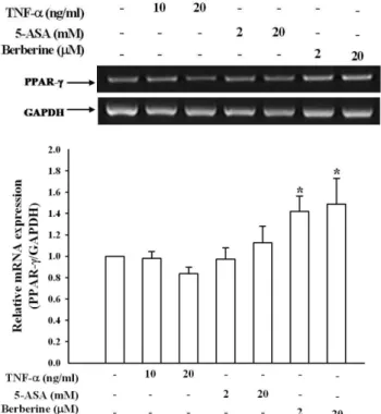
한편
, berberine
이PPAR-
γ를통해TNF-
α와IL-6
등의염 증성사이토카인의 발현을억제한다고도알려져있어, PCR
을통한
PPAR-
γ발현에미치는 영향을측정한 결과대조군으로사용한
5-ASA
의 경우는 큰 영향을 나타내지못하는 반면,
berberine
이PPAR-
γ발현을유의하게 증가시킴을확인하였다(Fig. 5).
Fig. 3 −
Inhibitory effects of berberine on TNF-
αinduced MCP-1 and IL-8 mRNA levels in HT29 cells. Serum-starved HT29 cells were
pretreated with berberine for 1 hr, and then, treated with 10 ng/m l of TNF-
αfor 3 hr. MCP-1 and IL-8 mRNA levels were measured
by using RT-PCR. The PCR products of MCP-1, IL-8, and GAPDH were separated in 2% agarose gel (A). The relative mRNA
expression of MCP-1 (B) and IL-8 (C) to GAPDH was quantitated by densitometry. Data are expressed as the mean±SEM of three
independent experiments. * P <0.05 compared to untreated control and
#P <0.05 compared to TNF-
αtreated group.
TNF-
α에의한백혈구와대장세포의부착에미치는berberine
의PPAR-
γ보다는NF-
κB
경로를통한억제효과Berberine
에의한NF-
κB
및PPAR-
γ전사활성이TNF-
α에의한단핵구세포와대장상피세포사이의부착을매개하는지확 인하기위해
NF-
κB
억제제인PDTC
및PPAR-
γ에대한효능제ciglitazone
과억제제GW9662
의처리에의한단핵구세포와대장상피세포부착능을조사하였다
.
그결과, TNF-
α에의한백혈구와대장상피세포의부착은
PDTC
에의해현저하게억제되었 으며, GW
의경우는어느정도억제효과를나타내었고, ciglitazone
은전혀억제효과를나타내지못하였다
(Fig. 6).
이러한결과는TNF-
α에의한단핵구세포와대장상피세포사이의부착을매개하는전사인자는
PPAR-
γ보다는NF-
κB
임을의미한다.
또한,
Fig. 6 −
Involvement of NF-
κB but not PPAR-
γon berberine- mediated adhesion of monocytes to HT29 cells.
Experiments were performed as described previously in Fig. 2. HT29 cells were lysed and the fluorescence was quantitated using a fluorescence detecting microplate reader. Data are expressed as a percentage of control, and the bar graph represents the mean±SEM from four independent experiments. * P <0.05 compared to untreated control and
#P <0.05 compared to TNF-
αtreated group.
Fig. 4 −
Berberine suppresses the TNF-
α-induced NF-
κB nuclear translocation and transcriptional activity in HT29 cells. (A) Immunocytochemical analysis of NF-
κB (p65) localization.
HT29 cells were first treated with berberine (20
µM) for 1 hr and then exposed to TNF-
α(10 ng/m l ) for 20 min.
After fixation with 3.7% paraformaldehyde/PBS, immunocy- tochemical analysis was performed as described in Materials and Methods. The results shown are represen- tative of two independent experiments. (B) NF-
κB luciferase activity. The NF-
κB gene transcription activity was measured by luciferase assay using manufacturer's protocol. Data are means±SEM from three independent experiments. * P <0.05 compared to untreated control and
#
P <0.05 compared to TNF-
αtreated group.
Fig. 5 −
Berberine alone increases the PPAR-
γmRNA level in
HT29 cells. Serum-starved HT29 cells were pretreated
with berberine for 1 hr, and then, treated with 10 ng/m l of
TNF-
αfor 3 hr. PPAR-
γlevel was measured by using the
RT-PCR. Data are expressed as the mean±SEM of three
independent experiments. * P <0.05 compared to untreated
control.
berberine
의효과가PDTC
처리의경우와유사한점은TNF-
α에의한단핵구세포와대장상피세포사이의부착을억제하는
berberine
의대장세포염증억제작용이NF-
κB
를경유함을암 시한다.
결 론
Berberine
은TNF-
α에의해유도된MCP-1
과IL-8
의발현및NF-
κB
의활성억제하여단핵구의대장상피세포에부착을억제 하였으며,
특히,
대조물질인5-ASA
와비교해볼때, berberine
은 훨씬낮은 농도에서 우수한억제효과를나타냈다
.
또한, berberine
에의해증가된PPAR-
γ의발현증가는단핵구의대장 상피세포에부착을 영향을미치지못하였다.
이상의결과는berberine
의대장세포염증억제작용이NF-
κB
를경유함을의 미한다.
감사의 말씀
이논문은
2005
년정부(
교육인적자원부)
의재원으로한국학술진흥재단의지원을받아수행된연구임
(KRF-2005-204-E00119).
참고문헌
1) Banks, C., Bateman, A., Payne, R., Johnson, P. and Sheron, N. : Chemokine expression in IBD. Mucosal chemokine expression is unselectively increased in both ulcerative colitis and Crohn's disease. Journal of Pathology
199, 28 (2003).
2) Groux, H. and Powrie, F. : Regulatory T cells and inflammatory bowel disease. Immunol Today
20, 442 (1999).
3) Rogler, G. and Andus, T. : Cytokine in inflammatory bowel disease. Wprle J. Surg.
22, 382 (1998).
4) Porath, D., Riegger, C., Drewe, J. and Schwager, J. : Epigallocatechin-3-gallate impairs chemokine production in human colon epithelial cell lines. J. Pharmacol. Exp. Ther.
315, 1172 (2005).
5) Banks, C., Bateman, A., Payne, R., Johnson, P. and Sheron, N. : Chemokine expression in IBD. Mucosal chemokine expression is unselectively increased in both ulcerative colitis and Crohn's disease. J. Pathol.
199, 28 (2003).
6) Richard, P. : Chemokines in the inflammatory bowel diseases.
Journal of Clinical Immunology
19, 5 (1999).
7) Izzo, R. S., Witkon, K., Chen, A. I., Hadjiyane, C., Weinstein, M. I. and Pellecchia, C. : Interleukin-8 and neutrophil markers in colonic mucosa from patients with ulcerative colitis. Am. J.
Gastroenterol.
87, 1447 (1992).
8) Spoettl, T., Hausmann, M., Herlyn, M., Gunckel, M., Dirmeier,
A., Falk, W., Herfarth, H., Schoelmerich, J. and Rogler, G. : Monocyte chemoattractant protein-1 (MCP-1) inhibits the intestinallike differentiation of monocytes. Clinical and Experimental Immunology
145, 190 (2006).
9) Tamaki, S., Yamauchi, M., Fukuoka, A., Makinodan, K., Koyama, N., Tomoda, K., Yoshikawa, M. and Kimura, H. : Production of inflammatory mediators by monocytes in patients with obstructive sleep apnea syndrome. Intern. Med.
48