Received: August 29, 2017 Revised: October 18, 2017 Accepted: October 22, 2017 TRAUMA AND INJURY
Correspondence to Jong-Keon Oh, M.D., Ph.D.
Department of Orthopaedic Surgery, Guro Hospital, Korea University College of Medicine, 148 Gurodong-ro, Guro-gu, Seoul 08308, Korea
Tel: +82-2-2626-3088 Fax: +82-2-851-3111 E-mail: [email protected]
http://www.jtraumainj.org Copyright © 2017 The Korean Society of Trauma
Proximal Tibia Fracture after Proxi- mal Tibia Autograft Harvest
Jin-Kak Kim, M.D.
1, Jong-Hyeop Song, M.D.
1, Kyungbum Lee, M.D.
2, Jae-Woo Cho, M.D.
1, Ki-Ho Moon, M.D.
1, Do-Hyun Yeo, M.D.
1, Beom-Soo Kim, M.D.
1, Jong-Keon Oh, M.D., Ph.D.
1Departments of
1Orthopaedic Surgery,
2General Surgery, Guro Hospital, Korea University College of Medicine, Seoul, Korea
Bone-grafting procedures are common in orthopedic trauma surgeries. There are only few reports on the morbidity after proximal tibia harvesting. Here, we report an expe- rience of complication after proximal tibia harvesting while treating subtrochanteric femoral osteomyelitis.
Keywords: Grafting, Bone; Tibial fractures
INTRODUCTION
Bone-grafting procedures are common in orthopedic surgeries for bone defects fol- lowing trauma, arthrodesis, and nonunion of fractures. Autologous bone graft has biologic advantages over heterogeneous or synthetic bone substitutes because of a combination of osteogenic, osteoinductive, and osteoconductive properties. These properties have not yet been achieved in heterogeneous or synthetic bone substitutes.
Several donor sites have been described for harvesting autologous cancellous bone such as the iliac crest, distal femur, proximal tibia, distal tibia, proximal humerus, olecranon, and distal radius [1]. There are some reports on donor site morbidity after iliac bone graft harvesting but only very few after proximal tibia harvesting [2,3]. Here, we report an experience of complication after proximal tibia harvesting while treating subtrochanteric femoral osteomyelitis.
CASE REPORT
A 55-year-old female visited our institute for the treatment of infected nonunion of
subtrochanteric femoral fracture. The patient did not have any other comorbidity, except for obesity. Her body mass index was 38.14 kg/m
2.
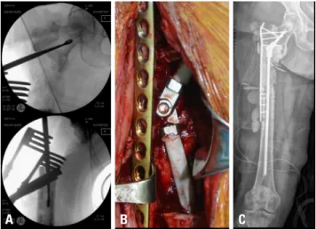
She underwent multiple surgeries for the subtrochan- teric femoral fracture at a local hospital, which resulted in non-union of the fracture. Furthermore, intraoperative samples for bacterial cultures were positive for methicil- lin-resistant staphylococcus aureus (Fig. 1)
We removed the previously placed broken plate and debrided the infected tissue. Then, we placed a plate for temporary fixation and inserted a cement rod with ce- ment spacer loaded with antibiotics. The 3.5 plate is not enough to maintain the fracture deformity when used alone; however, the intramedullary cement rod and spacer added stability until the next surgery (Fig. 2). Vancomy- cin was injected via intravenous for six weeks consulting to department of infectious disease. We set concentration to target for therapeutic drug monitoring of vancomycin.
After three weeks from 1st surgery, we converted the temporary fixative to definite femoral nail (Fig. 3). During each of the procedures, we performed microbiological cul- ture investigations during debridement and post-debride- ment. The post-debridement culture negative results let know us that the debridement was adequate.
During six months of follow up in the outpatient depart- ment, her levels of erythrocyte sedimentation rate (ESR)
and C-reactive protein (CRP) were not normalized, even though the intraoperative cultures were negative. Usually, we use bone grafts three months postoperatively after ESR and CRP have normalized, however, we could not do so in this case. There were no signs of infection such as fever, local warmth, redness, or tenderness. Additionally, we consulted the department of infectious diseases to identify the focus, which they could not. Therefore, we decided to perform debridement again. At that time, we changed all internal fixatives and inserted cement spacer with antibiot- ics. The tissue cultures were negative again.
Six months later, we performed bone grafting even though ESR/CRP had not fully normalized. We could not wait for longer because she had been walking with a crutch and partial weight bearing for almost four years. Fortunate- ly, a rheumatologist gave a diagnosis for her seropositive rheumatoid arthritis, which affect level of ESR and CRP.
The total size of the bone defect was 91 mm in the sub- trochanteric area. For this huge defect, we had to harvest the cancellous bones from the anterior superior iliac spine (ASIS) and bilateral proximal tibia. The other parts of ASIS were already used before. We had a choice of bilateral pos- terior superior iliac spine, but we thought it was not enough amounts for defects. We could add allograft bone chips or bone substitutes. But we wanted to avoid the chance of infection again even though it is rare. By creating a win-
Fig. 1. (A) Initial radiograph showed subtrochanteric femoral fracture. It was an atypical femoral fracture. (B) Primary treatment was open reduction
and nailing with cerclage wiring. A big incision (white arrow) was required. Biology of fracture site might be broken then. (C) Failure of implant 11 months postoperatively. There was no callus formation; however, progression of varus deformity of the proximal fragment was seen. (D) Revision treat- ment was plating. (E) However, after five months she underwent removal surgery again, because of infection. (F) Internal fixation with angled blade was tried but (G) failed again.
A B C D E F G
dow at Gerdy’s tubercle, we obtained the cancellous bones with curettes from the metaphysis of the proximal tibias.
The curettage was performed parallel to inferiorly but did not violate the medial cortex. The defect after harvesting did not require additional grafts. We obtained 44 g of can- cellous bone from the right proximal tibia, and 31 g from the left proximal tibia. We also obtained 20 g from the left ASIS. Bone grafting was performed in the subtrochanteric femoral defect (Fig. 4). Postoperatively, the patient was placed in bilateral lower extremity splints and instructed to not bear weight till postoperative 6 weeks. For her left leg, we planned to permit her tolerable partial weight bearing from 6 weeks. And for her right leg, we planned to permit after 3 months because of grafted site used to consolidate from 3 months.
On outpatient follow up two weeks later, her thigh pain was negligible but she complained of severe right knee pain. The radiographs showed cortical disruption from the medial to the lateral proximal metaphysis. However, there were no intra articular extensions noted on computed to- mography (Fig. 5). Although the time of the new fracture was unclear, it happened postoperatively because there was no fracture line in the immediate radiographs. She did not bear weight but her pain aggravated since three days after the surgery.
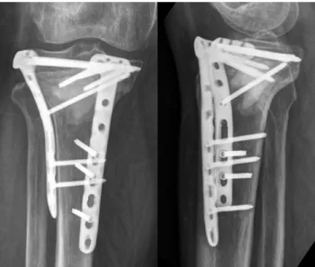
Open reduction and internal fixation was performed us- ing a 3.5 lateral proximal tibia plate and an additional medi- al plate was used. The bone graft substitute PRO-DENS
TM(combination of calcium sulfate with calcium phosphate, WRIGHT Medical Technology, Inc., Arlington, TN, USA) was used to fill the defects (Fig. 6). After 9 months fracture was united well.
A B C
Fig. 2. (A) We debrided the infected tissue including bone and soft
tissues. (B) Two antibiotics-loaded cement rods were used. They were connected by a hinge. (C) Immediate post-operative radiograph was taken. We used plate for temporary fixation.
Fig. 3. Conversion to definite fixation. We changed the previous plate
for a new one in view of the infection.
Fig. 4. For a huge defect in the subtrochanteric area, we had to harvest
from anterior superior iliac supine and bilateral proximal tibia.
DISCUSSION
Autologous cancellous bone graft has many benefits over allograft such as lower risk of immunologic complications and a combination of biologic properties. Anterior iliac crest is one of the standard harvest sites of autologous
cancellous bone. However, donor site morbidity following harvesting of bone graft from the ilium has been reported.
Many studies have reported that the iliac crest is associ- ated with a relatively high morbidity. The rate of major complications that resolve with intervention, including neurologic injury, vascular injury, deep infection, large hematoma, bowel herniation, ureteral injury, and fracture and pelvic instability are reported to range from 2.5% to 39%. The rate of minor complications that resolve with- out intervention such as persistent donor site pain, sero- mas, cosmetic defects, and temporary paresthesia range from 10 to 40% [4-6].
Due to the relatively high rate of complications with iliac crest bone grafts, other harvesting sites have gained popularity such as distal femur, proximal tibia, distal tibia, and local surgical site grafts. Of these, proximal tibia is relatively preferred over others because proximal tibia harvest site is near the surgical site within the same sterile drape usually and it can be under controlled with a tourniquet. Therefore, a graft may be obtained from the ipsilateral limb without re-draping. Additionally, a similar amount of bone graft may be obtained compared to the iliac crest. The reported volume of harvested graft from the proximal tibia is up to 30 cm
3[7].
Fig. 5. At the outpatient follow-up two weeks later, she complained of right knee pain. Anteroposterior (A) and lateral (B) radiographs showed trans-
verse cortical disruption of proximal tibia metaphysis. Additionally, coronal (C) and sagittal (D) cuts of computed tomography (CT) showed an extra-ar- ticular fracture. CT images show a huge vacant defect.
A B C D
Fig. 6. After bilateral plating, bone graft substitute was placed in the