79
http://dx.doi.org/10.4046/trd.2013.74.2.79 ISSN: 1738-3536(Print)/2005-6184(Online) Tuberc Respir Dis 2013;74:79-81
CopyrightⒸ2013. The Korean Academy of Tuberculosis and Respiratory Diseases. All rights reserved.
A Fatal Case of Acute Respiratory Failure Caused by Mycobacterium massiliense
Kyoung Hwa Choi, M.D., Ph.D.1, Hae Min Yu, M.D.2, Jae Seok Jeong, M.D.2,3, So Ri Kim, M.D., Ph.D.2,3,4, Yong Chul Lee, M.D., Ph.D.2,3,4
1Division of Pulmonology, Department of Internal Medicine, Presbysterian Medical Center, 2Department of Internal Medicine,
3Research Center for Pulmonary Disorders, Chonbuk National University Medical School, 4Research Institute of Clinical Medicine of Chonbuk National University−Biomedical Research Institute of Chonbuk National University Hospital, Jeonju, Korea
Few recent reports have indicated that Mycobacterium massiliense causes various infections including respiratory infection. However, there is scarce information on the clinical significance, natural history of the infection, and therapeutic strategy. This report describes a case of an immunocompetent old man infected by M. massiliense that causes acute respiratory failure. In light of the general courses of non-tuberculous mycobacterium infections, rapid progression and fatality are very rare and odd. In addition, we discuss the biological and pathological properties of M. massiliense with the review of cases reported previously including our fatal one.
Key Words: Mycobacterium; Mycobacterium Infections, Nontuberculous; Respiratory Insufficiency
Address for correspondence: Yong Chul Lee, M.D., Ph.D.
Department of Internal Medicine, Chonbuk National Univer- sity Medical School, 20, Geonji-ro, Deokjin-gu, Jeonju 561-712, Korea
Phone: 82-63-250-1664, Fax: 82-63-250-1633 E-mail: [email protected]
Received: Jun. 20, 2012 Revised: Jul. 9, 2012 Accepted: Jul. 25, 2012
CCIt is identical to the Creative Commons Attribution Non-Commercial License (http://creativecommons.org/licenses/by-nc/3.0/).
Introduction
Mycobacterium massiliense was originally classified as the Mycobacterium abscessus - chelonae complex
1. Although, to date, the knowledge of clinical manifes- tations of the infectious diseases caused by M. massi- liense is limited, M. massiliense is known to attack vari- ous organs in our body such as skin and lung
1,2. As for the pulmonary infection, according to one recent study, microbiologic response to the antibiotic therapy was more favorable in patients with M. massiliense (88%) than in those with M. abscessus lung disease (25%)
3. However, there are few reports describing the fatal case of pulmonary infection by M. massiliense .
Herein, we report a fatal case of acute respiratory fail- ure in an immunocompetent old man infected by M.
massiliense .
Case Report
A 75-year-old man visited our hospital to treat pulmo- nary non-tuberculous mycobacterium (NTM) disease.
He had complained productive cough for two months, and his diagnosis was confirmed by bacteriological anal- ysis using sputum specimen. He was non-smoker and had no specific history of chronic diseases. His vital signs were normal. On physical examination, inspiratory crackles were heard on the right lower lung field. The results of blood test showed no definitive abnormality.
A serologic test for human immunodeficiency virus was negative.
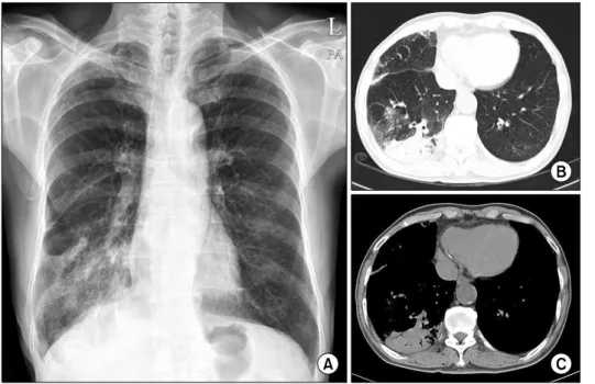
Chest X-ray revealed increased opacities in the right lower lobe (Figure 1A). Computed tomography scan of chest showed consolidation with air-bronchogram in the right lower lobe and focal centrilobular nodules on the right middle lobe (Figure 1B, C). On the bacteriological test, we verified the positive reactivity of acid fast stain-
Case Report
KH Choi et al: Acute respiratory failure by NTM
80
Figure 1. (A) Chest X-ray showed increased opac- ities in the right lower lobe.
(B, C) Contrast-enhanced computed tomography scan of chest demonstrated con- solidation within air-bron- chogram in the right lower lobe.
Figure 2. Chest X-ray during medical treatment in the in- tensive care unit showed diffuse bilateral infiltration.
ing in bronchial washing fluids. Moreover, we could find NTM DNA in the lung tissues obtained by percuta- neous transthoracic needle biopsy using polymerase chain reaction. Finally, M. massiliense was identified from a sputum specimen and a bronchial washing sam- ple, respectively, by a reverse blot hybridization assay.
However, there was no evidence of growth for other bacteria, fungi, and virus in microbiological examinat- ion.
According to the diagnositc criteria provided by the American Thoracic Society
4, he was diagnosed as M.
massiliense pulmonary infection, and the treatment was started with clarithromycin, amikacin, imipenem, and doxycycline. But he rapidly developed a severe respira- tory failure at 7 days after the initiation of medical treatment. Patient complained severe dyspnea with ta- chypnea, showing respiratory rate of 32 breaths per minute. Arterial blood gas analysis revealed that PO
2, PCO
2, pH, and PaO
2/FiO
2ratio were 49.9 mm Hg, 25.4 mm Hg, 7.465, and 81 mm Hg, respectively. Chest X-ray showed diffuse bilateral infiltrates (Figure 2). No improvement of gas exchange was obtained despite the support of mechanical ventilation, moreover, respiratory acidosis worsened to extreme levels. On the day 25 of diagnosis, the patient progressed to multi-organ failure
resulting in his death.
Discussion
NTM are found in the environment ubiquitously and
are formerly considered to be minor clinical signifi-
cance
4. Recently, the incidence of diseases caused by
NTM is increased and they relatively often cause pulmo-
Tuberculosis and Respiratory Diseases Vol. 74. No. 2, Feb. 2013