Response to Etoposide/Cisplatin Combination Chemotherapy in Small Cell Lung Cancer
Ji Woong Sohna, Shin Yup Leea, Su Jung Leeb, Hyo-Sung Jeonb, Jae Hee Leea, Jae Hyung Parka, Eun Jin Kima, Seung Ick Chaa, Chang Ho Kima, Young Mo Kanga, Jae-Tae Leec, Tae Hoon Junga, Jae Yong Parka,b*
aDepartment of Internal Medicine, School of Medicine, Kyungpook National University, Daegu, Korea, bCancer Research Center, Kyungpook National University, Daegu, Korea, cDepartment of Nuclear Medicine, School of Medicine, Kyungpook National University, Daegu, Korea
소세포폐암에서
Multidrug Resistance-1
유전자의 다형성과 Etoposide- cisplatin 항암화학요법 반응의 관계손지웅1, 이신엽1, 이수정2, 전효성2, 이재희1, 박재형1, 김은진1, 강영모1, 이재태3, 차승익1, 김창호1, 정태훈1, 박재용1,2
1경북대학교 의과대학 내과학교실, 2경북대학병원 암연구센터, 3경북대학교 의과대학 핵의학교실
배경 및 목적 : Multidrug Resistance-1 (MDR1) 유전자는 다약제내성에 관여하는 P-glycoprotein을 암호화한다. MDR1 유전자의 다형성은 P-glycoprotein의 발현과 기능의 차이를 일으켜 항암화학요법 반응에 영향을 미칠 수 있을 것이다.
저자들은 소세포폐암 환자에서 MDR1 유전자의 다형성과 일배체형에 따른 항암화학요법에 대한 반응을 조사하였다.
대상 및 방법 : 경북대학병원에서 병리적으로 소세포폐암으로 진단받고 etoposide–cisplatin 항암화학요법을 받은 54명 을 대상으로 하였다. 전혈 5cc에서 DNA를 추출하고 PCR-RFLP법을 통해 MDR1 유전자 엑손 21의 2677G>T 다형성과, 엑손 26의 3435C>T 다형성을 조사하고 다형성과 일배체형에 따른 항암화학요법의 반응을 조사하였다.
결 과 : 2677G>T 유전자형에 따른 항암화학요법의 반응은 유의한 차이가 없었다. 3435 CC 유전자형은 3435 CT+TT 형에 비해 치료 반응율이 유의하게 높았다 (P = 0.025). 유전자형 분석 결과와 일치되게 2677G/3435C 일배체형은 다른 일배체형에 비해 치료반응을 보이는 경우가 유의하게 많았다 (P = 0.015).
결 론 : 소세포폐암에서 MDR1 유전자의 2677G>T와 3435C>T 다형성 및 이들 다형성의 일배체형은 etoposide-cisplatin 항암화학요법의 반응을 예측할 수 있는 지표로 사용될 수 있을 것으로 생각된다.
(Tuberc Respir Dis 2005; 58:135-141)
Key words : MDR1, Polymorphisms, Chemotherapy Response, Small Cell Lung Cancer
Address for correspondence : Jae Yong Park, M.D.
*Corresponding author. Department of Internal Medicine, School of Medicine, Kyungpook National University, Samduk 2Ga 50, Daegu, 700-412, Korea Phone : 82-53-420-5536 Fax : 82-53-426-2046 E-mail : [email protected]
Received : Dec. 20. 2004 Accepted : Jan. 18. 2005
Introduction
Lung cancer is one of the major causes of cancer-related deaths worldwide. Small cell lung cancer (SCLC) represents approximately 20% of primary lung cancers, and it is characterized by rapid doubling time, high growth fraction and the early development of widespread metastasis1,2. Although
chemotherapy is the primary treatment for SCLC, intrinsic or acquired drug resistance is the major limiting factor for the effectiveness of chemotherapy.
Resistance to anticancer drugs happens through several mechanisms: decreased drug accumulation, drug inactivation, or enhanced DNA repair3.
The human multidrug-resistance (MDR)-1 gene encodes P-glycoprotein (PGP), which functions as an energy-dependent membrane efflux pump for a wide variety of lipophilic compounds. The PGP protein plays an important role in multidrug resistance by impairing the intracellular retention of anticancer drugs such as Vinca alkaloids, taxanes, anthracyclines and topoisomerase inhibitors4-6. There have been several studies showing that chemotherapy response
is inversely related with the level of PGP expression in various human cancers including SCLC 7-11, suggesting that variations in the PGP expression level or activity contribute to the therapeutic efficacy of chemotherapy.
Although the mechanism for altered MDR1 expression has not been clearly elucidated, hypome
thylation of the MDR1 promoter, altered activity of transcription factors, or gene rearrangements have been implicated in MDR1 regulation12-14. Several polymorphisms have been recently reported in the MDR-1 gene 15, and some of these variants [2677G
>T (Ala893Ser) at exon 21 and 3435C>T at exon 26) have been shown to affect the expression and function of PGP 16-18. Therefore, we have hypothesized that these two variants of MDR1 gene, and par
ticularly their haplotypes, could influence the response to chemotherapy. To test this hypothesis, we evalu
ated the association of 2677G>T and 3435C>T poly
morphisms and their haplotypes with the response to chemotherapy for SCLC patients treated with a com
bination chemotherapy of etoposide and cisplatin (EP).
Materials and methods 1. Study population
In the present study, we included 54 SCLC patients who were histologically diagnosed at Kyungpook National University Hospital, Daegu, Korea from January 2002 to June 2003. All these patients un
derwent complete staging procedures including chest radiograph, CT scan of the thorax and upper abdomen, brain MRI and bone scan. The clinical data for smo
king habits, weight loss and Eastern Cooperative Oncology Group performance status (ECOG PS) were collected prospectively. All the patients were received EP combination chemotherapy for more than two cycles as a first therapy. After two or
three cycles of chemotherapy, the response to che
motherapy was assessed according the WHO criteria19. Patients with a complete response or a partial res
ponse were defined as responders, and the patients having stable disease or progressive disease were defined as non-responders.
2. MDR1 genotyping
Genomic DNA was extracted from peripheral blood lymphocytes by proteinase K digestion and phenol/chloroform extraction. The MDR1 2677G>T (Ala893Ser) and 3435C>T (Ile1145Ile) genotypes were determined by PCR-RFLP assay. PCR primers were designed based on the GenBank reference sequence (accession no. M29440). The PCR primers for 2677G>T and 3435C>T polymorphisms were 5’-GGTTCCAGGCTTGCTGTAAT-3’ (forward) and 5’-TCACCTTCCCG(mutated A→G)G-3’ (reverse);
and 5’-GCTGCTTGATGGCAAAGA AA-3’ (forward) and 5’-ATTAGGCAGTGACTCG ATGATGA-3’ (re}
verse), respectively. PCR reactions were performed in a 20 ㎕ reaction volume containing 100 ng of genomic DNA, 10 pM of each primer, 0.2 mM dNTPs, 10mM Tris-HCl (pH 8.3), 50 mM KCl, 1.5mM MgCl2, and 1 unit of Taq polymerase (Takara Shuzo Co., Otsu, Shiga, Japan). The PCR cycle conditions consisted of an initial denaturation step at 94℃ for 5 min followed by 35 cycles of 30 s at 94℃; 30 s at 58℃ for 2677G>T and at 56℃ for 3435C>T; 30 s at 72℃; and a final elongation step at 72℃ for 10 min. The PCR products were digested overnight with the appropriate restriction enzymes (New England Biolabs, Beverly, MA, USA; BanI for 2677G>T and DpnII for 3435C>T) at 37℃. The digested PCR products were resolved on 6% acyl
amide gel. For quality control, the genotyping ana
lysis was repeated twice for all the subjects. To confirm the genotyping results, selected PCR-amplified
DNA samples (n = 2, respectively, for each genotype) were examined by DNA sequencing.
3. Statistical analysis
Chi-square test was used to evaluate the associa
tion between clinical variables and chemotherapy response. Hardy-Weinberg equilibrium of alleles at individual loci was tested with a goodness-of-fit x2 test with one degree of freedom to compare the observed genotype frequencies with the expected genotype frequencies among the subjects. Haplotypes and their frequencies were estimated based on the Bayesian algorithm using the Phase program20, which is available at http:// www.stat.washington.
edu/stephens/phase.html. Logistic regression analysis was performed to examine the association between genotypes/haplotypes and chemotherapy response with adjustment for possible confounders [age as a continuous variable, and sex, staging (limited vs extensive stage) and PS (ECOG 0-1 vs ECOG 2) as nominal variables]. Referent and 3 alternative models (codominant, dominant and recessive for the minor allele) were applied in the analyses. When multiple comparisons are made, the corrected P-values (Pc- values) were also calculated for multiple testing using Bonferroni’s inequality method. All analyses were performed using Statistical Analysis Software for Windows, version 6.12 (SAS institute, Gary, NC, USA).
Results 1. Patient characteristics
The Patients consisted of 46 men and 8 women, and their average age was 61.6 ± 7.9 years. The clinical staging was limited disease (LD) in 28 patients and extensive disease (ED) in 26 patients.
The ECOG PS was 0-1 in 35 patients and 2 in 19
patients. The overall response rate was 61% (complete response in 20% and partial response in 41%); 28%
of patients had stable disease; and 11% of patients had progressive disease. The overall response rate in the LD group tended to be higher than that in the ED group (75.0% vs 50.0%, P = 0.06), but age, sex and ECOG PS did not affect the response to chemotherapy.
2. MDR1 genotypes/haplotypes and chemo
therapy response
The frequencies of MDR1 2677 GG, GT and TT genotypes among the overall cases were 40.7%, 44.4% and 14.8%, respectively. The frequencies of the MDR1 3435 CC, CT and TT genotypes among the overall cases were 38.9%, 44.4% and 16.7%, res
pectively. The genotype distributions of both poly
morphisms among the overall cases were in Hardy- Weinberg equilibrium. No significant difference was observed in the genotype distributions of both polymorphisms between the patients with LD and with ED (data not shown).
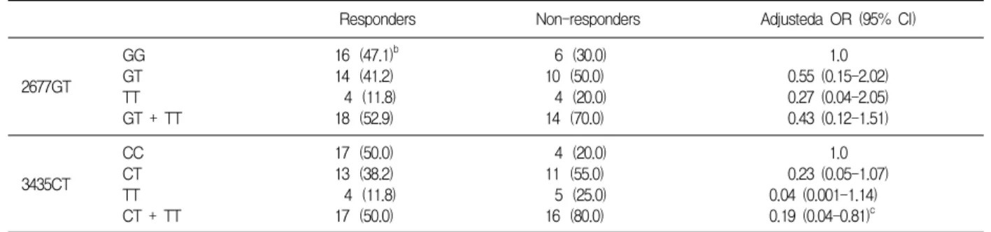
The distributions of MDR1 2677G>T and 3435C>
T genotypes among the responders and nonresponders are shown in Table 1. The 2677 GG genotype was more frequent in the responders (47.1%) than in the nonresponders (30.0%), and the 2677 GT and TT genotypes were less frequent in the responders (41.2%
and 11.8%, respectively) than in the nonresponders (50.0% and 20.0%, respectively), but these differences were not statistically significant. For the 3435C>T polymorphism, the 3435 CC genotype was associated with a significantly better chemotherapy response compared with the combined 3435 CT and TT genotype (P = 0.025).
The 2677G>T and 3435C>T polymorphisms were in linkage disequilibrium. The frequencies of the four haplotypes (G-C, G-T, T-C and T-T) among
Table 1. MDR1 2677GT and 3435CT genotypes and chemotherapy response
Responders Non-responders Adjusteda OR (95% CI)
2677GT
GG GT TT GT + TT
16 (47.1)b 14 (41.2)
4 (11.8) 18 (52.9)
6 (30.0) 10 (50.0) 4 (20.0) 14 (70.0)
1.0 0.55 (0.15-2.02) 0.27 (0.04-2.05) 0.43 (0.12-1.51)
3435CT
CC CT TT CT + TT
17 (50.0) 13 (38.2) 4 (11.8) 17 (50.0)
4 (20.0) 11 (55.0) 5 (25.0) 16 (80.0)
1.0 0.23 (0.05-1.07) 0.04 (0.001-1.14) 0.19 (0.04-0.81)c
a Adjusted for age, sex, stage and performance status.
b Numbers in parenthesis is percentage.
c P = 0.025.
Table 2. MDR1 haplotypes and chemotherapy response
Haplotypes Responders Non-responders Adjusteda OR (95% CI)
2677G-3435C 2677G-3435T 2677T-3435C 2677T-3435T 2677G-3435C Other haplotypes
44 (64.7)b 2 ( 2.9) 3 ( 4.4) 19 ( 27.9) 44 ( 64.7) 24 ( 35.3)
16 ( 40.0) 6 ( 15.0) 3 ( 7.5) 15 ( 37.5) 16 ( 40.0) 24 ( 60.0)
1.0 0.15 (0.02-0.97)c 0.30 (0.04-2.30) 0.44 (0.17-1.11)
1.0 0.34 (0.15-0.82)d
a Adjusted for age, sex, stage and performance status.
b Numbers in parenthesis is percentage.
c P = 0.047 and Bonferroni corrected P value = 0.188
d P = 0.015.
the overall cases were 55.6%, 7.4%, 5.6% and 31.5%, respectively. The haplotype distribution among the responders was significantly different from the ha
plotype distribution among the nonresponders (Table 2; G-C, G-T, T-C and T-T haplotype; 64.7%, 2.9%, 4.4% and 27.9% vs 40.0%, 15.0%, 7.5% and 37.5%, respectively; P = 0.03). Patients harboring the 2677G- 3435C haplotype had a significantly better response to chemotherapy compared with the chemotherapy response of the other haplotypes combined (P = 0.015).
Discussion
We investigated the association of MDR1 2677G
>T and 3435C>T polymorphisms and their haplotypes with the chemotherapy response in SCLC patients treated with a combination chemotherapy of EP. In the present study, the patients harboring the 2677G- 3435C haplotype responded to chemotherapy signi
ficantly better than those patients with other ha
plotypes. These results suggest that the MDR1 2677G>T and 3435C>T polymorphisms and their haplotype could be used to predict treatment response to etoposide-based chemotherapy.
As was stated in previous studies21,22, we also found that the 2677G>T and 3435C>T polymorphisms were in linkage disequilibrium. However, the allele frequencies of both polymorphisms among healthy Koreans (n = 371, the same subjects used as healthy controls in our previous study:Ref 23) differed from other ethnic populations. The frequency of the 2677T allele among healthy Koreans was 0.34, which was lower than the allele frequency in Chinese and Caucasians (0.44-0.50; and 0.38-0.46, respectively;
Ref. 15). The frequency of 3435T allele among healthy Koreans was 0.36, which was also lower than those in Chinese and Caucasians (0.40-0.54;
and 0.46-0.57, respectively; Ref. 15) but higher than
that in Africans (0.10-0.26; Ref. 15).
Several studies have reported that the MDR1 2677G>T and 3435C>T polymorphisms are associated with gene expression and function, but the results are inconsistent. Hoffmeyer et al.16reported that the 3435C>T polymorphism was associated with duodenal PGP levels in Caucasians. Individuals with the 3435 TT genotype had significantly lower duodenal MDR1 expression and higher plasma digoxin levels in comparison to individuals with the 3435 CC genotype. Johne et al.24also reported that the 3435 TT genotype was associated with higher digoxin levels. In contrast to these studies, Gerloff et al.25 reported no differences in digoxin levels among healthy Caucasian subjects carrying the 3435T allele or the 3435C allele. In addition, in a study that quantified MDR1 mRNA in duodenum, Nakamura et al.26 showed higher MDR1 mRNA levels in healthy Japanese subjects carrying the 3435T allele as compared to subjects with the 3435C allele. The controversy is not just limited to Asian populations:
a study performed by Illmer et al.27 found that the 3435 CC genotype was associated with lower MDR1 expression in acute myeloid leukemia blast samples. Conflicting data of a similar nature have also been reported for the 2677G>T polymorphism17,28. Kim et al.17 reported that the 2677T allele was associated with a 2-fold enhanced efflux of digoxin compared to the 2677C allele. In contrast, Tanabe et al.28 reported a non-significant opposite trend for PGP expression in placenta in relation to the 2677G
>T polymorphism (GG > GT > TT). In the present study, the 2677 GG genotype and 3435 CC genotype were associated with a better chemotherapy response.
In consistent with genotyping analyses, the 2677G- 3435C haplotype was significantly associated with a better chemotherapy response. These findings are in agreement with some reports17,26,27, but are disagree with others16,24,28. Although the reason for all these
discrepant results is currently unclear, it may be due to either that the regulation of PGP expression may be significantly different in different body tissues or that the methods used to measure PGP expression differed among different studies15,27. The variations in genetic backgrounds of the study subjects should also be taken into consideration.
MDR1 polymorphism may have an influence on disease risk, and/or disease progression29,30. In the present study, however, the frequencies of the 2677T and 3435T alleles among the SCLC cases were not significantly different from those among healthy Koreans (0.37 vs 0.34, and 0.39 vs 0.36, respectively). Moreover, no significant difference was observed in the genotype distributions of both polymorphisms between the patients with LD and with ED.
In conclusion, we found that the MDR1 2677G>T and 3435C>T polymorphisms and their haplotypes are associated with the response to EP combination chemotherapy in SCLC patients. Our finding suggests that these polymorphisms could be used as genetic markers for predicting treatment response to eto
poside-based combination chemotherapy in SCLC patients, although additional studies with a larger sample size are required to confirm our results.
Future studies for other MDR1 sequence variants and their biologic function are also needed to understand the role of MDR1 polymorphisms in determining the response to chemotherapy. Moreover, since genetic polymorphisms often vary significantly between different ethnic groups, further studies are warranted to clarify the association of MDR1 polymorphisms with chemotherapy response in diverse ethnic populations.
Acknowledgement
This study was supported in part by Medical
Research Institute grant, Kyungpook National University Hospital (1996).
References
1. Elias AD. Small cell lung cancer: state-of-the-art therapy in 1996. Chest 1997;112:251S-8S.
2. Simon GR, Wagner H. Small cell lung cancer. Chest 2003;123:259S-71S.
3. Gottesman MM, Fojo T, Bates SE. Multidrug resistance in cancer: role of ATP-dependent transporters. Nat Rev Cancer 2002;2:48-58.
4. Chen CJ, Chin JE, Ueda K, Clark DP, Pastan I, Gottesman MM, et al. Internal duplication and homology with bacterial transport proteins in the mdr1 (P-glycoprotein) gene from multidrug-resistant human cells. Cell 1986;47:381-9.
5. Dean M, Rzhetsky A, Allikmets R. The human ATP- binding cassette (ABC) transporter superfamily.
Genome Res 2001;11:1156-66.
6. Stouch TR, Gudmundsson O. Progress in under
standing the structure-activity relationships of P- glycoprotein. Adv Drug Deliv Rev 2002;54:315-28.
7. Holzmayer TA, Hilsenbeck S, von Hoff DD, Raninson IB. Clinical correlates of MDR1 (P-glycoprotein) gene expression in ovarian and small-cell lung carcinomas. J Natl Cancer Inst 1992;84:1486-91.
8. Poupon MF, Arvelo F, Goguel AF, Bourgeois Y, Jacrot M, Hanania N, et al. Response of small-cell lung cancer xenografts to chemotherapy: multidrug resistance and direct clinical correlates. J Natl Cancer Inst 1993;85:2023-9.
9. Campling BG, Young LC, Baer KA, Lam YM, Deeley RG, Cole SP, et al. Expression of the MRP and MDR1 multidrug resistance genes in small cell lung cancer. Clin Cancer Res 1997;3:115-22.
10. Kawasaki M, Nakanishi Y, Kuwano K, Yatsunami J, Takayama K, Hara N. Immunohistochemically detected p53 and P-glycoprotein predict the response to chemotherapy in lung cancer. Eur J Cancer 1998;
34:1352-7.
11. Hsia TC, Lin CC, Wang JJ, Ho ST, Kao A. Re
lationship between chemotherapy response of small cell lung cancer and P-glycoprotein or multidrug resistance-related protein expression. Lung 2002;180:
173-9.
12. Lutterbach B, Sun D, Schuetz J, Hiebert SW. The MYND motif is required for repression of basal transcription from the multidrug resistance 1 pro
moter by the t(8;21) fusion protein. Mol Cell Biol
1998;18:3604-11.
13. Nakayama M, Wada M, Harada T, Nagayama J, Kusaba H, Ohshima H, et al. Hypomethylation status of CpG sites at the promoter region and overexpression of the human MDR1 gene in acute myeloid leukemias. Blood 1998;92:4296-307.
14. Mickley LA, Spengler BA, Knutsen TA, Biedler JL, Fojo T. Gene rearrangement: a novel mechanism for MDR-1 gene activation. J Clin Invest 1997;99:1947- 57.
15. Marzolini C, Paus E, Buclin T, Kim RB. Polymor
phisms in human MDR1 (P-glycoprotein): recent advances and clinical relevance. Clin Pharmacol Ther 2004;75:13-33.
16. Hoffmeyer S, Burk O, von Richter O, Arnold HP, Brockmoller J, Johne A, et al. Functional polymor
phisms of the human multidrug-resistance gene:
multiple sequence variations and correlation of one allele with P-glycoprotein expression and activity in vivo. Proc Natl Acad Sci U S A 2000;97:3473-8.
17. Kim RB, Leake BF, Choo EF, Dresser GK, Kubba SV, Schwarz UI, et al. Identification of functionally variant MDR1 alleles among European Americans and African Americans. Clin Pharmacol Ther 2001;
70:189-99.
18. Fromm MF. The influence of MDR1 polymorphisms on P-glycoprotein expression and function in humans.
Adv Drug Deliv Rev 2002;54:1295-310.
19. World Health Organization. WHO handbook for reporting results of cancer treatment, WHO offset publication no. 48. Geneva: World Health Organization;
1979.
20. Stephens M, Smith NJ, Donnelly P. A new statistical method for haplotype reconstruction from population data. Am J Hum Genet 2001;68:978-89.
21. Tang K, Ngoi SM, Gwee PC, Chua JM, Lee EJ, Chong SS, et al. Distinct haplotype profiles and strong linkage disequilibrium at the MDR1 multidrug transporter gene locus in three ethnic Asian po
pulations. Pharmacogenetics 2002;12:437-50.
22. Kroetz DL, Pauli-Magnus C, Hodges LM, Huang CC, Kawamoto M, Johns SJ, et al. Sequence diversity and haplotype structure in the human ABCB1 (MDR1, multidrug resistance transporter) gene. Pharmacogenetics 2003;13:481-94.
23. Park SH, Lee GY, Jeon HS, Lee SJ, Kim KM, Jang SS, et al. -93G→A polymorphism of hMLH1 and risk of primary lung cancer. Int J Cancer 2004;112:
678-82.
24. Johne A, Kopke K, Gerloff T, Mai I, Rietbrock S, Meisel C, et al. Modulation of steady-state kinetics of digoxin by haplotypes of the P-glycoprotein
MDR1 gene. Clin Pharmacol Ther 2002;72:584-94.
25. Gerloff T, Schaefer M, Johne A, Oselin K, Meisel C, Cascorbi I, et al. MDR1 genotypes do not influence the absorption of a single oral dose of 1 mg digoxin in healthy white males. Br J Clin Pharmacol 2002;54:610-6.
26. Nakamura T, Sakaeda T, Horinouchi M, Tamura T, Aoyama N, Shirakawa T, et al. Effect of the mutation (C3435T) at exon 26 of the MDR1 gene on expression level of MDR1 messenger ribonucleic acid in duodenal enterocytes of healthy Japanese subjects. Clin Pharmacol Ther 2002;71:297-303.
27. Illmer T, Schuler US, Thiede C, Schwarz UI, Kim RB, Gotthard S, et al. MDR1 gene polymorphisms affect therapy outcome in acute myeloid leukemia patients. Cancer Res 2002;62:4955-62.
28. Tanabe M, Ieiri I, Nagata N, Inoue K, Ito S, Kanamori Y, et al. Expression of P-glycoprotein in human placenta: relation to genetic polymorphism of the multidrug resistance (MDR)-1 gene. J Pharmacol Exp Ther 2001;297:1137-43.
29. Siegsmund M, Brinkmann U, Schaffeler E, Weirich G, Schwab M, Eichelbaum M, et al. Association of the P-glycoprotein transporter MDR1(C3435T) poly
morphism with the susceptibility to renal epithelial tumors. J Am Soc Nephrol 2002;13:1847-54.
30. Drozdzik M, Bialecka M, Mysliwiec K, Honczarenko K, Stankiewicz J, Sych Z. Polymorphism in the P- glycoprotein drug transporter MDR1 gene: a possible link between environmental and genetic factors in Parkinson's disease. Pharmacogenetics 2003;13:259-63.