─ 201 ─ elSSN 2287-1683
plSSN 1738-8767
Journal of Trauma and Injury Vol. 27, No. 4, December, 2014
� Case Report �
� Address for Correspondence : Yang Bin Jeon, M.D.
Department of Thoracic and Cardiovascular Surgery, Gachon University Gil Hospital, Gachon University, 21 Namdong-daero 774 beon-gil, Namdong-gu, Incheon, Korea
Tel : 82-32-460-3010, Fax : 82-32-460-3019, E-mail : [email protected]
Submitted : August 11, 2014 Revised : August 31, 2014 Accepted : September 4, 2014
충돌 손상에 의한 첫 번째 늑골 골절에 동반된 Horner 증후군
가천대학교 의과대학 길병원 중증 외상센터 흉부외과, 1외과
마대성, 조현진, 이정남1, 전양빈
- Abstract -
Horner’s Syndrome after a Fracture of the First Rib Caused by a Crushing Injury
Dae Sung Ma, M.D., Hyun Jin Cho, M.D., Jung Nam Lee, M.D.
1, Yang Bin Jeon, M.D.
Department of Thoracic and Cardiovascular Surgery, Trauma Center, Gachon University Gil Hospital, Gachon University, Incheon, Korea
1
Department of Surgery, Trauma Center, Gachon University Gil Hospital, Gachon University, Incheon, Korea
Patients with Horner’s syndrome exhibit a variety of symptoms, including miosis, palpebral ptosis, and anhidrosis.
This syndrome is caused by interruptions of the sympathetic neural pathways.
This paper describes two cases of patients with Horner’s syndrome who experienced a first rib fracture after crushing injuries. [ J Trauma Inj 2014; 27: 201-3 ]
Key Words: Rib fracture, Horner’s syndrome, Trauma
I. Introduction
Patients with Horner’s syndrome exhibit a variety of symptoms, including miosis, palpebral ptosis, and anhidrosis. This syndrome is caused by interrup- tions of the sympathetic neural pathways. Horner’s syndrome-associated trauma is mostly known to affect second-order neurons with sympathetic innervation.(1)
First rib fractures, which are frequently docu- mented in cases of severe trauma, are rarely seen in patients with Horner’s syndrome.(2) Several cases of first rib fractures caused by motor vehicle acci- dents have been described in patients with Horner’s syndrome.(3-6)
This paper describes two cases of patients with Horner’s syndrome who experienced a first rib frac- ture after crushing injuries.
─ 202 ─
- Journal of Trauma and Injury Vol. 27, No. 4 -
II. Case
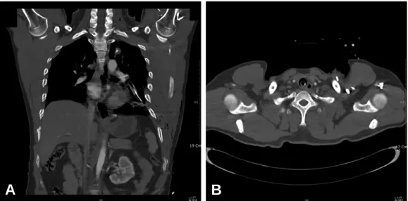
1. Case 1A 33-year-old man was transferred to the trauma center from a nearby hospital after his left shoulder was crushed to the waist area for 20 minutes by a press machine. Upon arrival at the hospital, the patient was intubated, wherein a chest tube was inserted into the left thorax. His vital signs were stable, but he presented in a stupor. He had subcu- taneous emphysema in the left chest wall and swelling in the right lower leg. A computed tomog- raphy (CT) scan of the brain did not reveal any abnormalities. A thoracic CT scan showed multiple bilateral rib fractures, including the left first rib (Fig. 1), and a left transverse process fracture in the thoracic spine from the 7th to 10thvertebrae. A radi- ograph of the right lower leg revealed distal tibia and fibular shaft fractures. He was admitted to the intensive care unit with intubation and sedation.
On the third day of admission, his mental status improved. The sedation was weaned, and the patient was extubated. He complained of anhidrosis on the left side of the face. A meticulous physical evalua- tion was performed, revealing additional signs of ptosis and miosis (Fig. 2). Horner’s syndrome after a first rib fracture was diagnosed. The patient was discharged 8 weeks later after orthopedic surgical treatment of the distal tibia and fibula and rehabili- tation. The symptoms of Horner’s syndrome were
managed conservatively, and they resolved over a period of 12 weeks.
2. Case 2
One month after a left brachial plexus injury, a 39-year-old man was transferred to our hospital through the outpatient department from a nearby hospital, as further evaluation was required. He had sustained a crushing injury to the upper thorax and pelvis from a forklift truck. He was conservatively managed for a pelvic bone fracture and multiple rib fractures, including a left first rib fracture. When he was transferred, he presented with paresthesia of the left hand, a limitation of the right ankle dorsi- flexion, and left-sided Horner’s syndrome. Magnetic resonance imaging of the brachial plexus revealed a probable rupture of the left T1 nerve root that was associated with the left first rib fracture and a pseudomeningocele at the left C9 nerve root. The symptoms of Horner’s syndrome resolved sponta- neously over a period of 8 weeks after the trauma.
Fig. 1. A computed tomographic scan of chest. Arrows indicate fracture of left first rib. (A) Coronal view (B) Axial view.
Fig. 2. Partial ptosis and miosis of left eye was presented.
A B
III. Discussion
Horner’s syndrome, which is also called ocu- losympathetic paresis, is a neurologic syndrome that consists of miosis, ptosis, and anhidrosis. It is caused by various conditions that affect the sympa- thetic pathway originating from the hypothalamus and supplying the head, eyes, and neck. The sympa- thetic innervation of the eye exhibits a three-order system.(1) The first-order neuron originates from the hypothalamus and descends to the ciliospinal center of Budge, which is located in the cervical spinal cord (levels C8-T2). The second-order neuron passes from the sympathetic trunk and terminates in the superior cervical ganglion, which is located near the bifurcation of the common carotid artery.
The third-order neuron travels from the superior cervical ganglion and innervates the iris dilator muscle and Mu
¨ller’s muscle. Among them, the sec- ond-order neuron, which crosses the area of the head and neck of the first rib posteriorly, is known to be vulnerable to upper thoracic trauma.(5,7)Traumatic Horner’s syndrome is not related to the severity of the trauma, but it may reveal associated injuries, including carotid artery dissection, or blunt injuries such as hematomas, cervical spine disloca- tion, and upper thoracic injuries such as first rib fracture or tension hemothorax.(7-9)
Cases with first rib fractures that are associated with Horner’s syndrome have rarely been reported.
Richardson et al. documented three cases of Horner’s syndrome in 55 patients with first rib fractures in 1972.
Among them, two cases exhibited complete recovery, while the symptoms persisted in the other case.(2)
Conservative management of Horner’s syndrome was possible in the patients with first rib fractures, although the period required for improvement of the symptoms differed from that described in previous literature.(3-6)
In Korea, several cases of Horner’s syndrome sec- ondary to injuries like cervical disc herniation, video-thoracoscopic surgery, or tube thoracostomy have been reported.(10-12) However, as per our knowledge, there have been no reports of Horner’s syndrome related to first rib fractures after a crushing injury.
All the cases sustained severe crushing injuries to the left upper thorax and presented symptoms of Horner’s syndrome that was associated with first rib fractures. During the follow up, the symptoms of Horner’s syndrome disappeared completely with conservative management.
Horner’s syndrome associated with first rib frac- tures caused by crushing injuries is a very rare con- dition. It may be managed conservatively if severe injuries often associated with it, such as carotid artery dissection and cervical spine dislocation are excluded.
REFERENCES