∙ Received: December 20, 2010. Accepted: February 28, 2011
∙ Corresponding author: Hoon Hee Park
Department of Nuclear Medicine, Severance Hospital, Yonsei University Health System, 250 Seongsanno, Seodaemun-gu, Seoul, 120-752, Korea
Tel: +82-2-2227-4481, Fax: +82-2-312-0578 E-mail: [email protected]
Original Article
PET/CT 장비 특성에 따른 방사성 의약품 주입량이방사선 종사자에게 미치는 영향
연세의료원 세브란스병원 핵의학과
박훈희⋅오기백⋅이승재⋅반영각⋅강천구⋅임한상⋅김재삼⋅이창호
A Study of Injection Dose for Patients and Exposure Dose for Technologists from the PET/CT Systems
Hoon Hee Park, Ki Beak Oh, Seung Jae Lee, Young Kag Bhan, Chun Goo Kang, Han Sang Lim, Jae Sam Kim and Chang Ho Lee
Department of Nuclear Medicine, Severance Hospital, Yonsei University Health System, Seoul, Korea
Purpose: It appears the different value when the injection dose is calculating for patients on each PET/CT systems. It directly affects the technologists’ radiation exposed dose. We studied the effect of the variable injection doses from several PET/CT systems to exposure dose for technologists. Materials and Methods: Six technologists have worked for 5 months through unit rotations with 3 PET/CT systems {Scanner 1 (S1): 0.15 mCi/kg, Scanner 2 (S2): 0.17 mCi/kg, Scanner 3 (S3): 0.12 mCi/kg}. Eighteen to 19 patients have had examinations per a day on each PET/CT systems. Examination parameters were adjusted to the same. TLDs were used for checking the exposure dose of technologists. Results: Each technologists’ the monthly average exposure dose was as follows; S1: 0.76 mSv, S2: 0.93 mSv, S3: 0.47 mSv. The maximum exposure dose was 1.12 mSv, and minimum was 0.42 mSv. The results showed significance in the correlation between the PET/CT system and the exposure dose (p<0.005). Conclusion: When the amount of injection dose was small, the exposure dose was decreased not only the patients but also the technologists. The exposure dose was decreased by the individual proficiency of technologists. However, the low injection dose can highly reduce the exposure dose for technologist so that there will be needed to following studies. (Korean J Nucl Med Technol 2011;15(1):45-50)
Key Words : PET/CT, Exposure Dose, Injection Dose
Introduce
Due to PET could measure the biological index quantitatively through imaginating many biochemical’s in vivo distribution in human body, it is using for determina- tion or diagnosis of biochemical or pathological phenomenon, prognosis after therapy, and therapy plan. The importance of
PET is increased recently. A period at the beginning of PET scan, it mainly used for brain examination.
1,2)However, it uses for diagnostic tool and evaluation of cancer. Although, PET is an appropriate diagnostic tool for the evaluation of a biological function and a highly applicative system. The image resolution is poor and hard to figure out the organs’
anatomical location.
3)These limitation could overcome through using with CT as PET/CT. PET/CT was developed in the late 1990s and has been successful in the early 2000s.
Each imaging devices set in parallel and CT scan performed
first and PET scan started. Therefore, the patients don’t have
to move and could have both imaging examination in the
A
B
C
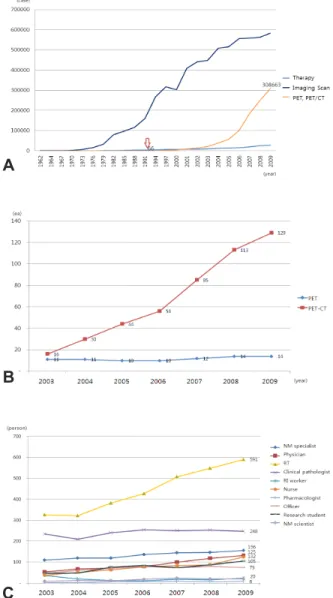
Fig. 1. Number of Annual Nuclear Medicine Examination Cases is on the graph A, PET and PET-CT have steadily grown from the late 1980. Annual Status of PET and PET/CT System is on the graph B, PET-CT have rapidly grown from the early 2000. Annual Status of Nuclear Medicine Worker is on graph C. As the status of PET-CT shows, the number of workers was heavily grown.
using
18F,
11C,
13N,
15O which have the less number of neutrons than the number of protons in the nucleus are an unstable radioactive isotopes. Radioisotope could explain as follows; when a proton converted to a neutron in nuclei, it emits positron to become in a stable state. The emitted positrons flow in a certain distance, and meet the electrons surrounding nuclei and destroyed. At this time, two gamma rays with the energy of 511 keV are emitted.
4)Radio- technologists had exposed from patients who had injections of radiopharmaceutical. Affecting factors are injected radio- pharmaceuticals volume and the energy, a bio-distribution of radiopharmaceuticals, the image acquisition time, the distance between patients and radio-technologists, and the high exposure of hands during the preparation of radiopharma- ceuticals.
5,6)However, the exposure dose from patient was the highest. Previously, and radiation exposure of technologists in nuclear medicine have been studied(Fig. 1).
As PET/CT sections are subdivided, there was a lack of the study, which kind of related factors of job evaluation and job function could affect radiation exposure.
7,8,9)An effort to reduce exposure dose for radio-technologists, and the matter of the heightened environmental awareness and safety are emerging.
10-12)Depending on the physical characteristics of PET/CT systems, the injected dose for patients were changed. In this study, the affection to radio-technologists from the injected dose of the patient in the different PET/CT systems was analyzed.
Materials and Methods
PET/CT systems
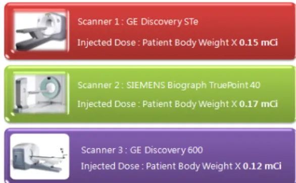
Three different PET/CT systems were enrolled; Scanner 1 [Discovery STe (General Electric Healthcare, Wisconsin, MI,
USA)], Scanner 2 [Biograph Truepoint 40 (Siemens Medical Systems, CTI, Knoxville, TN, USA)], Scanner 3 [Discovery STe (General Electric Healthcare, Wisconsin, MI, USA)]. The administrated radiopharmaceuticals were same(Fig. 2).
Injected dose
Based on the recommendation dose from each PET/CT
systems, the dose {Scanner 1 (S1) : 0.15 mCi/kg, Scanner 2
Fig. 3. Radiation workers were under the Unit rotation for 5 months, they divided into the group of ‘Senior’ and ‘Junior’.
Fig. 2. Scanner 1 [Discovery STe (General Electric Healthcare, Wisconsin, MI, USA)], Scanner 2 [Biograph Truepoint 40 (Siemens Medical Systems, CTI, Knoxville, TN, USA)], Scanner 3 [Discovery STe (General Electric Healthcare, Wisconsin, MI, USA)]
Works Sex Duration of Radiation Exposure Classification
A Male 12 year 4 month senior
B Male 7 year 2 month senior
C Male 7 year 1 month senior
D Male 3 year 6 month junior
E Male 1 year 9 month junior
F Male 1 year 1 month junior
Table 1. The number of Radiation workers were 6, who have the longest working experience was 12 years and 4 month, and the lowest was 1 year and 1 month.
(S2) : 0.17 mCi/kg, Scanner 3 (S3) : 0.12 mCi/kg} were injected to the patients.
Patients scanning
Patients were fasting at least 6 hours before the test. Takes approximately 10-15 minutes to relax before
18F-FDG injection and drink 500-1000 mL water and was injected intravenously. Motion was prohibited for preventing the uptake of muscle and then takes a rest for about 1 hour with lying before examination. Prior to examination, the patient urinated. And take a scout image in the supine position, After whole-body CT scan from skull base to the proximal femur, Average of 7 Bed PET emission testing were conducted during 1 Bed per 2 minutes 30 seconds.
Specificity of radiation workers
Six radiation-worker who had different experiences of radiation exposure were subjected, the radiation exposure dose were measured from August 2008 to December 2008(Fig. 3).
Rotation duty schedule
Six technologists (2 in 1 system) have worked for 6 months through unit rotations with 3 PET/CT systems. In rotation, Senior (A, B and C) and Junior (E, F and G) worked on the same month (Table 1).
Data analysis
A personal dosimetry of radio-technologists, TLD was measured monthly and the results were analyzed with on-parametric test (Kruskal-Wallis test) and multiple regression analysis (SPSS ver. 17).
Results
The individual exposure results were measured from monthly TLD data, there was a statistical significance (p : 0.003). The data were analyzed using the non-parametric test (Kruskal-Wallis test) (Fig. 4). On box plot, compare to the senior group (A, B and C), the radiation exposure dose of the junior group was higher and had various deviations.
Through this, we were confirmed a tendency that if the
worker had the long experience, the radiation exposure dose
Fig. 4. Individual radiation exposure : The individual exposure results were measured from monthly TLD data, there was a statistical significance (asym. p : 0.003). The data were analyzed using the non-parametric test (Kruskal-Wallis test).
Fig. 5. Box Plot : Compare to the senior group (A, B and C), the radiation exposure dose of the junior group was higher and had various deviations.
Fig. 6. Monthly exam case by PET/CT systems : The graph shows the number of examination cases on the different PET/CT systems. The examination cases were from 250 to 450. The deviation of examination cases showed a largest difference in Scanner 3.
Fig. 7. Monthly injection dose by PET/CT systems : The graph shows the amount of a monthly-consumed radiopharma- ceuticals. The injected dose was calculated with examination case and patients weight to have the whole amount of the used radiation dose. In Scanner 3, the examination cases were more than Scanner 1 and 2. But, it used the lower amounts of radiopharmaceuticals.
was lower(Fig. 5).
The number of examination cases were different on their schedule of PET/CT systems (250-450). The deviation of examination cases showed a largest difference in Scanner 3 (Fig. 6, 7). The injected dose was calculated with examination case and patients weight to have the whole amount of the used radiation dose. In Scanner 3, the examination cases were more than Scanner 1 and 2, it used the lower amounts of radiopharmaceuticals (Fig. 8).
Through a descriptive statistics, the average of monthly individual radiation exposure dose in each Scanner was obtained. The workers of Scanner 3 showed the lowest exposure rate, and Scanner 1 showed the highest exposure rate. However, these data just showed exposure rate so that
the many factors which could affect to exposure rate has not concerned. Therefore, Body Weight, Total Number of Examinations, and whole amount of injected dose were concerned and did a multiple regression analysis. Individual exposure dose in Scanner 1 increased 0.260 mSv compare to Scanner 3, and 0.399 mSv was decreased in Scanner 2. The factor in these calculation described 62.2 % of the individual radiation exposure dose(Fig. 9).
Conclusion
As the limitation of this study, only several workers were
on the rotation, and radiation-workers experience was not
Fig. 8. Monthly radiation exposure by PET/CT : Through a descriptive statistics, the average of monthly individual radiation exposure dose in each Scanner was obtained. The workers of Scanner 3 showed the lowest exposure rate, and Scanner 1 showed the highest exposure rate.
Fig. 9. Body Weight, Total Number of Examinations, and whole amount of injected dose were concerned and did a multiple regression analysis. Individual exposure dose in Scanner 1 increased 0.260 mSv compare to Scanner 3, and 0.399 mSv was decreased in Scanner 2. The factor in these calculation described 62.2% of the individual radiation exposure dose.
various so that it had limitations to include several meaningful factors. We focused on the analysis a tendency of working experience. Under the same working condition, radiation-workers who had comparatively long experience showed low radiation exposure rate. The exposure dose was decreased by the individual proficiency of radiation-workers, and the low amount of the injection dose can highly reduce the exposure dose for radiation-workers. Therefore, when the Nuclear Medicine examination performed, it is necessary to use certain amount of radiopharmaceuticals and to be recommended proper and quick use by radiation-workers.
요 약
PET/CT검사에서
18F-FDG가 가장 널리 이용되며, 장비의 물리적 특성에 따라 환자 주입
18F-FDG량이 다르게 권고되 고 있다. 또한, 검사 특성상 방사선종사자와 환자의 접촉으로 인하여 방사선의 피폭이 불가피하기에, 본 연구에서는 각기 다른 PET/CT 장비를 대상으로 환자에게 주입되는
18F-FDG 가 방사선 종사자에게 미치는 피폭선량과의 관계를 분석하 였다 . 총 3대의 각각 다른 PET/CT (Scanner 1 (S1) : 0.15 mCi/kg, Scanner 2 (S2) : 0.17 mCi/kg, Scanner 3 (S3) : 0.12 mCi/kg)를 대상으로 각 장비에 숙련도를 고려하여 총 6명의 방사선종사자를 5개월간 순환근무 하였고, 하루에 검사하는 환자수를 일정하게 유지하였다. 또한, 검사 진행 방법을 유사 하게 유지하고, 방사선종사자의 개인피폭선량계인 열형광유 리선량계 (TLD)를 매월 판독 하여 분석하였다. 개인의 월별 평균 피폭선량은 장비에 따라 S1은 0.76 mSv, S2는 0.93 mSv, S3는 0.47 mSv였다. 피폭선량은 개인 최대 1.12 mSv, 최저
0.42 mSv로 숙련도와 경험에 따라 유의한 차이를 보였고, 또 한 각 주입량에 따른 PET/CT의 종류에 따라 피폭선량은 유 의한 상관관계를 나타냈다. 본 연구를 통하여 주입
18F-FDG 가 적을수록 방사선종사자의 피폭선량이 낮았다. 또한, 개인 숙련도에 따라 피폭선량이 감소하였으나, 장비의 특성에 따 라 적은 방사성의약품 주입량의 영향이 방사선종사자의 피 폭선량을 현저하게 감소할 수 있기에 이에 대한 연구가 보다 활성화 되어야 할 것이다.
REFERENCE
1. Benatar NA, Cronin BF, O'Doherty MJ. Radiation dose rates from patients undergoing PET: implication for technologists and waiting areas. Eur J Nucl Med 2000;27(5):583-589.
2. Cherry SR, Sorenson JA, Phelps ME. Physics in nuclear medicine.
3rd ed. Saunders 2003. p. 325-329.
3. Gambhir SS. A tabulated summary of the FDG PET literature. J Nucl Med 2001;42(suppl 5):1S-79S.
4. Guillet B, Quentin P, Waultier S, Bourrelly M, Pisano P. Olivier Mundler. Techologist radiation exposure in routine clinical practice with 18F-FDG PET. J Nucl Med Technol 2005;33:175-179.
5. Hasegawa BH, Iwata K, Wong KH. Dual - modality imaging of function and physiology. Acad Radiol 2002;9:1305-21.
6. Kim HJ. Current status of imaging physics and instrumentation in nuclear medicine. Nucl Med Mol Imaging 2008;42(2):83-87.
7. Hendee WR. Real and perceived risks of medical radiation exposure.
West J Med 1998;11(1):380-386.
8. ICRP Report 60. Recommendation of the international commission on radiological protection. Pergamon Press, 1990.
9. Kearns WT, Urbanic JJ, Hampton CJ, McMullen KP, Blackstock AW, Stieber VW, Hinson WH. Radiation safety issues with positron-emission/computed tomography simulation for stereotactic