∙Received: July 2, 2013. Accepted: August 27, 2013.
∙Corresponding author : Yong Gwi Cho
Department of Nuclear Medicine, Inha University Hospital, 27 Inhang-ro, Jung-gu, Incheon 400-711, Korea
Tel: +82-32-890-3770, Fax: +82-32-890-3164 E-mail: [email protected]
Original Article 융합영상(Fusion image)에서 움직임을 줄이기 위한
보정기구의 개발
인하대학교병원 핵의학과
조용귀⋅표성재⋅김봉수⋅신채호⋅조진우⋅김창호
Development of Supplemental Equipment to Reduce Movement During Fusion Image Acquisition
Yong Gwi Cho, Sung Jae Pyo, Bong Su Kim, Chae Ho Shin, Jin Woo Cho and Chang Ho Kim Department of Nuclear Medicine, Inha University Hospital, Incheon, Korea
Purpose: Patients' movement during long image acquisition time for the fusion image of PET-CT (Positron Emission Tomography-Computed Tomography) results in unconformity, and greatly affects the quality of the image and diagnosis. The arm support fixtures provided by medical device companies are not manufactured considering the convenience and safety of the patients; the arm and head movements (horizontal and vertical) during PET/CT scan cause defects in the brain fundus images and often require retaking. Therefore, this study aims to develop patient-compensation device that would minimize the head and arm movements during PET/CT scan, providing comfort and safety, and to reduce retaking. Materials and Methods: From June to July 2012, 20 patients who had no movement-related problems and another 20 patients who had difficulties in raising arms due to shoulder pain were recruited among the ones who visited nuclear medicine department for PET Torso scan. By using Patient Holding System (PHS), different range of motion (ROM) in the arm (25
o, 27
o, 29
o, 31
o, 33
o, 35
o) was applied to find the most comfortable angle and posture. The manufacturing company was investigated for the permeability of the support material, and the comfort level of applying bands (velcro type) to fix the patient's head and arms was evaluated. To find out the retake frequency due to movements, the amount of retake cases pre/post patient-compensation were analyzed using the PET Torso scan data collected between January to December 2012. Results: Among the patients without movement disorder, 18 answered that PHS and 29
oarm ROM were the most comfortable, and 2 answered 27
oand 31
o, respectively. Among the patients with shoulder pain, 15 picked 31
oas the most comfortable angle, 2 picked 33
o, and 3 picked 35
o. For this study, the handle was manufactured to be adjustable for vertical movements. The material permeability of the patient-compensation device has been verified, and PHS and the compensation device were band-fixed (velcro type) to prevent device movements. A furrow was cut for head fixation to minimize the head and neck movements, fixing bands were attached for the head, wrist, forearm, and upper arm to limit movements. The retake frequency of PET Torso scan due to patient movements was 11.06% (191 cases/1,808 patients) before using the movement control device, and 2.65% (48 cases/1,732 patients) after using the device; 8.41% of the frequency was reduced. Conclusion: Recent change and innovation in the medical environment are making expensive medical image scans, and providing differentiated services for the customers is essential. To secure patient comfort and safety during PET/CT scans, ergonomic patient-compensation devices need to be provided. Therefore, this study manufactured a patient- compensation device with vertically adjustable ergonomic ROM according to the patient's body shape and condition during PET Torso scan. The defects in the basal ganglia images due to arm movements were reduced, and retaking was decreased. (Korean J Nucl Med Technol 2013;17(2):84-89)
Key Words : PET Torso, Patient compensation device
서 론
오늘날 핵의학는 Roentgen이 X-선을 발견한 이후 1913년 Hevesy와 Paneth에 의해 처음으로 방사성 추적자의 원리에 대하여 규명하였고, 1920년 Blumgart에 의해 방사성 추적자 를 이용 임상 연구를 시행하였다.1)
이처럼 핵의학적 영상 검사는 방사성의약품의 끊임없는 연구와 첨단 의료기기의 발달로 임상적 진단 및 치료 결정에 매우 중요한 역할을 담당하고 있다.
최근 PET (positron emission tomography)나 SPECT (single photon emission computed tomography)를 이용한 신체의 포 도당 대사나 심장의 혈류 같은 기능적 정보와 CT (computed tomography)나 MRI (magnetic resonance imaging)를 이용한 해부학적 정보를 얻어 이를 정합시킨 융합영상(fusion image) 을 통하여 각종 질환을 조기진단 및 정확한 진단이 가능해 졌다.
융합영상은 비교적 긴 영상 획득 시간에 의한 환자의 움직 임으로 CT 영상과 융합에서 부정합한 결과로 영상의 질과 진단에 큰 영향을 준다.
지금까지 의료장비 회사에서 제공하는 팔 지지대는 인체 의 체형과 팔을 올린 자세에서의 편의성과 안전성을 전혀 고 려하지 않아 PET/CT를 검사하는 동안 팔과 머리의 움직임 으로 재촬영이 많았다. 이러한 환자의 움직임은 PET/CT 융합 영상에서 뇌기저부와 안면부의 영상 결손으로 재촬영에 의 한 불필요한 검사시간과 환자의 피폭선량이 발생되고 있다.
최첨단 과학의 발달로 의료영상 기술이 향상되고 있는 가 운데 의료기관을 내원하는 환자 또는 보호자는 수준 높은 의 료서비스를 요구하고 있다.
따라서 이들이 원하는 최고의 서비스를 제공하기 위해서 는 의료 환경과 시설확충 뿐만 아니라 환자에게 안전성과 편 의성을 고려한 신속, 정확한 검사로 환자중심의 감동을 주는 의료서비스를 구축해야 한다.
이에 우리는 두경부와 팔, 다리의 움직임을 최소화 할 수 있는 환자보정기구를 개발하여 PET/ CT 촬영 시 환자에게 편의성과 안전성을 제공하고 환자의 움직임으로 인한 재촬 영을 줄여 불필요한 피폭선량을 줄이고자 한다.
실험재료 및 방법 1. 대 상
2012년 6월부터 7월까지 PET Torso 검사를 위해 본원을
내원한 환자 중 팔을 올릴 때움직임에 전혀 불편이 없는 환자 20명과 움직임에 불편이 있는 환자 20명을 대상으로 하 였다.
2. 실험장비 및 재료
1) Discovery 690 PET/CT (GE), Biograph Duo (SIEMENS) 2) 영보드(고경도 스폰지), 플라스틱 손잡이
3. 실험 방법
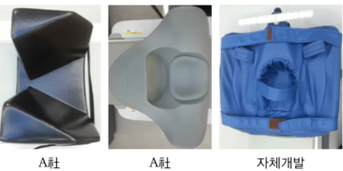
1) 장비회사별 PET Torso用 보정기구(Fig. 1)
A社 A社 자체개발
Fig. 1. Patient compensation device for PET Torso scan.
2) 검사대(PHS)와 환자 팔의 경사도(25o, 27o, 29o, 31o, 33o, 35o)를 변화시켜 가장 편한 자세의 경사도를 측정하 였다(Fig. 2).
Fig. 2. PHS and patient arm angle change.
3) PET Torso用 환자보정기구 제작 과정(Fig. 3)
Fig. 3. Making of Patient compensation device.
4) PET Torso用 & Lower extremity用 환자보정기구 구조 및 명칭(Fig. 4-1, 2)
Fig. 4-1. Structure of patient compensation device for PET Torso scan.
Fig. 4-2. Structure of patient compensation device for Lower ex- tremity scan.
5) PET/CT 검사 시 환자의 머리와 팔, 다리 등을 밴드(벨 크로 형태)로 고정하여 이에 대한 편의성과 안전성을 질의
하였다(Fig. 5-1, 2).
Fig. 5-1. Comfort of Patient head and arm position.
Fig. 5-2. Comfort of Patient Lower extremity position.
결 과
1. 환자보정기구 경사도 설문
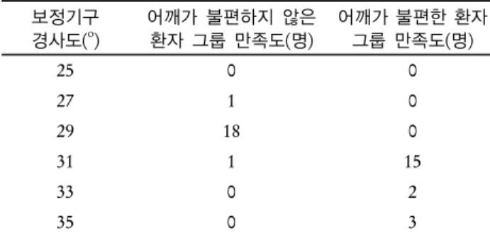
환자 어깨 상태에 따른 보정기구 경사도를 설문한 결과 움 직임에 불편이 전혀 없는 환자그룹은 PHS (Patient Holding System)와 팔의 경사도가 29o에서, 팔을 올리기가 불편한 환 자 그룹은 31o에서 대부분 편안하다고 답했다(Table 1).
Table 1. Patient compensation device angle change according to patient shoulder condition (N=40)
보정기구 경사도(o)
어깨가 불편하지 않은 환자 그룹 만족도(명)
어깨가 불편한 환자 그룹 만족도(명)
25 0 0
27 1 0
29 18 0
31 1 15
33 0 2
35 0 3
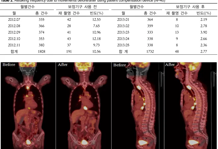
Table 2. Retaking frequency due to movements before/after using patient compensation device (N=40)
월별건수 보정기구 사용 전 월별건수 보정기구 사용 후
월 총 건수 재 촬영 건수 빈도(%) 월 총 건수 재 촬영 건수 빈도((%)
2012.07 335 42 12.53 2013.01 364 8 2.19
2012.08 366 28 7.65 2013.02 359 10 2.78
2012.09 374 41 10.96 2013.03 333 13 3.90
2012.10 353 43 12.18 2013.04 338 9 2.66
2012.11 380 37 9.73 2013.05 338 8 2.36
합계 1808 191 10.56 합 계 1732 48 2.77
Fig. 6. Head and neck images defected due to patient arm movements.
2. 재 촬영 빈도 조사
자체 제작한 환자보정기구 사용 전/후 움직임에 의한 재촬영 빈도를 조사한 결과 재 촬영 건수가 191건(10.56%)에서 48건 (2.77%)으로 사용 전 보다 143건(7.79%) 줄었다(Table 2).
3. 재 촬영 영상
1) PET/CT Torso 촬영 시 환자 팔의 움직임에 의한 두 경 부의 결손 영상(Fig. 6).
2) PET/CT Torso 촬영 시 환자 두 경부의 움직임에 의한 결손 영상(Fig. 7).
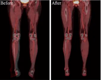
3) PET/CT Lower extremity 촬영 시 움직임에 의한 결손 영상(Fig. 8).
고찰 및 결론
최첨단 영상기술의 발달로 임상적 진단의 가치가 한층 높
아진 가운데 의료기관을 내원하는 환자들은 서비스에 대한 기대 수준이 점차 높아져 본인만의 아주 특별한 의료서비스 (편의성, 안정성 등)를 요구하고 있다.
이처럼 의료 환경의 변화와 혁신으로 고가의 영상검사 (PET/CT, SPECT/CT 등)가 늘어남에 따라 환자 또는 보호 자가 요구하는 차별화된 서비스 제공은 필수적이다. 따라서 환자에게 PET/CT 검사하는 동안 효율적이고 효과적인 서비 스를 제공하기 위해 최우선 과제는 안전성이 확보되어야 하 며 편안함을 도모할 수 있는 환경을 만들어야 한다.
PET/CT는 검사 고유의 특성상 방사성의약품을 투여 후 1시간 동안 안정을 취하고 촬영을 시행하므로 타 검사에 비 하여 비교적 장시간(약 1시간30분)을 한곳에 머물러야 하기 때문에 환자에게 체감되는 의료서비스의 질은 중요한 문제 이다.
PET/CT 검사 시 환자의 안정성과 편의성을 고려한 인체 공학적 보정기구를 개발하여 적용함으로서 검사하는 동안 불편함을 최소화하여 안전하고 정확한 검사의 결과를 얻어 야 한다.
Fig. 7. Images defected due to head and neck movements.
Fig. 8. Images defected due to Lower extremity movements.
그러나 이들 검사는 고가의 검사임에도 불구하고 검사과 정의 안전성과 편의성이 미흡한 현 시점에서 환자가 체감되 는 서비스는 현저히 저하되고 있다.
이러한 의료 환경에서 PET/CT를 검사하는 환자들의 체형 과 상황에 따라 인체공학적인 경사도(움직임에 불편이 없는 환자그룹; 29o, 팔을 올렸을 때 불편이 있는 환자그룹; 31o)를 고려한 환자보정기구를 개발하여 팔의 경사도를 상, 하 조절 하고 손잡이를 통한 팔의 움직임을 최소화 하였다.
또한 팔의 움직임에 의한 뇌기저부의 영상 결손을 줄이고, 머리와 다리의 움직임으로 인한 재촬영 빈도를 줄여 환자와 작업종사자의 불필요한 피폭선량 감소와 촬영 시간을 단축 시킬 수 있다.
현대의학에서 첨단장비 도입으로 각종 질환을 조기에 발 견함으로서 의료의 질적 향상을 이루고 있으나, 환자에게 차 별화된 최고의 서비스를 제공하기 위해서는 환자의 편의성
과 안전성이 확보되어야 하기 때문에 환자보정기구 개발은 그 시사점이 매우 크다.
요 약
PET-CT (Positron Emission Tomography-Computed Tomo- graphy)의 융합영상(Fusion image)에서 긴 영상 획득 시간에 의한 환자의 움직임은 융합영상에서 부적합한 결과로 나타 나 영상의 질과 진단에 큰 영향을 준다. 의료장비 회사에서 제공하는 팔 지지대는 환자 자세에서의 편의성과 안전성을 고려하지 않아 PET/CT를 검사하는 동안 팔과 머리의 움직 임 때문에 뇌기저부의 영상 결손이 생기고 머리의 좌우, 상 하 움직임으로 인하여 재촬영이 빈번하다. 이에 우리는 두경 부와 팔의 움직임을 최소화할 수 있는 환자보정기구를 개발 하여 PET/CT 촬영 시 환자에게 편의성과 안전성을 제공하고 환자의 움직임으로 인한 재촬영을 줄이고자 한다.
2012년 6월부터 7월까지 PET Torso 검사를 위해 핵의학 과에 내원하는 환자들 중 움직임에 불편함이 전혀 없는 환자 20명과 어깨 통증으로 팔을 올리기가 불편한 환자 20명을 대 상으로 Patient Holding System (PHS)와 팔의 경사도(25o, 27o, 29o, 31o, 33o, 35o)를 변화시켜 가장 편안한 자세와 경사 도를 선정 하였다. 또한 지지대 재질의 투과성에 대하여 유 관업체의 검증을 하였고, 환자의 머리와 팔을 밴드(벨크로 형태)로 고정하여 이에 대한 편의성을 질의하였다.
그리고 움직임에 의한 재촬영 빈도를 알아보기 위해 2012 년 1월부터 12월까지 PET Torso를 시행한 환자들 중 환자보 정기구 사용 전/후 재촬영 여부를 분석하였다.
움직임에 불편이 전혀 없는 환자에서는 18명이 PHS와 팔 의 각도가 29o에서 가장 편안함을 느낀다고 답하였으며, 2명
은 각각 27o, 31o이라고 답하였다. 어깨 통증을 느끼는 환자 에서는 15명이 31o에서 가장 편안함을 느낀다고 답하였고, 33o에서 2명, 35o에서 3명이었다. 이를 위해 손잡이 부분이 상, 하 움직임이 가능하도록 제작하였다. 환자보정기구 재질의 투과성은 검증된 상태이며, 환자보정기구 자체의 움직임을 방지하기 위하여 PHS와 보정기구를 밴드(벨크로 형태)로 고 정시켰다. 두경부의 움직임을 최소화할 수 있는 홈을 만들고 머리를 고정할 수 있게 고정밴드를 부착 하였고, 손목 및 전 완과 상완의 움직임을 제한할 수 있는 고정밴드를 부착하여 팔의 흔들림으로 인한 움직임을 최소화 하였다.
PET Torso 촬영 시 움직임에 의한 재촬영 빈도는 환자고 정기구 사용 전 10.56% (191건/1,808명), 사용 후 2.77% (48 건/1,732명)로 7.79%의 발생 빈도를 줄일 수 있었다.
최근 의료 환경의 변화와 혁신으로 고가의 영상검사가 늘 어남에 따라 고객이 요구하는 차별화된 서비스 제공은 필수 적이다. 따라서 PET/CT를 검사하는 동안 환자의 편안함과 안전성을 확보하기 위해 인체공학적인 환자보정기구를 제공
하여야 한다. 따라서 본 연구를 통해 PET Torso 검사 시 환 자의 체형과 상황에 따라 인체공학적 경사도를 상, 하 조절 할 수 있는 환자보정기구를 제작하였으며, 팔의 움직임에 의 한 뇌기저부의 영상결손을 줄이고 머리의 움직임으로 인한 재촬영을 줄일 수 있었다.
REFERENCES
1. 정준기, 고창순. 핵의학. 제3판. 고려의학 2008;1-4.
2. Martin A Lodge. Effect of patient arm motion in whole-body PET/CT. J Nucl Med 2011;52:1891-1897.
3. Tomas Beyer, Andreas Bockish. Whole body 18F- FDG PET/CT in the presence of truncation artifacts. J Nucl Med 2006;47:91-99.
4. Carlos A Garcia, Giuseppe Esposito. 18F-FDG hemispheric cerebral hypometabolic activity:patient motion artifact. J Nucl Med Technology 2006;34:86-87.
5. Waheeda Sureshbabu. CNMT, PET, Osama Mawlawi, PET/CT imaging artifacts. J Nucl Med Technology 2005;33:156-161.