∙Received: March 28, 2014. Accepted: April 21, 2014.
∙Corresponding author : Seung Woo Jung
Department of Nuclear Medicine, Dong-a University Hospital, Daesingongwon-ro, Seo-gu, Busan 602-715, Korea
Tel: +82-51-240-5633, Fax: +82-51-242-7237 E-mail: [email protected]
Original Article
PET/CT 저선량 적용 시 CT 감쇠보정법에 따른 피폭선량
저감효과
동아대학교병원 핵의학과
정승우⋅김홍균⋅권재범⋅박성욱⋅김명준⋅신영만⋅김영헌
Effect of the Dose Reduction Applied Low Dose for PET/CT According to CT Attenuation Correction Method
Seung Woo Jung, Hong Kyun Kim, Jae Beom Kwon, Sung Wook Park, Myeong Jun Kim, Yeong Man Sinand Yeong Heon Kim
Department of Nuclear Medicine, Dong-a University Hospital, Busan, Korea
Purpose: Low dose of PET/CT is important because of Patient’s X-ray exposure. The aim of this study was to evaluate the effectiveness of low-dose PET/ CT image through the CTAC and QAC of patient study and phantom study. Materials and Methods: We used the discovery 710 PET/CT (GE). We used the NEMA IEC body phantom for evaluating the PET data corrected by ultra-low dose CT attenuation correction method and NU2-94 phantom for uniformity. After injection of 70.78 MBq and 22.2 MBq of 18 F-FDG were done to each of phantom, PET/CT scans were obtained. PET data were reconstructed by using of CTAC of which dose was for the diagnosis CT and Q. AC of which was only for attenuation correction. Quantitative analysis was performed by use of horizontal profile and vertical profile. Reference data which were corrected by CTAC were compared to PET data which was corrected by the ultra-low dose. The relative error was assessed. Patients with over weighted and normal weight also underwent a PET/CT scans according to low dose protocol and standard dose protocol. Relative error and signal to noise ratio of SUV were analyzed. Results: In the results of phantom test, phantom PET data were corrected by CTAC and Q.AC and they were compared each other. The relative error of Q.AC profile was been calculated, and it was shown in graph. In patient studies, PET data for overweight patient and normal weight patient were reconstructed by CTAC and Q.AC under routine dose and ultra-low dose. When routine dose was used, the relative error was small. When high dose was used, the result of overweight patient was effectively corrected by Q.AC. Conclusion: In phantom study, CTAC method with 80 kVp and 10 mA was resulted in bead hardening artifact. PET data corrected by ultra- low dose CTAC was not quantified, but those by the same dose were quantified properly. In patients’ cases, PET data of over weighted patient could be quantified by Q.AC method. Its relative difference was not significant. Q.AC method was proper attenuation correction method when ultra-low dose was used. As a result, it is expected that Q.AC is a good method in order to reduce patient’s exposure dose. (Korean J Nucl Med Technol 2014;18(1):127-133)
Key Words : PET/CT, CTAC, Q.AC, Low dose
서 론
PET/CT 검사 시 발생하는 감마선은 매질을 통과할 때 에너지 영역에 따라 광전효과, 컴프턴 산란 등으로
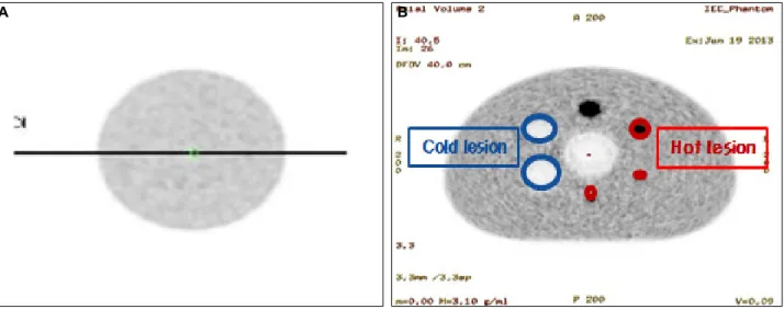
Fig. 2. Uniform NU2-94 phantom (A), NEMA IEC Body phantom (B) were used.
Fig. 3. (A), (B) Quantitative analysis was performed by use of horizontal profile and vertical profile.
Fig. 1. PET/CT Discovery 710 were used.
인해 검출기에 도달하기 전 감쇠되어 지는데 그 결과 실제 포도당 대사분포와 다른 양상을 보이며 정량적 계수가 이루어지지 않는다. 이로 인한 PET영상의 인 공물(artifact)를 없애기 위해 현재 대부분의 임상에서 CT 데이터를 이용하여 감쇠보정을 해주고 있다.1) 그 러나 환자의 피폭경감을 위한 저 선량에서의 감쇠보정 은 PET영상의 선속경화현상을 발생시킨다.2) 본 논문 에서는 현재 보편적으로 사용되고 있는 CT 데이터를 이용한 감쇠보정법인 CT감쇠보정(attenuation correc- tion : CTAC)와 이번에 박스카 알고리즘이3) 적용 된 PET/CT Discovery 710 (GE Healthcare, USA)의 감쇠 보정법인 Quantitative attenuation correction (Q.AC) 의 비교 실험를 통하여 임상에서의 피폭선량 저감방안 에 대하여 알아보고자 한다.
실험재료 및 방법 1. PET/CT장비 및 팬텀
이 실험에 사용한 장비는 PET/CT Discovery 710 (GE Healthcare, USA)이고(Fig. 1) 크리스탈(crystal)은 LBS (lutetium based scintillator)섬광체가 적용되었다. 팬텀은 두 가지로 NEMA IEC Body Phantom과 Uniform NU2-94 Phantom을 사용하였으며 각각 F-18 FDG를 70.78 MBq (1.91 mCi)와 70.2 MBq (0.6 mCi) 주입하였다(Fig. 2).
2. 실험 및 평가
실험의 신뢰성 향상을 위해 팬텀실험과 임상실험을 병행하였으며 선량에 따른 감쇠보정능력을 평가하기 위해 저 선량 조건과 일반선량 조건을 각각 설정하여 두 감쇠보정법을 비교하였다.
A B
A B
Fig. 4. PET/CT scans according to low dose protocol and standard dose protocol. Relative error and signal to noise ratio of SUV were analyzed.
1) 팬텀 실험
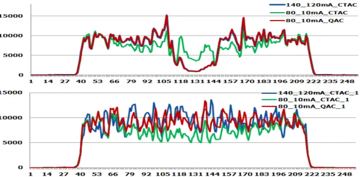
본 논문에서는 두 가지의 팬텀실험을 하였다. 첫 번째 균일성을 보기위해 Uniform NU2-94 Phantom내부에 F-18 FDG 22.2 MBq (0.6 mCi)를 물과 함께 주입한 뒤 CT스캔은 저 선량조건으로 80 kVp 10 mA로부터 일 반선량 조건으로 140 kVp 120 mA까지 단계별 투과스 캔을 진행하고 10분간 PET방출스캔 영상을 획득하였 다. 획득한 영상의 재구성 방법은 VUE point FX를 사 용하였으며 subset 16 iteration 3, 4 mm FWHM Gaussian post filter, matrix size 256×256 , DFOV 50 cm로 재구 성하였다. 영상의 분석은 일반선량 조건으로 촬영된 영상을 기준으로 잡고 저 선량조건에서의 두 감쇠보정 법이 적용된 PET 데이터를 profile한 뒤 기준값에 대한 상대적 오차를 비교하여 균일성 평가를 하였다. 두 번 째 정량성 평가를 위해 NEMA IEC Body phantom을 이용하여 구의 지름이 10, 13, 17, 22 mm는 열소(hot lesion)으로 설정하였고 28, 37 mm는 냉소(cold lesion) 으로 구분하였다. 내부에 물을 가득 채우고 동위원소 F-18 FDG를 70.78 MBq (1.91 mCi) 주입하고 열소 대 백그라운드 비율이 4:1로 팬텀을 채웠다. CT스캔 조건은 저 선량 조건으로 80 kVp 10 mA로부터 일반선량 조 건으로 140 kVp 120 mA까지 단계별 투과스캔을 진행 하고 20분간 PET방출스캔 영상을 획득하였다. 획득한 영상의 재구성 방법은 VUE point HD를 사용하였으며 subset 24, iteration 4, 2 mm FWHM Gaussian post filter, matrix size 256×256, DFOV 40 cm로 재구성하였다. 분 석 방법은 두 가지로 첫째, 획득한 PET영상에서 hori-
zontal profile과 vertical profile을 통해 location pixel당 count 수를 그래프화하여 정량성 평가를 하였다. 둘째, 일반선량조건에서의 선 profile값을 기준으로 잡고 저 선 량에서 두 감쇠보정법이 적용된 영상의 열소, 냉소, 백 그라운드의 상대적 오차(relative error %)를 비교하였다 (Fig. 3).
2) 환자 실험
대상 환자는 비교를 위하여 체질량지수(BMI) 30 이 상의 과체중군과 30 이하의 정상 체중군으로 나누어 촬영하였다. F-18 FDG를 몸무게(kg)×0.14로 계산하여 주입 후 1시간 뒤 Rotation speed: 0.50 s/HE, Pitch:
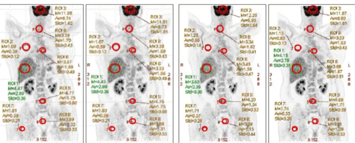
0.984:1, 일반선량 조건 120 kVp, automA 100-240 mA로 촬영하고 저 선량 조건에서는 100 kVp automA 10-20 mA 로 촬영하였으며 그 외의 조건들은 일반 PET/CT검사 방법과 동일하게 적용하였다. 환자데이터의 분석은 일반 선량와 저 선량으로 비교 촬영하여 재구성 된 PET영 상에서 GE사의 분석프로그램인 PET VCAR (GE Healthcare, USA)를 사용하여 뇌(brain), 간(liver), 폐(lung) 등의 주 요부위 8군데에 관심영역(resion of interest)를 그린 뒤 SUVmax, SUVmean에 대한 상대적 오차와 신호 대 잡음 비(SNR)을 비교 분석하였다(Fig. 4).
결 과
1. 팬텀 실험
Fig. 5. In the results of NU2-94 phantom test, phantom PET data were corrected by CTAC and Q.AC and they were compared each other. The relative error of Q.AC profile was been calculated, and it was shown in graph.
Fig. 6. In the results of NEMA IEC Body phantom test, phantom PET data were corrected by CTAC and Q.AC and they were compared each other. The relative error of Q.AC profile was been calculated, and it was shown in graph.
환자에게서 발생 된 감마선 계수의 균일도와 정량성 을 확인하기 위하여 두 가지의 팬텀 실험을 시행 후 결 과값을 산출하였다. 먼저 Lesion이 없어서 균일한 섭 취분포를 알 수 있는 Uniform NU2-94 Phantom으로 loca- tion pixel당 count 수를 profile한 그래프이다(Fig. 5). 두 감쇠보정법 중 Q.AC가 CTAC보다 일반선량 조건의 기 준값과 큰 차이를 보이지 않으며 균일한 분포를 보이 는 조직에서의 PET 데이터 정량의 균일성을 알 수 있 었다. 다음은 인체 내에서 감쇠 된 감마선의 계수 정량 성을 평가하기 위한 NEMA IEC Body phantom의 lo- cation pixel당 count 수를 profile한 그래프이다(Fig. 6).
섭취가 되지 않은 공기영역을 포함한 profile과 백그라 운드영역 profile의 그래프 양상을 보면 저 선량에서 Q.AC
는 CTAC보다 기준값인 일반선량 CTAC의 profile에 거의 비슷한 매칭을 보이는데 이것은 저 선량 조건의 검사에서도 일반선량검사에 근접한 감쇠보정능력을 보여주며 보정되어진 count양의 정량성를 알 수 있었 다. 특히 가운데 섭취가 없는 공기영역에서의 count양 은 CTAC가 Q.AC에 비하여 과한 획득이 이뤄진 것을 알 수 있으며 백그라운드 영역에서는 반대로 CTAC가 Q.AC 에 비하여 전반적으로 저하된 count양을 보이고 있는 데 이러한 PET영상의 정량적 데이터의 오류는 CTAC 보다 Q.AC가 더 보정이 잘 이뤄졌음을 알 수 있었다.
2. 환자 실험
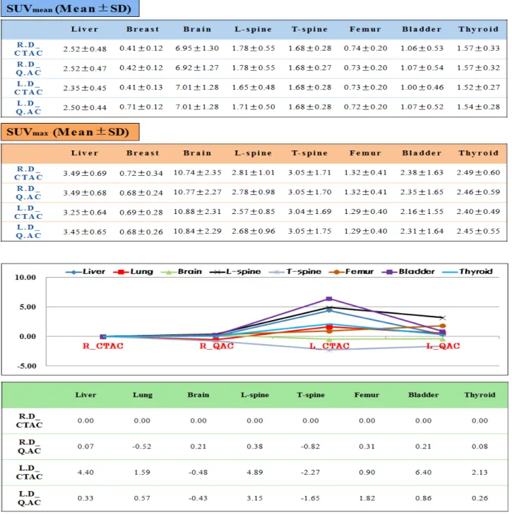
Table 1. Shown the SUVmean and SUVmax in the patient study
Fig. 7. Relative error % for SUVmean for the normal weight patient.
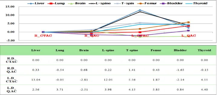
환자를 체질량지수(BMI) 기준으로 하여 과체중군과 정상 체중군으로 분류한 뒤 일반선량 조건과 저 선량 조건에서 각각의 감쇠보정법이 적용된 PET영상에서 GE사의 분석프로그램인 PET VCAR (GE Healthcare, USA)로 주요부위 8군데 SUVmean와 SUVmax 결과값을 구하였다(Table 1). 그런 다음 일반선량 조건에 CTAC 로 보정한 값을 기준값으로 잡고 다음과 같은 식으로
상대적 오차를 구하였다.
상대적 오차(relative error %)=(x0 - x)/x0 *100 x0=true value of a quantity
x=observed value of the quantity x0-x=absolute error
Fig. 8. Relative error % for SUVmean for the over weight patient.
Fig. 9. Relationship between CT mean error and PET quantita- tion error are linear. When the CT mean error is 0 (zero), there is no PET quantitation error.
구해진 SUVmean에 대한 상대적 오차값으로 그래프 를 그려보았다(Figs. 7, 8). 동일한 일반선량 조건에서 는 감쇠보정법에 따른 오차값의 변화는 ±0.1 이하로 미미한 수준이었다. 그러나 저 선량 조건에서는 기준 값에 대한 평균 오차값이 CTAC의 경우 주요부위 8군 데 수치가 4.40, 1.59, -0.48, 4.89, -2.27, 0.90, 6.40, 2.13 이고 Q.AC에서는 0.33, 0.57, -0.43, 3.15, -1.65, 1.82, 0.86, 0.26으로 CTAC에 비하여 Q.AC가 주요부위 모 두 전반적으로 기준값과의 오차가 적은 것을 알 수 있 었다. 그리고 과체중 환자에서는 CTAC의 경우 주요 부위 8군데 수치가 13.04, -2.97, -1.48, 13.34, 4.59, 5.58, 13.57, 7.99이고 Q.AC에서는 3.16, -0.42, -1.24, 5.46, 3.25, 4.83, 5.78, 5.04로 정상체중 환자보다 CTAC에 비하여 Q.AC가 상대적으로 기준값과의 오차가 적은 것을 확인할 수 있었다. 또한 과체중군과 정상체중군의 신호 대 잡음비(SNR)를 선량과 감쇠보정법을 다르게 적용하여 본 결과 저 선량에서는 CTAC에 비해 Q.AC 가 약간 낮은 값을 보이나 큰 변화는 없었으며 과체중 군과 정상 체중군 모두 그래프의 유의한 변화는 얻을 수 없었다. 기존의 CTAC나 Q.AC 모두 SNR값의 변 화에는 큰 영향이 없음을 알 수 있었다.
고찰 및 결론
PET/CT 촬영 시 CT 평균오차와 선량은 PET정량오 차와 비례관계를 가지며 CT평균오차가 0일 때 PET정
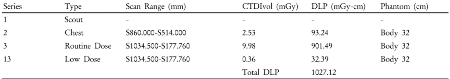
량오차가 발생하지 않는 것을 알 수 있다(Fig. 9).4) 본 논문의 연구 결과 기존의 CTAC에 비하여 Q.AC가 저 선량조건에서도 이러한 CT 평균오차의 변화를 줄여 PET정량의 일관성을 유지하게 하며 일반선량 조건에 가까운 이미지 질 구현이 가능하게 된다. 그 결과 기존의 CTAC를 사용할 때보다 낮은 선량의 적용이 가능하게 되는데 본원의 PET/CT Dose Report를 보면 환자의 유효선량 평가지표인 CTDIVol과 DLP값이 Q.AC를 적용하였을 때 SUV에 눈에 띄는 영향이 없는 정도에서 일반선량보다 저 선량에서 CTDIVol과 DLP가 30배가량 감소되는 효과를 볼 수 있었다(Table 2). 그러나 Q.AC
Table 2. Compared with the low dose and the routine dose protocols for CT effective dose reduction
Series Type Scan Range (mm) CTDIvol (mGy) DLP (mGy-cm) Phantom (cm)
1 Scout - - - -
2 Chest S860.000-S514.000 2.53 93.24 Body 32
3 Routine Dose S1034.500-S177.760 9.98 901.49 Body 32
13 Low Dose S1034.500-S177.760 0.36 32.39 Body 32
Total DLP 1027.12
적용 시 정상체중 환자에서는 유의한 차이를 얻을 수 없었고 과체중 환자에서 선량감소효과가 있는 것을 알 수 있었으며 CTAC와 비교하여 SNR값의 유효한 차이는 없었다. 따라서 반복적인 CT촬영을 요하는 임상실험 이나 저 선량조건에서의 소아환자검사 또는 호흡동기 (respiratory gating) 검사 시 감쇠보정지도(attenuation cor rection map)로 유효하게 적용할 수 있을 것이라 사료 된다.
요 약
PET/CT에서 환자피폭 문제로 인해 저 선량의 중요성이 강조되고 있다. 본 연구에서는 기존에 사용되던 CT 데 이터를 이용한 감쇠보정법인 CTAC와 새롭게 적용된 Q.AC를 환자실험과 팬텀 실험을 통해 저 선량으로 촬 영 시 PET 영상에 미치는 영향에 대해 알아보고자 한 다. 실험장비는 GE사의 PET/CT Discovery 710 (GE Healthcare, USA)를 사용하였으며 팬텀실험으로 감쇠보 정의 정량적 평가를 위한 NEMA IEC body phantom 과 균일성 평가를 위한 Uniform NU2-94 phantom을 사용하였다. 각각의 팬텀 내부에 동위원소 18-F FDG 를 70.78 MBq, 22.2 MBq 주입하고 CT조건은 저 선량 조건으로 80 kVp, 10 mA로부터 일반선량 조건으로 140 kVp, 120 mA 조건까지 스캔 후 CTAC와 Q.AC 두 감쇠보정법을 적용하여 재구성하였다. PET 영상에서 일반선량 조건을 기준값으로 정하고 horizomtal pro- file과 vertical profile을 통해 정량평가를 시행하고 기 준값과의 상대적 오차를 평가하였다. 또한 환자실험으 로 정상체중 환자와 과체중 환자를 구분하여 저 선량과 일반선량으로 비교 촬영한 뒤 CTAC와 Q.AC로 재구성 된 PET영상에서 주요장기별 SUV에 대한 상대적 오차 와 신호 대 잡음비를 비교분석하였다. 팬텀실험 결과 저
선량 조건에서 CTAC와 Q.AC로 각각 재구성한 PET 영상의 profile과 상대적 오차에서 CTAC보다 Q.AC 가 기준값과의 오차가 적은 그래프를 얻었다. 환자실 험의 경우 일반선량 조건에서는 정상체중 환자와 과체 중 환자 모두 감쇠보정법에 따른 상대적 오차값의 변 화가 적었으나 저 선량 조건에서는 정상체중 환자보다 과체중 환자에서 감쇠보정법의 변경에 의한 상대적 오 차의 감소폭이 커짐으로 기준값과 차이가 감소하였다.
기존의 감쇠보정법인 CTAC는 80 kVp, 10 mA의 저 선량 CT를 사용하는데 있어 PET 영상의 선속경화현 상이 발생한다. 이로 인해 CTAC를 이용하여 재구성 된 PET 데이터는 정량화하는데 문제가 될 수 있음을 확인했다. 반면에 새로운 알고리즘이 적용된 Q.AC는 과체중 환자의 경우 80 kVp, 10 mA 정도까지는 140 kVp, 120 mA 조건으로 촬영하여 재구성한 PET 데이터 결 과와 차이가 적음을 확인할 수 있었다. Q.AC를 이용 한 경우 기존보다 저 선량의 CT를 이용해 PET의 재구 성에 이용할 수 있으므로 환자의 피폭을 줄이는 데 큰 역할을 할 것으로 기대한다.
REFERENCES
1. Paul kinahan. PET/CT Issues: CT-based attenuation correc- tion(CTAC), Artifacts, and Motion Correction. Department of Radiologist University of Washington web site. Available at:
http://aapm.org/meetings/08SS/documents/Kinahan.pdf.
Accessed June 25, 2008.
2. 정준기, 이명철. 고창순 핵의학. 2008;102-104.
3. Colsher JG, Hsieh J, Thibault JB, Lonn A, Pan T, Stephen J, et al. Ultra Low Dose CT for Attenuation Correction in PET/CT.
IEEE Nuclear Science Symposium Conference Record. 19-25 Oct, 2008.
4. Lonn A. Q.AC: Quantitation Achieved Consistently. UK. GE Healthcare. Nov, 2012.