Received: 17 August, 2016 Revised: 8 September, 2016 Accepted: 8 September, 2016 Corresponding author: Wan-hee Lee
Department of Physical Therapy, College of Health Science, Sahmyook University, 815 Hwarang-ro, Nowon-gu, Seoul 01795, Republic of Korea Tel: 82-2-3399-1633 Fax: 82-2-3399-1639 E-mail: [email protected]
This is an Open-Access article distributed under the terms of the Creative Commons Attribution Non-Commercial License (http://creativecommons.org/licens es/by-nc/4.0) which permits unrestricted non-commercial use, distribution, and reproduction in any medium, provided the original work is properly cited.
Copyright © 2016 Korean Academy of Physical Therapy Rehabilitation Science
http://dx.doi.org/10.14474/ptrs.2016.5.3.155 pISSN 2287-7576
eISSN 2287-7584
Phys Ther Rehabil Sci 2016, 5 (3), 155-161 www.jptrs.org
Reliability and validity of a personal computer based muscle viewer for measuring upper trapezius and transverses abdominis muscle thickness
Ju-Ri Jeong
a, Ju Hee Han
b, Ji-Eun Cho
b, Wan-hee Lee
baMusculoskeletal Center, Samsung Seoul R&D Medical Clinic, Seoul, Republic of Korea
bDepartment of Physical Therapy, College of Health Science, Sahmyook University, Seoul, Republic of Korea
Objective: This study aimed to investigate the reliability and validity of a personal computer-based muscle viewer (PC-BMW) compared with that of a portable ultrasound (P-US) for measuring upper trapezius (UT) and transversus abdominis (TrA) muscle thickness at rest and during contraction.
Design: Observational inter-rater reliability study.
Methods: Fifty-five healthy participants (25 men, 30 women) participated in this study. PC-BMW and P-US were randomly measured at the UT and TrA muscles. Two examiners randomly obtained the images of all participants in 3 test sessions lasting 2 days. Intra-class correlation coefficients (ICCs), standard error of measurement, contraction ratio, and correlation were used to es- timate reliability and validity. Pearson’s correlation coefficients were used to analyze the relationship between muscle thickness measures taken from PC-BMW and P-US.
Results: The intra-rater reliability ICCs of UT and TrA muscle thickness for the PC-BMW were >0.995, indicating excellent reliability. Inter-rater reliability ICCs for the PC-BMW ranged from 0.963 to 0.987. The P-US also exhibited high reliability. A high correlation was found between the measurements of the two muscles in PC-BMW and P-US (p<0.01).
Conclusions: PC-BMW provides clear and excellent images, is pocket-sized and less expensive than a conventional ultrasound imaging system. PC-BMW can be utilized variously and has the advantage of rehabilitative ultrasound imaging. More research is needed to evaluate the utility of PC-BMW for rehabilitation.
Key Words: Muscle viewer, Reliability, Transversus abdominis, Ultrasonography, Upper trapezius
Introduction
The structure of the skeletal muscle is key to the perform- ance of functional human movement [1]. Recently, re- habilitative ultrasound imaging (RUSI) has become a com- mon method for evaluating skeletal muscle structure. In ad- dition, like magnetic resonance imaging (MRI), RUSI can clearly distinguish between muscle and tissue, and it is pos- sible to generate a high-quality image of the muscle struc- ture [2]. RUSI has the advantage of being relatively user- friendly, and can quantitatively measure various aspects of
muscle structure including muscle fiber size, thickness, length, cross-sectional area, and pennation angle [3-6].
These variables are highly correlated to muscle strength, ef- ficiency and muscle movement [7,8]. Muscle thickness and strength are closely related variables and have been used to identify the most useful structural changes in muscles during therapy [4,9]. Moreover, RUSI may provide real-time visual feedback enabling the proper performance of muscles dur- ing exercise [10].
Several studies have investigated the validity and reli-
ability of the ultrasound measurement of the limbs and trunk
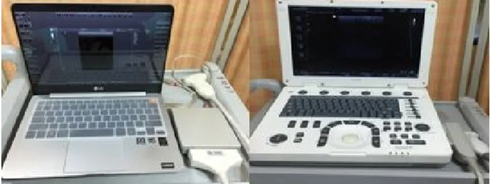
Figure 1. (A) Personal computer-based muscle viewer and (B) portable ultra- sound.
to that of the muscles of the trunk [13,21]. Although muscu- loskeletal ultrasound studies have been variously attempted, more research is needed regarding the measurement of dy- namic contraction in the posture that can be resolved in mus- cle activity.
Despite the many advantages of conventional ultrasound, it is heavy and expensive. Recently, a personal computer- based muscle viewer (PC-BMW) which addresses the dis- advantages of conventional ultrasound, has been developed.
PC-BMW has created Telemed
TM, which provides clear and excellent images, is pocket-sized, and less expensive than a conventional ultrasound imaging system. In addition, one more advantage of this device is that it can be used to down- load free PC software from anywhere. Unlike conventional portable ultrasound (P-US), not the process of storing and outputting measurement data require additional useful man- agement of data. Therefore, the purpose of the study was to establish the reliability and validity of PC-BMW against ul- trasound imaging for measuring UT and TrA muscle thickness.
Methods Subjects
Fifty-five healthy individuals (25 men, 30 women) with
Procedures
Muscle thickness measurements were performed with a PC-BMW (MicrUs EXT-1H; TELEMED, Vilnius, Lithuania) and a Medison Mysono P-US system (U5; Samsung Medison, Seoul, Korea) (Figures 1, 2). The PC-BMW is a new gen- eration of universal serial bus powered small-sized ultra- sound imaging equipment. In this study, the PC-BMW sys- tem used a 12-MHz linear transducer for both measurement conditions. A P-US was also used with a 12-MHz linear transducer to obtain images. The two imaging measure- ments via the two devices were conducted by two exam- iners, with more than 3 years’ experience in musculoskeletal ultrasound imaging.
The participants assumed two positions in rest and con- traction for the measurement of the UT muscle thickness.
For the resting position, the participants were sat upright in
a neutral position with the head straight-; the dominant side
was measured. For contraction, the participants held the arm
at 30
oabduction for 10 seconds [21]. In the two positions, the
participants were asked to fully extend the elbow and a goni-
ometer was used for the maintenance of the arm’s abduction
angle. To determine the transducer placement, the exam-
iners drew lines with a kohl pencil from the mid-line be-
tween C6 and the angle of the acromion. The probe was
placed parallel to the muscle fibers, and the examiners asked
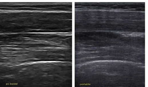
Figure 2. Resolution the difference between the two devices. (A) Personal computer-based muscle viewer. (B) portable ultrasound.
Table 1. Characteristics of the subjects (N=55)
Characteristic Value
Gender, male/female 25/30
Age (y) 28.41 (3.77)
Height (cm) 166.30 (7.13)
Weight (kg) 62.58 (12.75)
Body mass index (kg/m2) 22.45 (3.27)
Waist (cm) 74.42 (9.11)
Dominant side, left/right 4/51
Values are presented as number only or mean (SD).
the participants to maintain a fixed position. Scan images of the UT muscle thickness were calculated 2 cm lateral to the triangular myo-fascial junction at a direction of the muscle belly plane [13]. The TrA muscle thickness on the dominant side was measured during rest and in abdominal drawing-in maneuver (ADIM). Participants were examined in the hook- lying position at rest with their knees flexed at 90
oin the su- pine position [22]. The probe was transversely placed on the middle abdominal region between the border of the 11th cos- tal cartilage and the anterior superior iliac crest [15]. To per- form ADIM for TrA contraction, participants were in- structed to “take in a deep breath, draw your belly button up and in towards your lumbar spine” [14,23]. For the quantifi- cation of the ADIM, we used a pressure biofeedback unit (PBU) (Stabilizer; Chattanooga Group Inc., Hixson, TN, USA) [24]. After setting a pressure of 40 mmHg in the PBU, the ability to contract the TrA muscle resulted in a pressure reduction from 4 to 10 mmHg [25,26].
The participants rested for 1 to 2 minutes after ADIM to reduce the influence of fatigue. The image was measured by drawing a line 1.5 cm apart from the myo-fascial junction and a vertical line was drawn for the 3 muscles layers (external oblique, internal oblique, transverse abdominis) [27]. All images were measured by the two examiners for two days.
Data analysis
All demographic data were analyzed for descriptive statistics. To describe the intra- and inter-rater reliability of UT and TrA muscle thickness at rest and during contraction, intra-class correlation coefficients (ICCs) and 95% con- fidence intervals (CIs) were calculated. ICCs of the type
(3,1) were used to evaluate the reliability of the data. ICCs
<0.50 were considered poor; 0.50 to 0.75, moderate to good;
and >0.75, excellent reliability [28]. Based on the reliability coefficients, SEM was calculated as standard deviation×
. To investigate the linear relationship between the two methods, Pearson’s correlation coefficient (r) and the r
2value were used. The statistical analyses were per- formed using PASW Statistics ver. 18.0 for Windows (IBM Co., Armonk, NY, USA).
Results
Demographic characteristics
Participants characteristics were as follows: 55 healthy participants (male=25, female=30) with mean age 28.41±
3.77 years, mean weight 62.58±12.75 kg, and mean height
166.30±7.13 cm. The mean body mass index and waist cir-
cumference were 22.45±3.27 kg/m
2and, 74.42±9.11 cm, re-
spectively (Table 1).
Table 3. Intra-rater between repeated measures PC-BMW and P-US for the transverses abdominis MT (2 days unit: mm) (N=55)
MT Subheading 1st test 2nd test 3rd test ICC 95% CI SEM
E1 1st day Rest PC-BMW 3.02 (1.31) 3.05 (1.28) 3.09 (1.33) 0.998 0.996-0.999 0.058
P-US 2.87 (0.81) 2.94 (0.86) 2.95 (0.85) 0.980 0.969-0.988 0.119
ADIM PC-BMW 4.22 (1.68) 4.26 (1.69) 4.16 (1.69) 0.997 0.996-0.998 0.092
P-US 4.09 (1.15) 4.15 (1.21) 4.19 (1.20) 0.991 0.986-0.994 0.113
2nd day Rest PC-BMW 3.15 (1.05) 3.20 (1.08) 3.18 (1.07) 0.997 0.995-0.998 0.058
P-US 2.95 (0.80) 3.00 (0.79) 3.08 (0.87) 0.963 0.942-0.977 0.158
ADIM PC-BMW 4.25 (1.33) 4.25 (1.34) 4.28 (1.36) 0.971 0.954-0.982 0.229
P-US 4.13 (1.03) 4.19 (1.07) 4.17 (1.02) 0.992 0.988-0.995 0.093
E2 1st day Rest PC-BMW 3.02 (1.27) 3.06 (1.25) 3.06 (1.24) 0.954 0.928-0.971 0.269
P-US 2.85 (0.93) 2.92 (0.89) 2.93 (0.90) 0.978 0.966-0.987 0.135
ADIM PC-BMW 4.15 (1.49) 4.18 (1.46) 4.18 (1.47) 0.977 0.964-0.986 0.223
P-US 4.02 (1.16) 4.04 (1.17) 4.01 (1.08) 0.990 0.984-0.994 0.114
2nd day Rest PC-BMW 3.07 (1.08) 3.08 (1.06) 3.10 (1.04) 0.973 0.957-0.983 0.175
P-US 2.95 (0.80) 3.00 (0.79) 3.08 (0.87) 0.984 0.976-0.990 0.100
ADIM PC-BMW 4.21 (1.36) 4.21 (1.34) 4.23 (1.34) 0.924 0.881-0.953 0.373
P-US 4.13 (1.03) 4.19 (1.07) 4.17 (1.02) 0.991 0.985-0.994 0.090
Values are presented as mean (SD).
PC-BMW: personal computer-based muscle viewer, P-US: portable ultrasound, MT: muscle thickness, ICC: intraclass correlation coefficient, 95% CI: 95% confidence interval, SEM: standard error of the mean, E1: examiner 1, E2: examiner 2, ADIM: abdominal draw-in maneuver.
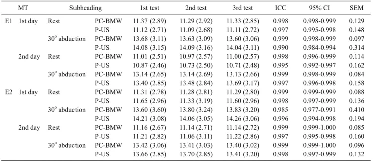
E1 1st day Rest PC-BMW 11.37 (2.89) 11.29 (2.92) 11.33 (2.85) 0.998 0.998-0.999 0.129 P-US 11.12 (2.71) 11.09 (2.68) 11.11 (2.72) 0.997 0.995-0.998 0.148 30o abduction PC-BMW 13.68 (3.11) 13.63 (3.09) 13.60 (3.06) 0.999 0.998-0.999 0.097 P-US 14.08 (3.15) 14.09 (3.16) 14.04 (3.11) 0.990 0.984-0.994 0.314 2nd day Rest PC-BMW 11.01 (2.51) 10.97 (2.57) 11.00 (2.57) 0.998 0.996-0.999 0.114 P-US 10.87 (2.46) 10.73 (2.50) 10.71 (2.48) 0.995 0.992-0.997 0.162 30o abduction PC-BMW 13.14 (2.65) 13.14 (2.69) 13.13 (2.66) 0.999 0.998-0.999 0.084 P-US 13.40 (2.85) 13.48 (2.84) 13.69 (3.17) 0.997 0.996-0.998 0.158 E2 1st day Rest PC-BMW 11.31 (2.78) 11.28 (2.81) 11.29 (2.80) 0.999 0.999-0.999 0.088 P-US 11.65 (2.96) 11.33 (3.19) 11.60 (2.96) 0.998 0.997-0.999 0.136 30o abduction PC-BMW 13.60 (3.60) 13.80 (3.24) 13.83 (3.20) 0.985 0.977-0.991 0.410 P-US 14.21 (3.08) 14.06 (3.05) 14.26 (3.06) 0.996 0.994-0.998 0.194 2nd day Rest PC-BMW 11.16 (2.67) 11.14 (2.71) 11.14 (2.72) 0.999 0.999-1.000 0.085 P-US 11.21 (2.82) 11.06 (3.11) 11.22 (2.86) 0.997 0.995-0.998 0.160 30o abduction PC-BMW 13.42 (3.06) 13.41 (3.03) 13.40 (3.02) 0.999 0.999-1.000 0.096 P-US 13.66 (2.85) 13.70 (2.85) 13.41 (3.20) 0.998 0.997-0.999 0.132 Values are presented as mean (SD).
PC-BMW: personal computer-based muscle viewer, P-US: portable ultrasound, MT: muscle thickness, ICC: intraclass correlation coefficient, 95% CI: 95% confidence interval, SEM: standard error of the mean, E1: examiner 1, E2: examiner 2.
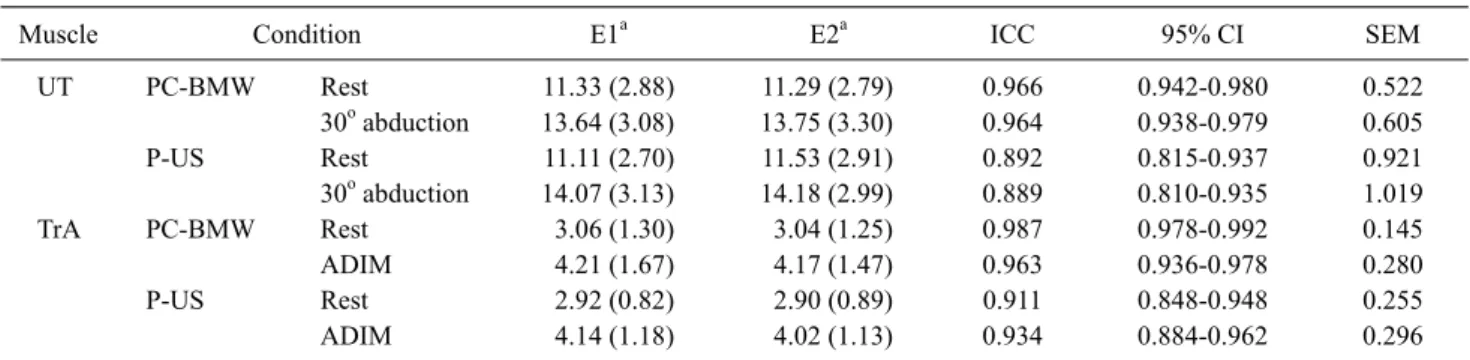
Table 4. Inter-rater between repeated measures on PC-BMW and P-US for the two muscles (unit: mm) (N=55)
Muscle Condition E1a E2a ICC 95% CI SEM
UT PC-BMW Rest 11.33 (2.88) 11.29 (2.79) 0.966 0.942-0.980 0.522
30o abduction 13.64 (3.08) 13.75 (3.30) 0.964 0.938-0.979 0.605
P-US Rest 11.11 (2.70) 11.53 (2.91) 0.892 0.815-0.937 0.921
30o abduction 14.07 (3.13) 14.18 (2.99) 0.889 0.810-0.935 1.019
TrA PC-BMW Rest 3.06 (1.30) 3.04 (1.25) 0.987 0.978-0.992 0.145
ADIM 4.21 (1.67) 4.17 (1.47) 0.963 0.936-0.978 0.280
P-US Rest 2.92 (0.82) 2.90 (0.89) 0.911 0.848-0.948 0.255
ADIM 4.14 (1.18) 4.02 (1.13) 0.934 0.884-0.962 0.296
Values are presented as mean (SD).
PC-BMW: personal computer-based muscle viewer, P-US: portable ultrasound, E1: examiner 1, E2: examiner 2, ICC: intraclass correlation coefficient, 95% CI: 95% confidence interval, SEM: standard error of the mean, UT: upper trapezius muscle, TrA: transverses abdominis muscle ADIM: abdominal draw-in maneuver.
aMeasurement are mean (SD) based on three images taken by the examiner on the same day (day 1).
Table 5. Correlation between muscle thickness measurements
taken from PC-BMW and P-US (N=55)
Examiner Muscle Condition
Pearson’s correlation coefficient (r)
p r2
E1 UT Rest 0.842 <0.001 0.708
30o abduction 0.786 <0.001 0.618
TrA Rest 0.690 <0.01 0.477
ADIM 0.727 <0.01 0.529
E2 UT Rest 0.890 <0.001 0.792
30o abduction 0.905 <0.001 0.819
TrA Rest 0.719 <0.01 0.517
ADIM 0.805 <0.01 0.648
PC-BMW: personal computer-based muscle viewer, P-US: portable ultrasound, E1: examiner 1, E2: examiner 2, UT: upper trapezius muscle, TrA: transverses abdominis muscle, ADIM: abdominal draw-in maneuver.
Inter-rater reliability analysis
For the muscles, the ICCs for inter-rater reliability ranged from 0.889 to 0.987 (Table 4). The SEM values of UT ranged from 0.522 to 1.019 cm, and the SEM values of TrA ranged from 0.145 to 0.296.
Correlation between muscle thickness measurements taken from PC-BMW and P-US
The results showed a good correlation between PC-BMW and P-US measurements of UT (p<0.001) and TrA (p<0.01) muscle thickness for examiner 1 (E1) and 2 (E2) (Table 5).
Discussion
The aim of this study was to investigate the reliability and validity of PC-BMW compared to that of conventional P-US. PC-BMW is convenient to use and may be employed to quantify muscle structure non-invasively like conven- tional ultrasound, but is considerably lighter in weight and less expensive.
In this study, PC-BMW of the UT muscle had a good in- tra-rater reliability (E1 rest ICC=0.998, E1 contraction ICC=0.999, E2 rest ICC=0.999, E2 contraction ICC=0.985- 0.999) and a good inter-rater reliability (rest ICC=0.966, contraction ICC=0.964). In the study by Leong et al. [21], which involved UT measurement, the reliability of rest and 30
oshoulder abduction position tissue stiffness (supersonic shear imaging) was very good (intra-rater rest ICC=0.97, contraction ICC=0.93, inter-rater rest ICC=0.78, con- traction ICC=0.83). Similarly, in this study, we used pre- vious research methods, which had shown high reliability.
Both devices showed a high reliability for UT measurement, but PC-BMW showed slightly higher values than P-US.
When using the two devices during the experiment, the im- age output of PC-BMW more clear than that of P-US for dis- tinguishing the boundaries of the fascia. The difference in clarity between the two devices is considered to have af- fected the results.
We found a good intra-rater and inter-rater reliability in the PC-BMW measurement of the TrA muscle (ICC=
0.924-0.998, and 0.963-0.987, respectively). In previous studies, the reliability of ultrasound measurement of TrA during rest and ADIM was very high with most values
>0.900 [12,15,20]. Properly performing ADIM, depends on
of TrA muscle thickness was higher (rest E1 r=0.690, E2 r=0.719, contraction E1 r=0.727, E2 r=0.805), and UT showed higher correlation values. In a previous validity study comparing MRI and US, UT showed good correlation values (r=0.52, p<0.001) [13]. The results were used as the proper method to measure the muscle structures. PC-BMW is not determined by anything on the image output as com- pared to conventional P-US. PC-BMW used proprietary software, the Echo Wave II ver. 3.5, and calculated the length measured at the same time as image viewing. In the case of P-US, after moving image files to the PC, the Sante Dicom viewer program (Dicom Softwere, Santesoft Ltd., Athenes, Greece) was used to view the output. We believe that we were able to observe differences in length and make measurements based on the PC resolution and type of program. Further research evaluating the utility of PC-BMW for rehabilitation and measurement of other muscle con- traction conditions is needed. The PC-BMW device is con- sidered as a clinically useful method for assessing muscle structure.
Conflict of Interest
The authors declared no potential conflicts of interest with respect to the authorship and/or publication of this article.
References
1. Neumann DA. Kinesiology of the musculoskeletal system. St.
Louis: Elsvier Mosby; 2002.
2. Scott JM, Martin DS, Ploutz-Snyder R, Caine T, Matz T, Arzeno NM, et al. Reliability and validity of panoramic ultrasound for muscle quantification. Ultrasound Med Biol 2012;38:1656-61.
3. Esformes JI, Narici MV, Maganaris CN. Measurement of human muscle volume using ultrasonography. Eur J Appl Physiol 2002;
87:90-2.
8. McNee AE, Gough M, Morrissey MC, Shortland AP. Increases in muscle volume after plantarflexor strength training in children with spastic cerebral palsy. Dev Med Child Neurol 2009;51:429-35.
9. Gruther W, Benesch T, Zorn C, Paternostro-Sluga T, Quittan M, Fialka-Moser V, et al. Muscle wasting in intensive care patients:
ultrasound observation of the M. quadriceps femoris muscle layer. J Rehabil Med 2008;40:185-9.
10. Henry SM, Westervelt KC. The use of real-time ultrasound feed- back in teaching abdominal hollowing exercises to healthy sub- jects. J Orthop Sports Phys Ther 2005;35:338-45.
11. Nabavi N, Mosallanezhad Z, Haghighatkhah HR, Mohseni Bandpeid MA. Reliability of rehabilitative ultrasonography to measure transverse abdominis and multifidus muscle dimen- sions. Iran J Radiol 2014;11:e21008.
12. Ishida H, Hirose R, Watanabe S. Comparison of changes in the contraction of the lateral abdominal muscles between the ab- dominal drawing-in maneuver and breathe held at the maximum expiratory level. Man Ther 2012;17:427-31.
13. O'Sullivan C, Meaney J, Boyle G, Gormley J, Stokes M. The val- idity of rehabilitative ultrasound imaging for measurement of trapezius muscle thickness. Man Ther 2009;14:572-8.
14. Koppenhaver SL, Hebert JJ, Fritz JM, Parent EC, Teyhen DS, Magel JS. Reliability of rehabilitative ultrasound imaging of the transversus abdominis and lumbar multifidus muscles. Arch Phys Med Rehabil 2009;90:87-94.
15. Teyhen DS, Miltenberger CE, Deiters HM, Del Toro YM, Pulliam JN, Childs JD, et al. The use of ultrasound imaging of the abdominal drawing-in maneuver in subjects with low back pain.
J Orthop Sports Phys Ther 2005;35:346-55.
16. Koppenhaver SL, Hebert JJ, Parent EC, Fritz JM. Rehabilitative ultrasound imaging is a valid measure of trunk muscle size and activation during most isometric sub-maximal contractions: a systematic review. Aust J Physiother 2009;55:153-69.
17. Kim MK, Ko YJ, Lee HJ, Ha HG, Lee WH. Ultrasound imaging for age-related differences of lower extremity muscle architec- ture. Phys Ther Rehabil Sci 2015;4:38-43.
18. Ko YJ, Ha HG, Jeong JR, Lee WH. Variations in lateral abdomi- nal muscle thickness during abdominal drawing-in maneuver in three positions in a young healthy population. Phys Ther Rehabil Sci 2014;3:101-6.
19. Lee HJ, Shin KH, Byun SM, Jeong HS, Hong JS, Jeong SJ, et al.
The changes of rectus abdominis muscle thickness according to the angle during active straight leg raise. Phys Ther Rehabil Sci 2013;2:44-8.
20. Ishida H, Watanabe S. Changes in lateral abdominal muscles'
thickness immediately after the abdominal drawing-in maneuver and maximum expiration. J Bodyw Mov Ther 2013;17:254-8.
21. Leong HT, Ng GY, Leung VY, Fu SN. Quantitative estimation of muscle shear elastic modulus of the upper trapezius with super- sonic shear imaging during arm positioning. PLoS One 2013;8:
e67199.
22. Jung DE, Kim K, Lee SK. Comparison of muscle activities using a pressure biofeedback unit during abdominal muscle training performed by normal adults in the standing and supine positions.
J Phys Ther Sci 2014;26:191-3.
23. McGalliard MK, Dedrick GS, Brismée JM, Cook CE, Apte GG, Sizer PS Jr. Changes in transversus abdominis thickness with use of the abdominal drawing-in maneuver during a functional task.
PM R 2010;2:187-94; quiz 226.
24. de Paula Lima PO, de Oliveira RR, Costa LO, Laurentino GE.
Measurement properties of the pressure biofeedback unit in the
evaluation of transversus abdominis muscle activity: a system- atic review. Physiotherapy 2011;97:100-6.
25. Chattanooga G. Stabilizer pressure bio-feedback. Operating instructions. Hixson: Chattanooga Group Inc.; 2005.
26. Lima PO, Oliveira RR, Moura Filho AG, Raposo MC, Costa LO, Laurentino GE. Concurrent validity of the pressure biofeedback unit and surface electromyography in measuring transversus ab- dominis muscle activity in patients with chronic nonspecific low back pain. Rev Bras Fisioter 2012;16:389-95.
27. Hodges PW, Pengel LH, Herbert RD, Gandevia SC. Measure- ment of muscle contraction with ultrasound imaging. Muscle Nerve 2003;27:682-92.
28. Portney LG, Watkins MP. Foundations of clinical research:
Applications to practice. Upper Saddle River, NJ: Prentice Hall Health; 2000.