컴퓨터 단층촬영을 이용한 요로결석 검사에서 저선량 CT의 적용에 대한 유용성 평가
Usability Evaluation of Applied Low-dose CT When Examining Urinary Calculus Using Computed Tomography
김현진, 지태정 가야대학교 방사선학과
Hyeon-Jin Kim([email protected]), Tae-Jeong Ji([email protected]) 요약
본 연구는 컴퓨터단층촬영을 이용한 요로결석 검사에서 저선량 CT(Low dose Computed Tomography) 프로토콜의 적용에 따른 유용성을 평가하였다. 연구대상은 2016년 6월–12월까지 부산지역 일개 의료기관을 내원한 비뇨기계 환자이며 연구에 이용된 프로토콜은 통계적 반복 재구성법(Adaptive Statistical Iterative Reconstruction, ASIR)을 50% 적용한 저선량 CT 프로토콜이다. 정량적 분석 결과 횡단면상(Axial image)의 신장의 관심 영역(region of image. ROI) 내 평균 화소 값과 표준편차는 복부촬 영 프로토콜 26.21±7.08, 저선량 CT 프로토콜 20.03±8.16 이며 관상면(Coronal imalge) 영상에서 신장 의 관심영역 내 평균 화소 값과 표준편차는 복부촬영 프로토콜 22.07±7.35, 저선량 CT 프로토콜 21.67± 6.11 이었다. 정성적 분석결과 인공물에 대한 4명의 관찰자 평균값은 복부촬영 프로토콜 19.14±0.36, 저선 량 CT 프로토콜 19.17±0.43 이며 해상도 및 대조도의 평균값은 복부촬영 프로토콜 19.35±0.70, 저선량 CT 프로토콜 19.29±0.58 이었다. 피폭선량 분석 결과 복부촬영 프로토콜의 CTDIvol 평균값은 18.02 mGy, DLP 평균값은 887.51 mGy·cm 이며 저선량 CT 프로토콜 CTDIvol 평균값은 7.412 mGy, DLP 평균값은 361.22 mGy·cm 이었다. 이로 인한 선량의 감소율은 각 58.82%, 59.29% 이었다.
■ 중심어 :∣CT 선량 지표∣저선량 CT∣요로결석∣선량보고서∣
Abstract
The aim of this study was to evaluate the usability of applied Low dose Computed Tomography(LDCT) protocol in examining urinary calculus using computed tomography. The subjects of this study were urological patients who visited a medical institution located in Busan from June to December 2016 and the protocol used in this study was Adaptive Statistical Iterative Reconstruction: low-dose CT with 50% Adaptive Statistical Iterative Reconstruction (ASIR). As results of quantitative analysis, the mean pixel value and standard deviation within kidney region of image(ROI)of the axial image were 26.21±7.08 in abdomen CT pre scan and 20.03±8.16 in low-dose CT. Also the mean pixel value and standard deviation within kidney ROI of the coronal image were 22.07±7.35 in abdomen CT pre scan and 21.67±6.11 in low dose CT. The results of qualitative analysis showed that four raters’ mean values of observed kidney artifacts were 19.14±0.36 when using abdomen CT protocol and 19.17±0.43 in low-dose CT, and the mean value of resolution and contrast was 19.35±0.70 when using abdomen CT protocol and 19.29±0.58 in low-dose CT. Also the results of a exposure dose analysis showed that the mean values of CTDIvol and DLP in abdomen CT pre scan were 18.02 mGy and 887.51 mGy·cm respectively and the mean values of CTDIvol and DLP when using low-dose CT protocol were 7.412 mGy and 361.22 mGy·cm respectively. The resulting dose reduction rate was 58.82% and 59.29%, respectively.
■ keyword :∣Computed Tomography Dose Index∣Low Does CTl∣Dose Report∣Urinary Calculus∣
접수일자 : 2017년 02월 02일 수정일자 : 2017년 03월 06일
심사완료일 : 2017년 03월 06일
교신저자 : 김현진, e-mail : [email protected]
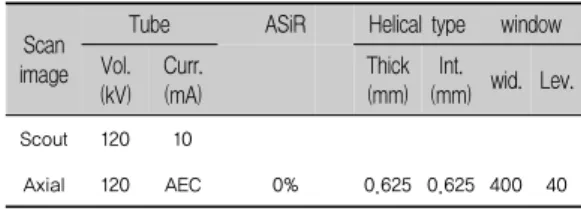
Scan image
Tube ASiR Helical type window Vol.
(kV) Curr.
(mA)
Thick (mm)
Int.
(mm) wid. Lev.
Scout 120 10
Axial 120 AEC 0% 0.625 0.625 400 40
Ⅰ. 서 론
요로결석이란 어떤 원인에 의해 요로에 돌이 형성되 어 감염이나 요관폐색 등의 합병증을 유발하는 질환이 다. 요로결석은 비교적 흔한 비뇨기과 질환으로 미국에 서는 5-10%의 국민이 요로결석을 경험하며 우리나라 에서도 식생활의 서구화로 인해 꾸준히 증가하고 있다 [1][2]. 요로결석의 진단을 위해서 고식적인 방법으로 경정맥 요로조영술(Intravenous urography, IVU)이 이 용되었으나 조영제 주입으로 인한 알러지 반응이나 신 장 독성을 포함한 여러 가지 합병증을 유발하며 진단 시 민감도 52%, 특이도 94%로 결석의 평가에 제한점을 갖고 있다[3]. 컴퓨터단층촬영(CT)은 비뇨기계의 횡단 면을 나타내므로 요관 내에 위치한 결석을 나타낼 수 있으며 결석의 정확한 위치와 크기를 알 수 있다. 김봉 수 등의 연구결과[4] 컴퓨터단층촬영을 이용한 요로결 석의 진단 시 민감도 98%, 특이도 95%로 경정맥 요로 조영술에 비해 정확도가 매우 높게 나타났다. 그러나 컴퓨터단층촬영은 매우 높은 피폭선량이 조사되는 단 점을 지닌다. 특히 비교적 젊은 환자의 경우 고유방사 선 피폭증상이 발생하므로 컴퓨터단층촬영을 이용한 진단은 우려를 불러일으키고 있다[5][6]. 요로결석은 특 성상 한번 결석이 발생한 환자는 반복적으로 결석이 발 생할 확률이 높은 질환이며 이로 인해 환자가 받는 전 체 피폭선량은 증가되는 결과를 초래한다[7]. 소량의 방 사선에 노출되더라도 암 발병률이 증가될 확률이 높아 지는 것으로 보고되고 있으므로[8] 컴퓨터단층촬영을 이용한 요로결석 진단에서는 선량의 관리가 무엇보다 필요하다. 일반적으로 컴퓨터단층촬영을 통한 요로결 석의 진단 시 이용되는 프로토콜은 복부CT의 Pre scan 프로토콜을 이용하는 경우가 적지 않다. 하지만 요로결 석 진단을 목적으로 하는 검사에서는 선량을 감소시켜 스캔하더라도 결석의 위치나 크기를 판단하는데 문제 되지 않는다. 그러므로 본 연구의 목적은 폐암의 선별 적 진단 시 일반적으로 이용 되는 저선량CT(Low dose Computed Tomography, LDCT)방식을 이용하여 요로 결석 진단에 적용 가능한 프로토콜을 제시하고 피폭선 량의 저감화 대책을 마련하여 방사선 방호의 최적화를
달성하고자 한다.
Ⅱ. 실험 및 방법
1. 연구 대상
2016년 6월-12월까지 부산지역의 일개 의료기관을 내원한 비뇨기계 환자 중 요로결석이 의심되어 컴퓨터 단층촬영을 시행한 환자 총 79명을 대상으로 검사를 시 행하였다. 본 연구를 위해 IRB 승인(kaya IRB-162)을 얻어서 연구를 진행하였으며 검사 이전에 환자의 동의 를 구하고 시행하였다.
2. 연구방법
본 연구에 이용된 CT 장비는 GE(USA)사의 Optima 660 CT 이며 동일한 환자를 대상으로 두 번 촬영 하였 다. 첫 번째 촬영에서는 기존 요로결석 검사 시 이용되 는 프로토콜을 사용 하였으며[Table 1] 두 번째 촬영에 서는 적응식 통계적 반복 재구성법(Adaptive Statistical Iterative Reconstruction, ASIR)을 50% 적 용한 저선량CT 프로토콜을 사용하여 동일한 위치를 추 가적으로 스캔하였다[Table 2]. 스캔에서 얻어진 영상 을 통하여 신장부위의 신호대 잡음비(SNR) 및 결석의 위치와 크기를 비교하였으며 저선량CT의 요로결석 적 용의 유용성을 평가하였다.
Table 1. Exposure and scan conditions base on the Abdomen CT examination.
Vol. and Curr.; tube voltage and current, Wid.and Lev.; window width and level, Stand.; recon type of standard., AEC;
autoexposurecontrol
Values
Pixel
Axial Coronal
1 2 1 2
Min. 21.88 11.66 19.60 16.92
Max. 28.43 24.46 26.86 23.42
Aver. 26.21 20.03 22.07 21.67
SD 7.08 8.16 7.35 6.11
*P-val. 0.023 0.017 0.012 0.024
Scan image
Tube ASiR Helical type window Vol.
(kV) Curr.
(mA)
Thick (mm)
Int.
(mm) wid. Lev.
Scout 100 10
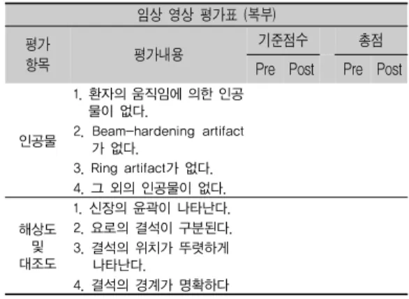
Axial 100 160 50% 0.625 0.625 400 40 임상 영상 평가표 (복부)
평가
항목 평가내용 기준점수 총점
Pre Post Pre Post
인공물
1. 환자의 움직임에 의한 인공 물이 없다.
2. Beam-hardening artifact 가 없다.
3. Ring artifact가 없다.
4. 그 외의 인공물이 없다.
해상도 및 대조도
1. 신장의 윤곽이 나타난다.
2. 요로의 결석이 구분된다.
3. 결석의 위치가 뚜렷하게 나타난다.
4. 결석의 경계가 명확하다 Table 2. Exposure and scan conditions base on the
Abdomen LDCT examination.
Vol. and Curr.; tube voltage and current, Wid.and Lev.; window width and level, Stand.; recon type of standard., AEC;
autoexposurecontrol
3. 영상의 정량적 분석방법
영상의 정량적 분석 방법으로 신장의 횡단면상(Axial image)과 관상면(Coronal imalge)에서 일정 크기의 관 심영역(region of image, ROI)을 설정하여 해당 관심영 역내 모든 화소(pixel)에 대한 최대값, 최소값, 평균 및 표준 편차를 구하고 설정된 관심영역 값을 구하였다 [Fig. 1].
Axial image coronal image
Fig. 1. Setting up the ROI for quantitative analysis in axial, coronal images.
4. 영상의 정성적 분석방법
영상의 정성적 분석 방법으로 CT 분야 임상 영상 평 가지의 영상 정보 항목을 참조하여 별도의 요로결석 영 상 평가표를 작성하였으며 결석의 크기와 위치, 경계면 의 선예도, 결석부위의 해상도 및 대조도 부분만을 평 가하였다[Table 3]. 영상의학과 전문의 2명과 전공의 2 명이 평가하였으며 각 문항 당 5점 척도로『매우 좋음, 좋음, 보통, 나쁨, 매우 나쁨』으로 구분하여 각 항목의 총점은 20점으로 하였다. 정성적 평가의 정확도를 위해 판독용 모니터 및 영상의 묘출 조건은 동일 조건으로 하였으며 통계방법(SPSSver. 18.0)으로 비모수적 검정
을 이용하였고 p 값이 0.05 미만일 때 통계적으로 유의 한 차이가 있는 것으로 해석하였다.
Table 3. The clinical image evaluation table non- enhanced Abdominal CT& low-dose CT.
Ⅲ. 연구결과
1. 영상의 정량적 분석
[Table 4]는 프로토콜 차이에 따른 요로결석 부위의 횡단면상과 관상면상에서의 관심영역 값을 나타내었 다. 횡단면상에서 복부촬영 프로토콜로 스캔 시 신장의 관심영역 내 평균 화소 값과 표준편차는 26.21±7.08, 저선량 CT 프로토콜로 스캔한 경우 평균 화소 값과 표 준편차는 20.03±8.16 이었다. 관상면상에서 복부촬영 프로토콜로 스캔 시 신장의 관심영역 내 평균 화소값과 표준편차는 22.07±7.35, 저선량 CT 프로토콜로 스캔 한 경우 평균 화소값과 표준편차는 21.67±6.11 이었다.
Table 4. Results of pixel and HU values in the ROI for quantitative analysis in axial images of the Abominal CT.
1; Abdomen CT pre scan protocol. 2; Abdomen LDCT scan protocol, SD; standard deviation, *p-value was less than 0.05.
Item Observer*
Avg. SD P
1 2 3 4
Abdomen CT pre scan
Artifact 19.35 18.89 19.20 19.12 19.14 0.36 0.035 Res.& Cont. 19.08 19.20 19.52 19.76 19.35 0.70 0.042 LDCT scan
Artifact 19.16 19.36 18.64 19.52 19.17 0.43 0.027 Res.& Cont. 18.64 18.76 19.48 19.48 19.29 0.58 0.033
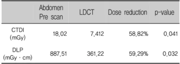
Abdomen
Pre scan LDCT Dose reduction p-value CTDI
(mGy) 18.02 7.412 58.82% 0.041
DLP
(mGy·cm) 887.51 361.22 59.29% 0.032
2. 영상의 정성적 분석
영상의 정성적 분석을 위하여 횡단면상과 관상면상 에서 저선량 CT 프로토콜 사용에 따른 영상의 인공물 과 해상도 및 대조도와 관련된 항목만을 평가 하였다.
[Table 5]는 각 문항당 5점 척도를 기준으로 4개 문항에 대한 각 항목의 총점을 20점으로 평가한 결과를 나타내 었다. 인공물에 대한 4명의 관찰자 평균값은 복부촬영 프로토콜로 스캔 시 19.14±0.36, 저선량 CT 프로토콜 로 스캔 시 19.17±0.43 이었으며 해상도 및 대조도의 평균값은 복부촬영 프로토콜로 스캔 시 19.35±0.70, 저 선량CT 프로토콜로 스캔 시 19.29±0.58 이었다.
3. 저선량CT 프로토콜 이용 시 피폭선량 분석 [Table 6]은 저선량 CT 프로토콜 사용 유‧무에 따른 환자의 CTDIvol과 DLP의 평균을 나타내었다. 복부촬 영 프로토콜로 스캔 시 CTDIvol의 평균값은 18.02 mGy, DLP의 평균값은 887.51 mGy·cm 이었으며 저선 량 CT 프로토콜을 이용한 경우 CTDIvol 평균값은 7.412 mGy, DLP 평균값은 361.22 mGy·cm 이었다. 이 로 인한 선량의 감소율은 각 58.82%, 59.29% 이었다.
Table 5. Results of evaluation by the clinical image evaluation table.
*, The average of clinical image evaluation values with 79 patients.
Res. & cont.; resolution and contrast, Avg.; average, SD; standard deviation. p-value was less than 0.05.
Tabel 6. Comparative Study on the Patient Exposure of Abdomen CT prescan and LDCT.
Ⅳ. 고찰 및 결론
컴퓨터단층촬영을 이용한 요로결석 진단의 유용성은 이미 다양한 연구를 통해 입증되어져 있다[9][10]. 매우 짧은 검사시간으로 환자의 불편함이 최소화 되었으며 조영제 사용에 따른 부작용이 없으므로 신장기능이 저 하되어진 환자에서도 검사의 적용이 가능하다. 또한 석 회화를 포함한 결석뿐만 아니라 경정맥 요로조영술에 서 보여주기 힘든 요산결석, 시스틴결석 등과 같은 방 사선 투과성이 높은 결석들도 주위 연부조직보다 현저 히 높은 음영으로 나타난다. 그러나 컴퓨터단층촬영은 방사선량이 매우 높은 단점을 지니고 있다. 그러므로 여러 방면에서 선량의 감소를 위한 노력들이 이루어지 고 있으며 본 연구도 그 중 하나이다. 임상에서 실제 CT 검사를 하는 방사선사들은 프로토콜의 적용 시 영 상의 화질저하를 방지하기 위한 방법으로 선량을 증가 시켜 검사하는 경우가 적지 않다. 모든 방사선 검사는 검사 과정에서 정당화(Justification)가 이루어져야 한 다. 하지만 정당화만을 강조하면 불필요한 피폭으로 인 하여 환자를 위험에 빠뜨리는 결과를 얻을 수 있다. 그 러므로 최적의 임상정보 제공를 제공하기 위한 ALARA(As Low As Reasonably Achievable) 원칙을 기본으로 하여 피폭선량과 영상의 화질에 대한 최적화 가 이루어져야 한다. 그러므로 장치의 특성을 이해하고 정확한 정보를 전달하는 것이 중요하다. 본 연구에서는 통계적 반복 재구성법을 이용하고 선량지표에 영향을 주는 관전압과 관전류를 조절하여 선량 저감화 프로토 콜을 제시하였다. 분석 결과 복부촬영 프로토콜과 저선 량CT 프로토콜을 각각 적용하여 스캔 시 노이즈 값의 증가는 발생하였지만 판독에는 영향을 미치지 않는 정 도의 차이를 나타내었고 환자의 피폭선량 감소치는 각 58.82, 59.29%로 나타났다. 그러므로 환자의 피폭선량 저감화를 위하여 요로결석 진단을 목적으로 하는 CT 촬영에서는 반드시 저선량 CT 프로토콜의 적용이 필요 할 것이라 생각된다.
참 고 문 헌
[1] R. Bartoletti, T. Cai, N. Mondaini, F. Melone, F.
Travaglini, M. Carini, and M. Rizzo,
“Epidemiology and risk factors in urolithiasis,”
Urologia Internationalis, Vol.79, Suppl.1, 2007.
[2] G. C. Curhan, “Epidemiology of stone disease,”
Urologic Clinics of North America, Vol.34, Issue.3, pp.287-293, 2007.
[3] S. Yilmaz, T. Sindel, G. Arslan, C. Özkaynak, K.
Karaali, A. Kabaalioğlu, and E. Lüleci, “Renal colic: comparison of spiral CT, US and IVU in the detection of ureteral calculi,” European radiology, Vol.8, Issue.2, pp.212-217, 1998.
[4] B. S. Kim, S. N. Kung, H. C. Kim, W. C.
Hwang, I. S. Lee, I. K. Hwang, H. C. Kim, S. H.
Bae, S. K. Lee, and S. H. Lee, “Usefulness of Unenhanced Helical CT in Patients with Suspected Ureteral Colic,” Journal of the Korean Society of Radiology, Vol.47, No.1, pp.61-67, 2002.
[5] B. S. John, U. Patel, and K. Anson, “What radiation exposure can a patient expect during a single stone episode?,” Journal of endourology, Vol.22, No.3, pp.419-422, 2008.
[6] D. J. Brenner and E. J. Hall, “Computed tomography an 14 increasing source of radiation exposure,” The New England ournal of medicine, Vol.357, No.22, pp.2277-2284, 2007.
[7] M. Lopez and B. Hoppe, “History, epidemiology and regional diversities of urolithiasis,” Pediatric nephrology, Vol.25, No.1, pp.49-59, 2010.
[8] National Research Council, Health Risks from Exposure to Low Levels of Ionizing Radiation:
BEIR VII Phase 2, The National Academies Press, 2006.
[9] V. P. Sukumar, J. Collingwood, T. Crawley, D.
Schofield, J. Henson, K. Lakin, D. Connolly, and
J. Giles, “Low dose computed tomography in suspected acute renal colic,” Clin Radiol, Vol.56, No.11, pp.873-876, 2001
[10] P. Rogalla, C. Klüner, and M. Taupitz,
“Ultra-low dose CT to search for stones in kidneys andcollecting system [in German],”
AktuelleUrol, Vol.35, No.4, pp.307-309, 2004.
저 자 소 개
김 현 진(Hyeon-Jin Kim) 정회원
▪2016년 8월 : 동의대학교 보건의 과학과(방사선학박사)
▪2016년 3월~ 현재 : 가야대학교 방사선학과 교수
<관심분야> : 의료영상정보학, 방사선 영상학, 컴퓨터 단층촬영
지 태 정(Tae-Jeong Ji) 정회원
▪2006년 12월 : 안동대학교 생물 학과(이학박사)
▪2004년 3월 ~ 현재 : 가야대학교 방사선학과 교수
<관심분야> : 초음파영상학, 방사선생물학, 인체생리 학, 투시조영영상학