Concurrent Chemoradiotherapy in Locally Advanced Esophageal Cancer
Sang Jun Byun, M.D.*, Jin Hee Kim, M.D.*, Ok Bae Kim, M.D.* and Hong Suk Song, M.D.
†Departments of *Radiation Oncology and
†Internal Medicine, Dongsan Medical Center,
Keimyung University School of Medicine, Daegu, Korea
Purpose: This study was designed to evaluate the results of local control, survival rate, prognostic factors, and failure pattern in locally advanced esophageal cancer.
Materials and Methods: We retrospectively studied 50 patients with locally advanced esophageal cancer treated with concurrent chemoradiotherapy at Keimyung University Dongsan Medical Center from June of 1999 to August of 2008. Seven patients with inappropriate data were excluded, and 43 patients were analyzed. There were 39 males and four female patients ranging in age from 43 to 78 years (median, 63 years). There were seven patients with stage IIA and 36 with stage III. Irradiation from 46 Gy to 63 Gy (median, 54 Gy) was carried out 5 days per week, 1.8 Gy once a day. There were eight patients with neo-adjuvant chemotherapy, and we mostly used 5-fluorouracil, cisplatin with 3 cycles for concurrent chemotherapy. The range of follow up periods was from 2 to 82 months (median, 15.5).
Results: There were nine patients that exhibited a complete response, 23 that exhibited a partial response, 9 that exhibited no response, and 2 that exhibited disease progression. The median survival time was 15 months.
Two-year and 5-year survival rates were 36.5% and 17.3%, respectively. Two-year and 5-year disease-free survival rates were 32.4% and 16%, respectively. Treatment failure occurred in 22 patients (51.2%). Patterns of failure were categorized as local failure in 18 patients and distant metastasis in four patients. In a univariate analysis for prognostic factors related to overall survival and disease-free survival, the hemoglobin levels during chemoradiotherapy (≥12 vs. <12, p=0.02/p=0.1) and the response to the treatments (CR/PR vs. NR/PD, p=0.002/p<0.0001) were statistically significant. In a multivariate analysis, only response to the treatments was revealed to be statistically significant. There was no statistical significance associated with patient age, gender, disease stage, T-stage, smoking history, tumor location, or neo-adjuvant chemotherapy.
Conclusion: Our survival rate was similar to those of other institutions. Local recurrence was the main reason for failure. It is suggested that further prospective studies should be performed to improve local control.
Key Words: Esophageal cancer, Concurrent chemoradiotherapy, Survival rate, Prognostic factor, Failure
Submitted November 17, 2010, accepted January 19, 2011 Reprint requests to Jin Hee Kim, M.D., Department of Radiation Oncology, Dongsan Medical Center, Keimyung University, 194 Dongsan-dong, Jung-gu, Daegu 700-712, Korea
Tel: 053)250-7665, Fax: 053)250-7984 E-mail: [email protected]
Introduction
Squamous cell carcinoma in the esophagus is a lethal disease with poor prognosis. It is related to a lymphatic spreading pattern with defects in serosal lining, frequent direct extension into surrounding structures, and even metastasis.
1)In the past decades, surgery after diagnosis of the disease in a locally-advanced state was a standard therapeutic option and
reported to have low curative rates. There were also several reports to demonstrate better outcomes with postoperative radiotherapy than those with surgery alone.
2∼4)DeMeester and Barlow
2)suggested that improvement of survival rates might be expected because microscopic tumor after surgery could be eradicated by radiotherapy. The use of chemotherapy followed by surgery had proven to be not effective for preventing distant metastasis or local failure in esophageal cancer.
5)There were also several trials assessing the effect of preoperative radiotherapy, but there was no clear benefit regarding survival in case of locally-advanced esophageal cancer.
6)Recently, concurrent chemoradiotherapy has been associated
with more favorable outcomes for locally-advanced esophageal
Table 1. Patients Characteristics
Characteristics Values
Age Gender Male Female
Pathology - Squamous cell carcinoma Well differentiated
Moderately differentiated Poorly differentiated Unknown
Stage IIA III
Performance status ECOG* 0 ECOG 1
Radiation therapy dose (Gy) <54
≥54 T-stage T2 T3 T4 N-stage Nx N0 N1
Tumor location Cervical Upper thoracic Middle thoracic Lower thoracic
43∼78 (median 63) 39 4 4 16 5 18 7 36 22 21 6 37 5 17 21 1 10 32 2 2 26 13
*Eastern Cooperative Oncology Group.
cancer because it can improve local control and combat micrometastases simultaneously. Chemotherapeutic agents like cisplatin and 5-fluorouracil (5-FU) are typically used as radiosensitizers and impart anti-tumor effects.
7,8)A phase III trial in the United States, Radiation Therapy Oncology Group (RTOG) 85-01
9)revealed that the addition of chemotherapy to radiation therapy is superior to radiation therapy alone. Furthermore, concurrent chemoradiotherapy has become a standard of treatment for locally-advanced esopha- geal cancer.
This study was designed to retrospectively evaluate the local control, survival rate, prognostic factors and failure patterns in locally-advanced esophageal cancer treated with concurrent chemoradiotherapy in our institution.
Materials and Methods
We retrospectively studied 50 patients with locally-advanced esophageal cancer treated with concurrent chemoradiotherapy at Keimyung University Dongsan Medical Center from June of 1999 to August of 2008. Patients underwent physical examination, complete blood cell count, chest X-ray, chest high resolution computed tomography, positron emission tomography computed tomography (PET-CT), and endoscopic biopsies to confirm the presence of disease and evaluate the clinical stage. PET-CT scan was performed since March of 2007 in our institution and only eight patients underwent PET-CT for clinical staging. The patients were grouped according to the AJCC TNM 6th edition staging method. The eligibility criteria for locally-advanced disease were stage T1N1M0 or T2-4, any N, M0. Periods for evaluating treat- ment outcomes were calculated from the first day of radio- therapy.
Two patients without regular follow-up after completion of concurrent chemoradiotherapy and five patients for whom we could not confirm treatment results due to the absence of appropriate follow-up examination were excluded from this study; in total, 43 patients were analyzed in our study. The patients’ characteristics are presented in Table 1. There were 39 males and four female patients with a range of age from 43 to 78 years old (median, 63 years). There were seven patients with stage IIA and 36 with stage III. All patients were squamous cell carcinoma in pathology. The performance
status before the treatments was Eastern Cooperative Oncology Group (ECOG). 0 in 22 patients and ECOG 1 in 21 patients.
In radiation therapy planning, a margin of 5 cm above and below the GTV and 1.5-cm radial margin are usually applied to cover subclinical disease. The margin was reduced to 2 cm in above and below the GTV and 1 cm in radial margin after 45 Gy. Two-dimensional technique was applied for 28 patients until December of 2004 with anteroposterior (AP)-posteroan- terior (PA) bilateral opposing fields up to 41.4 Gy and more than two fields to shield spinal cord for remaining dose.
Three-dimensional conformal technique was used since Jan of 2005 for 15 patients. Forty-six Gy to 63 Gy of radiation was delivered with 6/10/15 MV photons (median, 54 Gy) for 5 days per week and 1.8 Gy once a day.
There were eight patients who underwent concurrent
chemoradiotherapy 4 weeks after completion of neo-adjuvant
chemotherapy. Six patients were treated with 5-FU/cisplatin
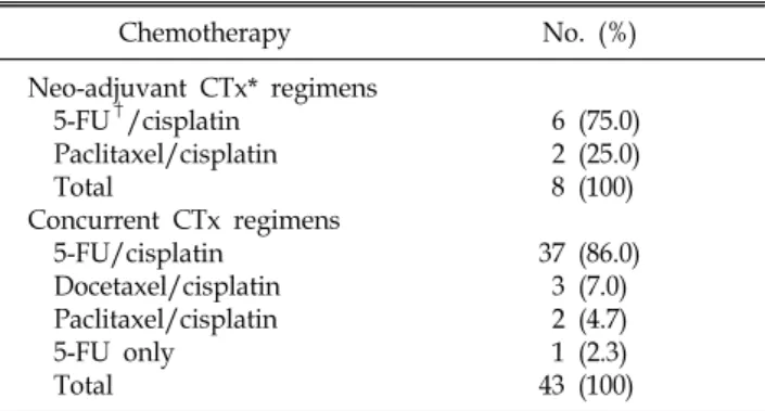
Table 2. Chemotherapeutic Regimens
Chemotherapy No. (%)
Neo-adjuvant CTx* regimens 5-FU†/cisplatin
Paclitaxel/cisplatin Total
Concurrent CTx regimens 5-FU/cisplatin
Docetaxel/cisplatin Paclitaxel/cisplatin 5-FU only Total
6 (75.0) 2 (25.0) 8 (100) 37 (86.0)
3 (7.0) 2 (4.7) 1 (2.3) 43 (100)
*chemotherapy, †5-fluorouracil.
Table 3. Treatment Response
Response No. (%)
Complete response 9 (20.9)
Partial response 23 (53.5)
No response 9 (20.9)
Progression of disease 2 (4.7)
Total 43 (100)
Fig. 1. Overall survival curve.
and two patients were given paclitaxel/cisplatin before concurrent chemoradiotherapy. Concurrent chemoradiotherapy was administered, mainly 5-FU/cisplatin-based regimens with continuous infusion. 5-FU 1,000 mg/m
2with 5% dextrose in water 500 mL was delivered, starting on days 1 to 4 as a continuous intravenous infusion. Cisplatin 75 mg/m
2with 0.9% normal saline 500 mL was delivered during 1 day with 3-week interval. Thirty-seven patients were treated with 5-FU/cisplatin and two of them were administered additional chemotherapy. One was given additional docetaxel/cisplatin, and another one patient was treated with paclitaxel/cisplatin.
Three patients were treated with docetaxel/cisplatin and one of them received additional dose of 5-FU/cisplatin. Two patients were treated with paclitaxel/cisplatin and one of them received additional 5-FU/cisplatin treatment. The remaining 1 patient was administered only 5-FU (Table 2).
Follow-up chest CT scans or endoscopic examinations were performed at 1 to 3 months after completion of concurrent chemoradiotherapy. We used World Health Organization (WHO) criteria including complete response (CR), partial response (PR), no response (NR), and progression of disease (PD) for assessment after completion of concurrent chemora- diotherapy.
10)The range of follow-up periods was from 2 to
82 months with a median of 15.5 months. The time to local failure and distant metastases were analyzed from the starting day of any treatment modalities including neo-adjuvant chemotherapy and concurrent chemoradiotherapy after diagnosis.
The Kaplan-Meier method was used to estimate overall survival rates (OS) and disease-free survival rates (DFS).
Univariate analysis evaluating factors associated with OS was performed by a log-rank test. Factors found to influence survival on univariate analysis were then analyzed by Cox proportional hazard regression analysis. Statistical analyses were performed with SPSS ver. 17.0 (SPSS Inc., Chicago, IL, USA).
Results
1. Local control and survival
As shown in Table 3, there were nine patients showing complete response (20.9%), 23 presenting partial responses (53.5%), nine showing no response (20.9%), and two with disease (4.7%). Two-year and 5-year overall survival rates were 36.5% and 17.3%, respectively (Fig. 1). Two-year and 5-year disease-free survival rates were 32.4% and 16%, respectively (Fig. 2). The median survival of all patients was 15 months.
2. Prognostic factor
We analyzed several factors that may impact disease
prognosis including patient age, gender, disease stage, smoking
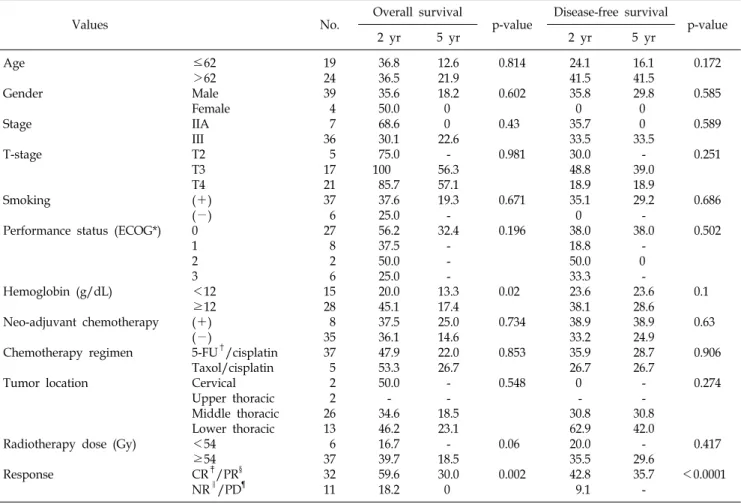
Table 4. Univariate Analysis of Prognostic Factors Related to Overall Survival
Values No. Overall survival
p-value Disease-free survival
p-value
2 yr 5 yr 2 yr 5 yr
Age Gender Stage T-stage
Smoking
Performance status (ECOG*)
Hemoglobin (g/dL) Neo-adjuvant chemotherapy Chemotherapy regimen Tumor location
Radiotherapy dose (Gy) Response
≤62
>62 Male Female IIA III T2 T3 T4 (+) (−) 0 1 2 3
<12
≥12 (+) (−)
5-FU†/cisplatin Taxol/cisplatin Cervical Upper thoracic Middle thoracic Lower thoracic
<54
≥54 CR‡/PR§ NR∥/PD¶
19 24 39 4 7 36 5 17 21 37 6 27 8 2 6 15 28 8 35 37 5 2 2 26 13 6 37 32 11
36.8 36.5 35.6 50.0 68.6 30.1 75.0 100
85.7 37.6 25.0 56.2 37.5 50.0 25.0 20.0 45.1 37.5 36.1 47.9 53.3 50.0 - 34.6 46.2 16.7 39.7 59.6 18.2
12.6 21.9 18.2 0 0 22.6
- 56.3 57.1 19.3 - 32.4
- - - 13.3 17.4 25.0 14.6 22.0 26.7 - - 18.5 23.1 - 18.5 30.0 0
0.814 0.602 0.43 0.981
0.671 0.196
0.02 0.734 0.853 0.548
0.06 0.002
24.1 41.5 35.8 0 35.7 33.5 30.0 48.8 18.9 35.1 0 38.0 18.8 50.0 33.3 23.6 38.1 38.9 33.2 35.9 26.7 0
- 30.8 62.9 20.0 35.5 42.8 9.1
16.1 41.5 29.8 0 0 33.5
- 39.0 18.9 29.2 - 38.0
- 0 - 23.6 28.6 38.9 24.9 28.7 26.7 - - 30.8 42.0 - 29.6 35.7 -
0.172 0.585 0.589 0.251
0.686 0.502
0.1 0.63 0.906 0.274
0.417
<0.0001
*Eastern Cooperative Oncology Group, †fluorouracil, ‡complete response, §partial response, ∥no response, ¶progression of disease.
Fig. 2. Disease free survival curve.
history, total radiation dose, prior treatments before concurrent chemoradiotherapy, hemoglobin level at the time of chemoradiotherapy, tumor location in the esophagus, response to treatment and absence or presence of neo-adjuvant
chemotherapy.
As shown in Table 4, on univariate analysis for identifying potential prognostic factors related to overall survival, hemoglobin level at the time of chemoradiotherapy (<12 vs.
≥12, p=0.02) and response to treatments (CR/PR vs. NR/PD, p=0.002) were statistically significant (Fig. 3, 4) and the total dose of radiation therapy (<54 Gy vs. ≥54 Gy, p=0.06) was marginally significant. On multivariate analysis, response to treatments was only found to be statistically significant (95%
confidence interval [CI], 1.289 to 7.595; hazard ratio [HR], 3.129; p=0.012).
On univariate analysis for identifying potential prognostic
factors related to disease-free survival, response to treatments
(CR/PR vs. NR/PD, p<0.0001) was statistically significant
and hemoglobin level at the time of chemoradiotherapy (<12
vs. ≥12, p=0.1) was marginally significant. On multivariate
analysis, response to treatments was also found to be
Fig. 3. Overall survival curves according to hemoglobin level.
Fig. 4. Overall survival curves according to response after
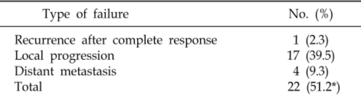
treatments. *complete response, †partial response, ‡no respon- se, §progression of disease.Table 5. Patterns of Failure
Type of failure No. (%) Recurrence after complete response 1 (2.3)
Local progression 17 (39.5)
Distant metastasis 4 (9.3)
Total 22 (51.2*)
*percentage in all patients.
Table 6. Complications
Complication No. (%)
Acute complication Esophagitis 5/43 (11.6)
Ulceration 1/43 (2.3)
Late complication Stricture/obstruction 12/43 (27.9)
statistically significant (95% CI, 1.836 to 9.841; HR, 4.251;
p=0.001). There was no statistical significance associated with patient age, gender, disease stage, T-stage, smoking history, tumor location, or neo-adjuvant chemotherapy.
3. Patterns of treatment failure
There were 22 patients with recurring disease (51.2%) and the patterns of the recurrence were separated to local failure in one patient among nine with complete response (2.3%), local progression in 17 patients (39.5%), and distant metastasis in four individuals (9.3%), as shown in Table 5. Disease recur- rence in the one patient with a complete response occurred 42 months after completion of concurrent chemoradiotherapy.
There was no patient who had both local treatment failure and
distant metastasis.
4. Complications
Endoscopic examination was performed 1 to 3 months after completion of concurrent chemoradiotherapy and five patients (11.6%) developed esophagitis. Three patients were diagnosed at 2 months and two patients at 1 month following chemo- radiotherapy. The one patient (2.3%) with esophagitis after 1 month also suffered from ulcers.
Esophageal strictures and obstructions occurred in 12 patients (27.9%) with dysphagia. Six patients were found to have strictures 2 months after treatment and two patients developed dysphagia after 1 month. Dysphagia was found in the four patients at 5, 6, 7, and 22 months following treatment by follow-up endoscopic examination (Table 6).
Discussion and Conclusion
Although there have been several trials conducted with the
goal of improving survival of locally-advanced esophageal
cancer, it is still challenging to expect better outcomes in
these patients. Herskovic et al.
11)reported a 50% 1-year
overall survival rate and 38% 2-year overall survival rate, with
a median survival period of 12.5 months in 121 locally-advan-
ced esophageal cancer patients who received concurrent
chemoradiotherapy. Smith et al.
12)also reported a 27% 2-year
overall survival among 119 patients with stage I and II after
concurrent chemoradiotherapy with a median survival period
of 14.8 months. They also found improved overall survival in
patients with locally-advanced esophageal cancer compared to those who received radiation therapy alone. Two-year and 5-year overall survival rates were 27% and 9%, respectively, in chemoradiotherapy group, and 12% and 7%, respectively, in the group that only received radiation.
RTOG 85-01 showed that treatment with concurrent chemo- radiotherapy resulted in significantly increased overall survival rates compared to radiotherapy alone in cases of locally-adva- nced esophageal cancer (T1-3 N0-1 M0). The overall survival rate for the concurrent chemoradiotherapy group at 5 years was 26% and 0% for the group that only received radiothe- rapy.
9)In our study, the overall survival rates at 2 years and 5 years were 36.5% and 17.3%, respectively, with a median survival periods of 15 months, similar to outcomes of other trials. However, the distribution of patients according to disease stage was relatively uneven, seven patients were stage IIA and 36 were stage III. More reliable results would be obtained if the study had been conducted with a more uniform patients group.
There are several factors related to prognosis in esophageal cancer patients including disease stage, tumor location, tumor size, histologic type, gender, ethnicity, patient age, and response to treatment. Generally, tumor oxygenation is also considered to be of prognostic value and effect local control of tumors in locally-advanced cancers.
13∼17)Many investigators also have reported that hemoglobin level before treatment is a valuable prognostic factor.
18∼21)In our study, the pre-treatment hemoglobin level higher than 12 g/dL is a significant progno- stic factor in survival. Neuhof et al.
21)have reported that patients with hemoglobin concentrations >13.4 g/dL had significantly better overall survival rates than ones with lower hemoglobin concentrations. Rades et al.
20)also reported that the best overall survival rate was achieved when the hemoglobin concentration level was between 12.1 and 14.0 g/dL followed by levels >14.1 g/dL and <12 g/dL suggest- ing the existence of an optimal range of hemoglobin concent- rations. Therefore, assessment of hemoglobin level before concurrent chemoradiotherapy and regular follow-up during the treatments are proposed to enhance the effect on patient prognosis.
Response to treatments has been also reported to be a prognostic factor related to survival rates.
22,23)Stahl et al.
22)reported that patients with tumors responding to treatment had
a better probability of surviving, whereas the outcomes of non-responders was generally poor. We divided patients in our study into two response groups: patients with complete responses or partial responses and patients with no response or disease progression. Given the results of our study, patient prognosis in patients may be predicted according to the response to treatment.
Stage is one of the most meaningful prognostic factors in estimating survival rates including depth of tumor invasion, nodal involvement, and distant metastases. Results related to staging in our institution did not show any significance to expect the prognosis for esophageal cancer patients. However, the number of patients in our study was relatively small compared to other institutional studies. Furthermore, the TNM classification with 6th edition is simply assorted compared with recent staging system.
Esophageal cancers in men tend to have a more aggressive nature with poorer outcomes.
1)Result in our study did not show any statistical significance according to gender. How- ever, the relatively small number of female patients in our study causes a little significance for expecting prognosis.
Herskovic et al.
11)reported a 39% rate of local recurrence after concurrent chemoradiotherapy, 7% were distant metasta- sis only and 5% were local recurrences with distant metasta- sis. In our study, local recurrence rate after complete response to treatment was 2.3%, local progression was found in 39.5%
of the patients, and distant metastases were reported in 9.3%
of the patients. There were no patients with local recurrence and distant metastasis at the same time. The patterns of treatment failure in our study are similar with other institutional studies.
Generally, it has been found that acute complications in patients undergoing chemoradiotherapy are more severe than those of patients receiving radiation alone. These complica- tions include esophagitis along with other common problems like nausea, vomiting, epidermitis, fatigue, and ulceration.
Khurana et al.
24)reported a 6% rate of ulceration and 21%
rate of esophageal stricture among 68 patients with esophageal
cancer after concurrent chemoradiotherapy. Our results showed
11.6% of esophagitis with 2.3% of ulceration and 27.9% of
esophageal stricture and obstruction. In our study, small
number of patients and irregular follow-up periods might
effect on the difference in the results compared with other
trials. Future studies need to examine at appropriate time with many patients group to get more reliable result.
In conclusion, the survival rates of patients with locally-ad- vanced esophageal cancer treated with concurrent chemora- diotherapy in our study were similar to those of other institutions. Local recurrence was a main cause of treatment failure. It is suggested that further prospective studies need to be performed to improve local control.
References
1. Halperin EC, Perez CA, Brady LW. Perez and Brady's principles and practice for radiation oncology. 5th ed. New York: Lippincott Williams & Wilkins, 2008:1131-1153
2. DeMeester TR, Barlow AP. Surgery and current management for cancer of the esophagus and cardia. Curr Probl Cancer 1988;12:243-328
3. Harrison LB, Fogel TD, Picone JR, Fischer DB, Wei- ssberg JB. Radiation therapy for squamous cell carcinoma of the esophagus. J Surg Oncol 1988;37:40-43
4. Pearson JG. The value of radiotherapy in the management of squamous oesophageal cancer. Br J Surg 1971;58:794-798 5. Kelsen DP, Ginsberg R, Pajak TF, et al. Chemotherapy followed by surgery compared with surgery alone for localized esophageal cancer. N Engl J Med 1998;339:1979-1984 6. Arnott SJ, Duncan W, Gignoux M, et al. Preoperative
radiotherapy in esophageal carcinoma: a meta-analysis using individual patient data (Oesophageal Cancer Collaborative Group). Int J Radiat Oncol Biol Phys 1998;41:579-583 7. Rich T. Chemoradiation in conservation therapy for esopha-
geal cancer. Hematol Oncol Clin North Am 2001;15:291-302 8. Hennequin C, Maylin C. Continuous radiosensitizing che-
motherapy. Pathol Biol (Paris) 1999;47:279-281
9. Cooper JS, Guo MD, Herskovic A, et al. Chemora- diotherapy of locally advanced esophageal cancer: long-term follow-up of a prospective randomized trial (RTOG 85-01).
Radiation Therapy Oncology Group. JAMA 1999;281:1623-1627 10. James K, Eisenhauer E, Christian M, et al. Measuring response in solid tumors: unidimensional versus bidimensional measurement. J Natl Cancer Inst 1999;91:523-528
11. Herskovic A, Martz K, al-Sarraf M, et al. Combined chemotherapy and radiotherapy compared with radiotherapy alone in patients with cancer of the esophagus. N Engl J Med 1992;326:1593-1598
12. Smith TJ, Ryan LM, Douglass HO Jr, et al. Combined
chemoradiotherapy vs. radiotherapy alone for early stage squamous cell carcinoma of the esophagus: a study of the Eastern Cooperative Oncology Group. Int J Radiat Oncol Biol Phys 1998;42:269-276
13. Zenda S, Hironaka S, Boku N, et al. Impact of hemo- globin level on survival in definitive chemoradiotherapy for T4/M1 lymph node esophageal cancer. Dis Esophagus 2008;21:195-200
14. Knocke TH, Weitmann HD, Feldmann HJ, Selzer E, Potter R. Intratumoral pO2-measurements as predictive assay in the treatment of carcinoma of the uterine cervix.
Radiother Oncol 1999;53:99-104
15. Menon C, Fraker DL. Tumor oxygenation status as a prognostic marker. Cancer Lett 2005;221:225-235
16. Vaupel P. Prognostic potential of the pre-therapeutic tumor oxygenation status. Adv Exp Med Biol 2009;645:241-246 17. Nordsmark M, Bentzen SM, Rudat V, et al. Prognostic
value of tumor oxygenation in 397 head and neck tumors after primary radiation therapy: an international multi-center study.
Radiother Oncol 2005;77:18-24
18. Grigiene R, Aleknavicius E, Kurtinaitis J. Prognostic value of anemia for patients with cervical cancer treated with irradiation. Medicina (Kaunas) 2005;41:916-924
19. Langendijk H, de Jong J, Wanders R, Lambin P, Slotman B. The importance of pre-treatment haemoglobin level in inoperable non-small cell lung carcinoma treated with radical radiotherapy. Radiother Oncol 2003;67:321-325 20. Rades D, Schild SE, Bahrehmand R, Zschenker O,
Alberti WA, Rudat VR. Prognostic factors in the nonsur- gical treatment of esophageal carcinoma with radiotherapy or radiochemotherapy: the importance of pretreatment hemoglobin levels. Cancer 2005;103:1740-1746
21. Neuhof D, Neumayer F, Einbeck W, et al. Retrospective evaluation of combined modality treatment and prognostic fac- tors in patients with esophageal cancer. Acta Oncol 2005;44:
168-173
22. Stahl M, Stuschke M, Lehmann N, et al. Chemoradiation with and without surgery in patients with locally advanced squamous cell carcinoma of the esophagus. J Clin Oncol 2005;23:2310-2317
23. Noh OK, Je HU, Kim SB, et al. Results of definitive chemoradiotherapy for unresectable esophageal cancer. J Korean Soc Ther Radiol Oncol 2008;26:195-203
24. Khurana R, Dimri K, Lal P, et al. Factors influencing the development of ulcers and strictures in carcinoma of the esophagus treated with radiotherapy with or without concurrent chemotherapy. J Cancer Res Ther 2007;3:2-7
국문초록
국소적으로 진행된 식도암에서 동시항암화학방사선치료의 결과