Spinal Accessory Neuropathy Associated With the Tumor Located on the Jugular Foramen
Soyoung Lee, MD, Shimo Yang, MD, Jieun Lee, MD, Inhwan Kim, MD
Department of Rehabilitation Medicine, Dongsan Medical Center, Keimyung University School of Medicine, Daegu, Korea
Spinal accessory neuropathy is commonly caused by iatrogenic injury or secondary to trauma or infection.
Nevertheless, the tumor related palsy is rare. We present a case of an 18-year-old male patient suffering from paralysis of his right trapezius and sternocleidomastoid muscle. An electrophysiologic diagnostic study confirmed the spinal accessory neuropathy of the proximal segment. In addition, magnetic resonance imaging showed the location of tumor on the jugular foramen. However, the type of the tumor was not confirmed through biopsy because the patient refused surgical procedure. Based on the study, it is hypothesized that the tumor located on the jugular foramen should be considered as a cause of the spinal accessory nerve of the proximal segment.
Keywords Accessroy nerve, Glomus jugulare, Neoplasms
Annals of Rehabilitation Medicine
Ann Rehabil Med 2013;37(1):133-137 pISSN: 2234-0645 • eISSN: 2234-0653 http://dx.doi.org/10.5535/arm.2013.37.1.133
INTRODUCTION
The spinal accessory nerve (SAN) is the pure motor innervation to the trapezius and sternocleidomastoid (SCM) muscles. The trapezius muscle is a major stabi- lizer of the scapula. On the other hand, SCM muscle is an ipsilateral flexor or contralateral rotator of the neck. The SAN causes weakness of the trapezius, which leads to scapular winging. The occurrence of SCM muscle palsy is rare, which leads to weakness of contralateral rotation of the neck. Most of the reported cases of SAN are associ-
ated with iatrogenic injury caused during various surgical procedures in the posterior triangle of the neck, such as tumor resection, cervical lymph node biopsy, and radi- cal neck dissection [1]. Other causes such as traction in- jury, blunt trauma, infection have also been reported as the causes of SAN [2]. But, the SAN related to the tumor located on the jugular foramen is very rare. The tumors located on the jugular foramen have caused the SAN by invasion or compression of SAN. We report in this paper an unusual case of a patient presented with SAN, which was secondary to the tumor located on the jugular fora- men.
CASE REPORT
An 18-year-old male visited the clinic with complaints of constant dull ache located in the right shoulder girdle, associated with sudden difficulty with overhead activi- ties and heavy lifting. There was no report of history of neck trauma or manipulation that would predispose the patient to injury. A week later, the pain was worsened.
There were no associated episodes of headaches, nausea
Received February 1, 2012; Accepted June 5, 2012 Corresponding author: Shimo Yang
Department of Rehabilitation Medicine, Dongsan Medical Center, Keimyung University School of Medicine, 56 Dalseong-ro, Jung-gu, Daegu 700-712, Korea
Tel: +82-53-250-7264, Fax: +82-53-250-7268, E-mail: shimo.yang@gmail.
com
This is an open-access article distributed under the terms of the Creative Commons Attribution Non-Commercial License (http://creativecommons.
org/licenses/by-nc/3.0) which permits unrestricted noncommercial use, distribution, and reproduction in any medium, provided the original work is properly cited.
Copyright © 2013 by Korean Academy of Rehabilitation Medicine
and vomiting or numbness in right upper extremity.
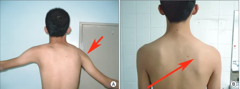
Physical examination revealed winging of the patient’s right scapula and severe atrophy of the right trapezius muscle (Fig. 1). Moreover, there was weakness in contra- lateral rotation and ipsilateral flexion of the neck on the right side. Passive range of motion of his right shoulder joint was normal. Manual muscle test grade was poor plus in flexion and abduction; and normal in external and internal rotation on his right shoulder. The patient was unable to abduct and flex the right arm forwardly to more than 70 to 80 degrees (Fig. 1). There were normal muscle tone, normal sensory function and symmetrical
upper extremity reflexes. Hawkins’, Neer’s, Roos’, Wright’s, and Adson’s provocative tests were all negative.
The magnetic resonance imaging (MRI), nerve conduc- tion study (NCS), and electromyography (EMG) on the right shoulder were performed to determine the cause of weakness and pain, on the basis of history and physical examination.
The MRI demonstrated increased signal intensity at the supraspinatus muscle. This finding was suggestive of par- tial tear of supraspinatus muscle on the articular surface.
NCS and EMG were carried out almost 10 days after the onset of symptoms. NCS of the SANs showed low ampli-
Fig. 1. Weakness of the shoulder abduction (A) and winging of the right scapula (B).
Table 1. Nerve conduction studies
Nerve
Motor conduction study Recording site Distal latency
(ms) Distal amplitude
(mV) Conduction
velocity (m/sec)
Left median nerve Abductor pollicis brevis 2.45 12.7 58.8
Right median nerve Abductor pollicis brevis 2.60 15.7 59.1
Left ulnar nerve Abductor digiti minimi 2.25 9.7 57.8
Right ulnar nerve Abductor digiti minimi 2.35 9.3 54.3
Left axillary nerve Deltoid 3.35 9.0 -
Right axillary nerve Deltoid 3.40 8.5 -
Left spinal accessory nerve Upper trapezius 1.95 3.9 -
Left spinal accessory nerve Middle trapezius 3.65 5.1 -
Left spinal accessory nerve Lower trapezius 5.75 2.4 -
Right spinal accessory nerve Upper trapezius 1.95 1.8 -
Right spinal accessory nerve Middle trapezius No response
Right spinal accessory nerve Lower trapezius No response
tude and prolonged latency of nerve potential of the right side when compared with the left side (Table 1). EMG studies revealed positive sharp waves at rest (Fig. 2) and reduced recruitment, normal morphology, regular firing pattern, and stable stability on volition in the right upper trapezius and SCM muscle. The other examined muscles in both the upper extremities showed no denervation potentials. These findings were consistent with the SAN, involving proximal to the innervation site of SCM. The proximal segment of SAN was considered the following causes, such as the space-occupying lesion in the head and neck, trauma injury or infection. Cervical MRI re- vealed no abnormal findings. However, MRI of the brain showed a 19×19×20 mm-sized mass in the right jugular foramen. The tumors located on the jugular foramen
were most commonly paragangliomas, schwannomas, and meningiomas. The lesion was hyperintense to brain parenchyma on T1-weighted series and of low signal on T2-weighted series. Subsequently, computed tomog- raphy (CT) of the temporal bone was performed where axial images revealed relatively smooth expansion of the right jugular foramen (Fig. 3). Cerebral angiography re- vealed no vascular abnormalities.
Because of presumed nerve injury associated with the tumor located on the jugular foramen, surgical approach was needed potentially. However, operation was ad- journed due to patient’s personal reasons. During the fol- low-up, the patient was treated intermittently for the SAN such as with electrical stimulation therapy and exercise.
After 10 months, he visited our clinic for follow-up study.
Fig. 2. Electromyography of the right sternocleidomastoid and trapezius muscles. Denervation in the examined muscles with posi- tive sharp waves.
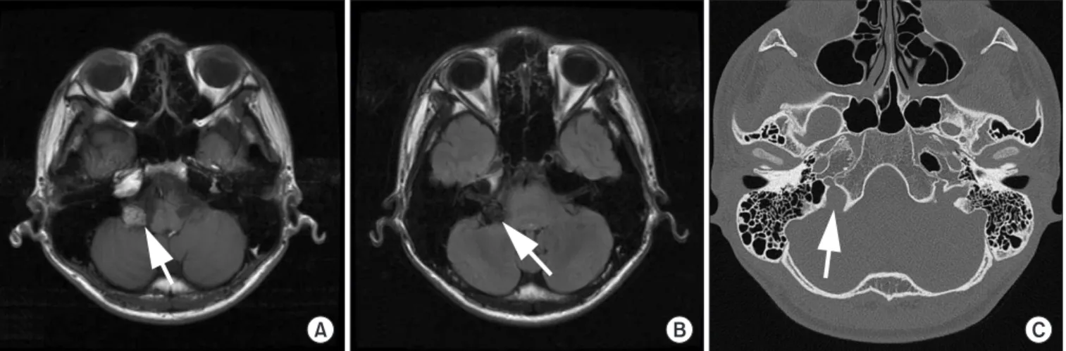
Fig. 3. Magnetic resonance imaging of the tumor located on the jugular foramen. (A) On T1-weighted image, the tu- mor (arrow) shows high signal intensity when compared with brain matter. (B) On T2-weighted image, the tumor (ar- row) has low signal intensity when compared with brain matter. (C) Computed tomographic image of the same tumor (arrow) shows smooth expansion of the right jugular foramen.
There were no gross interval changes on the previous findings on brain MRI of the 19×19×22 mm-sized tumor (Fig. 4). And there was no aggravation of the symptoms.
DISCUSSION
The SAN is a pure motor nerve that derives from the C1—C4 cervical segments and ascends through the fora- men magnum, to return through the jugular foramen. Af- ter it supplies motor innervation to SCM muscle, it enters the posterior triangle of the neck before it innervates the trapezius muscle. Moreover, the nerve travels caudally and dorsally in the subcutaneous tissue along the pos- terior triangle of the neck. The SAN is damaged along a course of the nerve by either compression or other injury.
In the SAN, the patient often initially presents with winged scapula. This condition can develop due to the nerve injury to the long thoracic nerve, the SAN, or the dorsal scapular nerve. In the present case, the lower me- dial border and inferior angle of the patient’s right scapu- la were more prominent and laterally located. Also, there was shoulder drooping that would suggest trapezius at- rophy. Based on these findings, paralysis of the trapezius muscle was suspected.
The SAN has occurred predominantly in the distal seg- ment, involving the trapezius muscle because the spinal accessory nerve is susceptible to traumatic injury in the posterior triangle of the neck. The SAN of proximal seg- ment involving the trapezius and SCM muscle is uncom- mon, especially when associated with the tumor.
The common tumors to develop in the jugular foramen are paragangliomas, schwannomas, meningiomas, and metastatic tumors. It is difficult to identify the type of
tumor on the basis of clinical presentation alone. There- fore, the definitive preoperative diagnosis is made with radiographic studies.
Typically, on MRI studies, a schwannoma located on the jugular foramen will frequently show hypointense on T1 images and hyperintense on T2 series when compared with the brain white matter. On bone algorithm CT im- ages, a smooth enlargement of the jugular foramen will be vivid.
A paraganglioma located on the jugular foramen will commonly show isointense or hypointense on T1- weighted images and hyperintense on T2-weighted im- ages when compared with the brain. A distinctively more aggressive appearance on bone algorithm CT images, with permeative and destructive osseous changes sur- rounding the jugular foramen will also be demonstrated.
A meningioma located on the jugular foramen will be presented with permeative and sclerotic changes on the bone algorithm CT studies. The signal intensity with MRI varies on T1 and T2-weighted images, depending on the histological pattern. They may be indistinguishable from other lesions on the jugular foramen. It has been report- ed that when compared with schwannomas, meningio- mas usually have lower T2-weighted signal [3].
In the present case, the tumor located on the jugular fo- ramen showed smooth expansion on bone algorithm CT studies and high signal intensity on T1 images and low signal intensity on T2 images. It is inferred that the type of the tumor was meningiomas rather than paragangliomas or schwannomas. However, the type of the tumor located on the jugular foramen could not be confirmed before taking a biopsy.
The most common presenting clinical symptoms and Fig. 4. Follow-up magnetic reso- nance imaging (MRI) of the tumor (both arrow) located on the jugu- lar fo ramen. (A) T1-weighted im- ages and (B) T2-weighted images demonstrate no interval changes in the previous findings of the MRI.
signs include loss of hearing, middle ear mass, dyspha- gia, and tinnitus in patients with the tumor located on the jugular foramen. Moreover, two or more lower cranial nerves might be affected, resulting in the classic jugular foramen syndrome. The presence of isolated spinal ac- cessory neuropathy, as observed in our case, is very rare in cases of the tumor located on the jugular foramen [4].
For SAN due to the tumors located on the jugular fora- men, the treatment is operative. Ideal treatment of the tumors is total removal, with preservation of cranial nerves and vessels. However, clinical total resection may be associated with unacceptable complications. The choice of the surgical approach implies the surgeon with full knowledge and understanding of the anatomy of the region.
The common complication that appears after the re- moval of the tumor is lower cranial nerve injury [4].
Therefore, we should consider physiotherapy and con- servative treatment for postoperative complication.
The presence of lesion should be considered in the different diagnosis of the tumor located on the jugular foramen and further confirmed by biopsy. Diagnosis of precise location and progress of the lesion is necessary to ensure adequate long-term follow-up.
In conclusion, the location of tumor on the jugular fo-
ramen is of very rare occurrence, and it predominantly affects spinal accessory nerve. Although it is a rare dis- ease, the SAN of the proximal segment should be studied to determine the tumor located on the jugular foramen through CT and MRI for management plan.
CONFLICT OF INTEREST
No potential conflict of interest relevant to this article was reported.
REFERENCES
1. Friedenberg SM, Zimprich T, Harper CM. The natural history of long thoracic and spinal accessory neuropa- thies. Muscle Nerve 2002;25:535-9.
2. Vandeweyer E, Goldschmidt D, de Fontaine S. Trau- matic spinal accessory nerve palsy. J Reconstr Micro- surg 1998;14:259-61.
3. Kaplan RD, Coons S, Drayer BP, Bird CR, Johnson PC.
MR characteristics of meningioma subtypes at 1.5 tesla. J Comput Assist Tomogr 1992;16:366-71.
4. Bakar B. Jugular foramen meningiomas: review of the major surgical series. Neurol Med Chir (Tokyo) 2010;50:89-96.